

doi: 10.3174/ajnr.A8093.
Discrimination of Hemorrhage and Contrast Media in a Head Phantom on Photon-Counting Detector CT Data
Affiliations
- PMID: 38164551
- PMCID: PMC11285985
- DOI: 10.3174/ajnr.A8093
Item in Clipboard
Discrimination of Hemorrhage and Contrast Media in a Head Phantom on Photon-Counting Detector CT Data
AJNR Am J Neuroradiol.
.
Abstract
In this anthropomorphic head phantom study, samples containing blood and contrast agent with concentrations ranging from 0 to 6 mg iodine per milliliter and another set of samples without blood for reference were scanned with a photon-counting detector CT using a standard cranial protocol. It was demonstrated that photon-counting detector CT can reliably distinguish hemorrhage and contrast media, including density determination of the latter. The technology promises to add value in several neuroimaging applications.
© 2024 by American Journal of Neuroradiology.
Figures
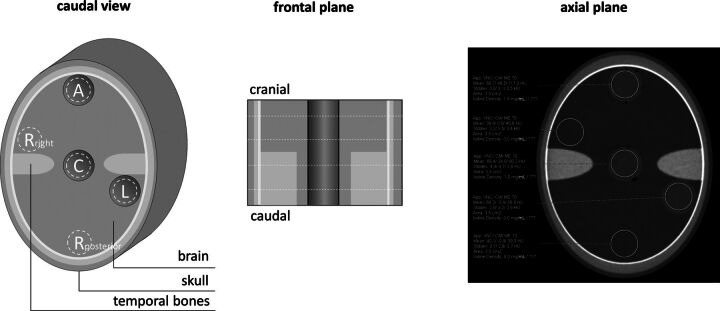
The schematic structure of the phantom and the respective positions of measurement. A indicates anterior; C, central; L, left; Rposterior, posterior reference; Rright, right reference).
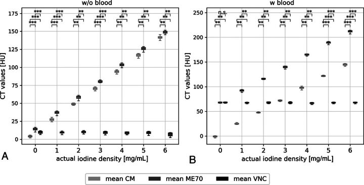
Measured CT values within the samples without (A) and with (B) blood, at all positions (anterior, center, left) and on all slices (next and above the temporal bone structures) presented in boxplots. CT values are compared among ME70, the CM map, and VNC. Statistically significant differences are marked. n.s. indicates P > . 05; *, P < . 05; **, P < . 01; ***, P < . 001).
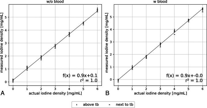
Linear regression of initial and measured iodine concentration of the samples. Reference samples without (A) and with (B) blood including all positions (anterior, center, left) and the compared between-sections above and next to temporal bone (tb) structures.
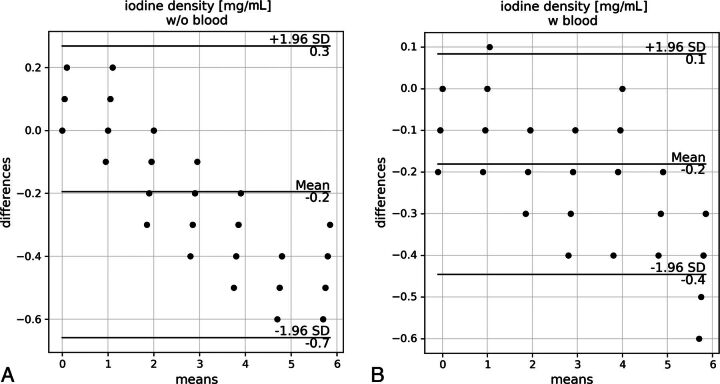
Bland-Altman plots showing the means and differences between the measured and actual iodine concentrations in samples without (A) and with (B) blood. All positions (anterior, center, and left) and sections (above and next to temporal bone structures) are considered. W/o indicates without; w, with.
Similar articles
-
Feasibility of multi-contrast imaging on dual-source photon counting detector (PCD) CT: An initial phantom study.Med Phys. 2019 Sep;46(9):4105-4115. doi: 10.1002/mp.13668. Epub 2019 Jul 5. Med Phys. 2019. PMID: 31215659 Free PMC article.
-
Evaluating spectral performance for quantitative contrast-enhanced breast CT with a GaAs based photon counting detector: a simulation approach.Biomed Phys Eng Express. 2024 Jul 17;10(5). doi: 10.1088/2057-1976/ad5f96. Biomed Phys Eng Express. 2024. PMID: 38968931
-
Photon-Counting Detector CT: Potential for 75% Reduction in Contrast Medium Amount: A Phantom Study.Acta Med Okayama. 2024 Apr;78(2):135-142. doi: 10.18926/AMO/66916. Acta Med Okayama. 2024. PMID: 38688831
-
Clinical applications of photon counting detector CT.Eur Radiol. 2023 Aug;33(8):5309-5320. doi: 10.1007/s00330-023-09596-y. Epub 2023 Apr 5. Eur Radiol. 2023. PMID: 37020069 Free PMC article. Review.
-
[CT technology: photon-counting detector computed tomography].Radiologie (Heidelb). 2023 Jul;63(7):497-506. doi: 10.1007/s00117-023-01166-z. Epub 2023 Jun 8. Radiologie (Heidelb). 2023. PMID: 37289254 Free PMC article. Review. German.
Cited by
-
Establishing a Foundation for the In Vivo Visualization of Intravascular Blood with Photon-Counting Technology in Spectral Imaging in Cranial CT.Diagnostics (Basel). 2024 Jul 19;14(14):1561. doi: 10.3390/diagnostics14141561. Diagnostics (Basel). 2024. PMID: 39061698 Free PMC article.
-
Photon counting CT vs. flat-panel CT in the evaluation of enhancement patterns in chronic subdural hematoma after middle meningeal artery embolization.Front Neurol. 2025 May 16;16:1608308. doi: 10.3389/fneur.2025.1608308. eCollection 2025. Front Neurol. 2025. PMID: 40452768 Free PMC article.
-
In Vivo Discrimination of Iodine and Tantalum-Based Liquid Embolics After Intracranial or Spinal Embolization Using Photon-Counting Detector CT.Clin Neuroradiol. 2025 Feb 6. doi: 10.1007/s00062-025-01502-x. Online ahead of print. Clin Neuroradiol. 2025. PMID: 39915306
-
Assessing anemia in stroke patients through virtual non-contrast imaging with photon-counting detector CT: validation on supra-aortic vessel CT-Angiography.Neuroradiology. 2025 Apr 24. doi: 10.1007/s00234-025-03620-2. Online ahead of print. Neuroradiology. 2025. PMID: 40272466
References
-
- Eskey CJ, Meyers PM, Nguyen TN, et al. ; American Heart Association Council on Cardiovascular Radiology and Intervention and Stroke Council. Indications for the performance of intracranial endovascular neurointerventional procedures: a scientific statement from the American Heart Association. Circulation 2018;137:e661–89 10.1161/CIR.0000000000000567 - DOI - PubMed
MeSH terms
Substances
LinkOut - more resources
Full Text Sources