. 2024 Jan 11;11(1):ENEURO.0315-23.2023.
doi: 10.1523/ENEURO.0315-23.2023. Print 2024 Jan.

Striatal Neurons Are Recruited Dynamically into Collective Representations of Self-Initiated and Learned Actions in Freely Moving Mice

Affiliations

Striatal Neurons Are Recruited Dynamically into Collective Representations of Self-Initiated and Learned Actions in Freely Moving Mice

Lior Tiroshi et al. eNeuro. .

Abstract

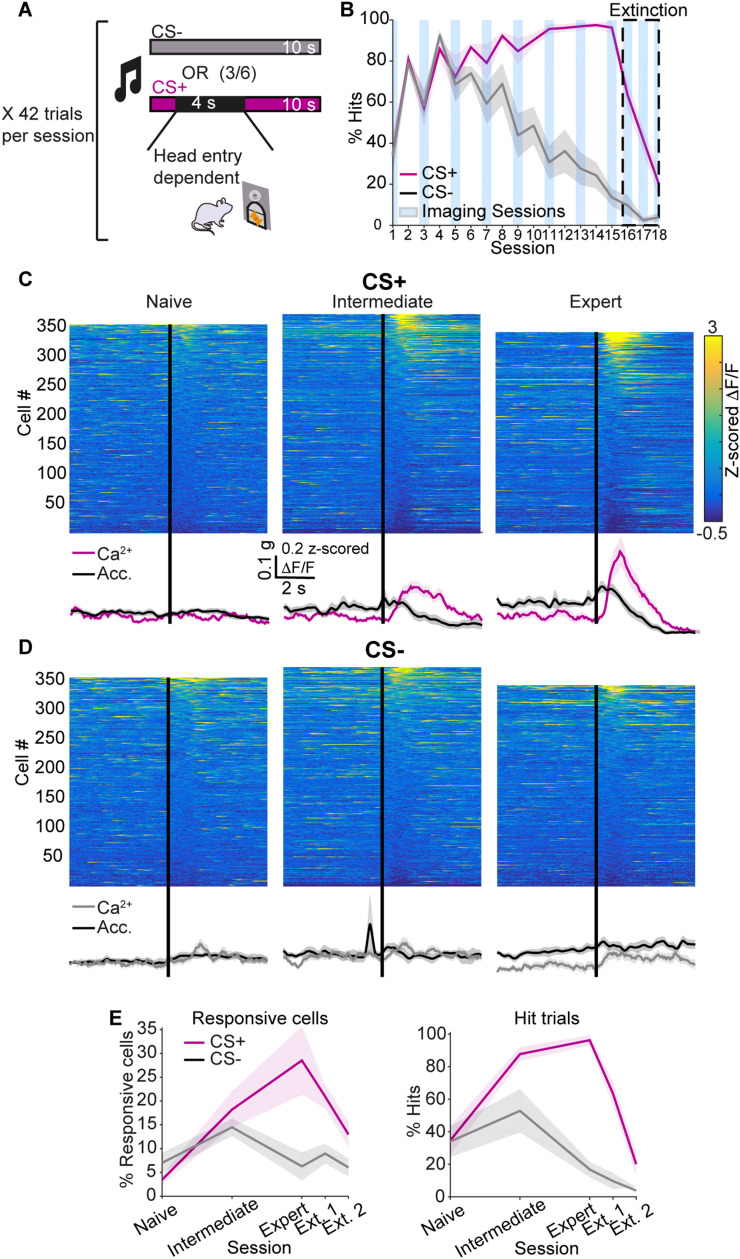
Striatal spiny projection neurons are hyperpolarized-at-rest (HaR) and driven to action potential threshold by a small number of powerful inputs-an input-output configuration that is detrimental to response reliability. Because the striatum is important for habitual behaviors and goal-directed learning, we conducted a microendoscopic imaging in freely moving mice that express a genetically encoded Ca2+ indicator sparsely in striatal HaR neurons to evaluate their response reliability during self-initiated movements and operant conditioning. The sparse expression was critical for longitudinal studies of response reliability, and for studying correlations among HaR neurons while minimizing spurious correlations arising from contamination by the background signal. We found that HaR neurons are recruited dynamically into action representation, with distinct neuronal subsets being engaged in a moment-by-moment fashion. While individual neurons respond with little reliability, the population response remained stable across days. Moreover, we found evidence for the temporal coupling between neuronal subsets during conditioned (but not innate) behaviors.

Keywords: basal ganglia; calcium imaging; correlations; parvalbumin-positive fast-spiking interneurons; population coding; spiny projection neurons.


Figures

Figure 1.
Figure 1.
The collective neuronal activity is strongly modulated by self-initiated movement in freely moving mice. A, A 1-mm-diameter GRIN lens is implanted into the dorsolateral striatum. B, Implanted mouse with a microendoscope and an accelerometer mounted on its head moves freely in a behavior chamber. C, Image via lens in freely moving mouse reveals signals from dozens of striatal neurons (see Fig. 1-1 for distribution of striatal subtypes). D, Ca2+ signals of two individual neurons (top, green), the average Ca2+ signal across all imaged neurons (56 neurons, middle, green) and the simultaneously recorded total body acceleration of a representative mouse (bottom, black). The two individual neuron exhibited were chosen because they were both shown to respond significantly to changes in the acceleration: with the top one reducing its Ca2+ signal and bottom one increasing its Ca2+ signal. E, Color-coded matrix showing activity around movement onset (Fig. 1-2), averaged across all movement onset events (top). Average Ca2+ activity across the population of imaged neurons and average total body acceleration across movement onset events are represented by green and black traces, respectively (middle). PSTH of Ca2+ events centered around the movement onset (bottom). Bin width: 67 ms. See Fig. 1-3 for description of positively and negatively modulated neurons. F, Same as E but around movement offset. G, Rates of detected neurons that did (green) or did not (white) significantly modulate their Ca2+ signals following movement onset (Fig. 1-2). Shaded areas represent S.E.M. g in scale bar is the gravitational constant 9.81 m/s2.
Figure 2.
Figure 2.
Responses to task-related cues develop with training. A, Mice were trained in an operant conditioning paradigm to associate an auditory CS with a sucrose–water mixture reward. Each session consisted of 42 trails. In each trial, a cue (CS+ or CS−) was presented for 10 s in a pseudo-random manner. If the mouse entered its head into a designated port during the 10 s time window following CS+ (but not CS−) onset, reward was available for 4 s. B, The learning rate quantified as the portion of cue presentations during which the mouse entered its head into the port (% hits). Blue rectangles mark imaging sessions and the dashed box marks extinction sessions. C, Color-coded matrix showing neuronal activity around CS+ presentation averaged across all CS+ presentation events for naive (left), intermediate level (middle) and expert (right) mice. Average Ca2+ activity across the population of imaged neurons and average total body acceleration across CS+ presentation events are represented by pink and black traces, respectively (bottom). See Fig. 2-1 for the development of the total body acceleration profile around CS+ presentation as training progresses. D, Same as C, for CS− presentation. Average Ca2+ activity across the population is represented by gray traces. E, Rate of responsive neurons (left) and “hit” rate (right, same data as in panel B) around CS+ (pink) and CS− (gray) presentation on various stages of training. Shaded areas represent the S.E.M. g in scale bar is the gravitational constant 9.81 m/s2. See Fig. 2-2 for the relationship between the neural responses around CS+ presentation and total body acceleration or reward delivery.
Figure 3.
Figure 3.
Responses around movement onset and cue presentation are dynamic across imaging sessions. A, Four example cells that were detected on 3 imaging sessions. The images on the bottom represent the footprints produced by CNMF-E of one example cell across the various sessions. B, Average Ca2+ signal of an example neuron on the first (top) and second (bottom) free movement imaging sessions that exhibited significant responses (according to our bootstrapping criterion, see Materials and Methods) around movement onset on both sessions. C, Same as A, around CS+ presentation. D, Percentage of neurons significantly responsive on the first (light green) or second (medium green) free movement imaging sessions, both of them (dark green) or neither (white). See Table 3-2 for the time between the two free movement sessions for each mouse. E, Percentage of neurons from mouse 1 (left) and mouse 2 (right) significantly responsive on the first (light green) or second (medium green) free movement imaging sessions, both of them (dark green) or neither (white), that were at most 3 d apart. F, Percent of neurons significantly responsive on one (light), two (medium) or three (dark) advanced conditioning sessions around CS+ (left, pink) and CS− (right, gray) presentation. See Fig. 3-1 for analysis of the reliability around CS+ presentation within a single session. G, PDF of the percent of movement onset (green) and CS+ presentation (pink) events after which the neuron produced a Ca2+ event for positively modulated neurons. H, PDF of the percent of neurons that responded with a significant Ca2+ transient following each movement onset (green) of CS+ presentation (pink). I, Examples of neurons exhibiting significant and nonsignificant responses around movement onset and CS+ presentation. Cell 1 responded significantly only to CS+ presentation. Cell 2 responded significantly only to movement onset. Cell 3 responded significantly to both. J, Percentage of neurons significantly responsive around movement onset (green), CS+ presentation (pink), both (dark purple) or neither (white) out of the neurons that were detected both in a free movement session and in an advanced conditioning session.
Figure 4.
Figure 4.
Responses around ipsi- and contralateral turns and around grooming are dynamic across imaging sessions. A, Color-coded matrix showing neuronal activity around the onset of contra- (left) and ipsilateral (right) turns averaged across all turning events for all mice. Average Ca2+ activity across the population of imaged neurons is represented by green and purple traces for contra- and ipsilateral turns, respectively. The average total body acceleration across the relevant turning events are represented by black traces (bottom). B, Same as A, around grooming initiation. C, Percentage of positively (light) and negatively (dark) modulated neurons out of the neurons significantly responsive around contra- (green) and ipsilateral (purple) turns on the two free movement sessions. D, Percentage of neurons significantly responsive around ipsilateral turns only (purple), contralateral turns only (green), both turning direction (gray) or neither (white) on the two free movement sessions. E, PDF of the percent of contra- (green) and ipsilateral (purple) turns and grooming initiation (orange) events after which the neuron produced a Ca2+ event for positively modulated neurons. F, Average Ca2+ signal of an example neuron on the first (top) and second (bottom) free movement imaging sessions that exhibited a significant response around contralateral turns on the second session but not on the first. G, Same as F, for ipsilateral turns. H, Same as F, for grooming initiation. I, Percentage of neurons significantly responsive around contralateral turns (green), ipsilateral turns (purple) and grooming initiations (orange) on the first (light) or second (medium) free movement session only, both (dark) or neither (white) out of the neurons that were detected on both sessions. g in scale bar is the gravitational constant 9.81 m/s2.
Figure 5.
Figure 5.
Pairwise correlations are not distance dependent. A, Pairwise correlations as a function of the distance between the neurons’ centers for all pairs of co-imaged neurons during rest. Each point is the average correlation across all neuronal pairs belonging to the relevant distance bin. Dashed line is the linear fit to the data points. B, Same as A, divided into positively (left) and negatively (right) modulated pairs. C, Same as (A), around movement onset. D, Same as (B), during movement. E, Distribution of neuronal pairs for the various distances for all pairs (left), positively (middle) and negatively modulated pairs (right). Bin width is 10 µm. F, Same as (B), around CS+ presentation on intermediate training session. G, Same as (F), divided into positively (left) and negatively (right) modulated pairs. H, Same as (F), on expert training session. I, Same as (G), on expert training session. See Fig. 5-1 for analysis of the correlations in the microendoscopy background neuropil signal and their dependence on distance.
Figure 6.
Figure 6.
Behavioral variability more strongly affects trial-by-trial correlations around spontaneous movement than during learned behaviors. A, Population joint JPSTH, averaged across all neuronal pairs, centered around movement onset (top). Bar plot shows values of the JPSTH diagonal (bottom). Solid red line represents the mean value of the JPSTH diagonal in a 10 s time window far removed from movement onset events. Dashed red lines represent the mean ± the SD. B, Same as (A), for pairs of positively modulated neurons. C, Same as (B), for contralateral turns. D, Same as (B), for ipsilateral turns. E, Same as (B), for movement onset events with a small acceleration change. Thick lines mark the time windows for calculation of baseline (black) and post-event (red) maximal values. F, Same as (B), for movement onset events with a small acceleration change. (G) Boxplot of baseline (black) and post-event (red) maximal values of population JPSTH diagonals around movement onset for small acceleration change movements. Each circle represents one mouse (N = 6 mice). H, Same as (G), for large acceleration change movements. I Boxplot of the difference between the maximal value of the population JPSTH diagonal in the post-event and baseline time windows for movement onset with small (left) and large (right) acceleration changes. The bold line is the median and the whiskers are the 25th and 75th percentiles. Red crosses represent outliers. J, Same as (B), for CS+ presentations on expert training session (top). K, Same as (J), for first 10 trials of the first extinction session. L, Same as (E), for CS+ presentations. M, Same as (F), for CS+ presentations. N, Same as (G), for CS+ presentations (N = 5 mice). O, Same as (H), for CS+ presentations. P, Same as (I), for CS+ presentations. The bold line is the median and the whiskers are the 25th and 75th percentiles. Red crosses represent outliers.

Similar articles

References

    1. Aertsen AMHJ, Gerstein GL, Habib MK, Palm G (1989) Dynamics of neuronal firing correlation: modulation of “effective connectivity”. J Neurophysiol 61:900–917. - PubMed
    1. Albin RL, Young AB, Penney JB (1989) The functional anatomy of basal ganglia disorders. Trends Neurosci 12:366–375. 10.1016/0166-2236(89)90074-X - DOI - PubMed
    1. Alexander GE, Crutcher MD (1990) Functional architecture of basal ganglia circuits: neural substrates of parallel processing. Trends Neurosci 13:266–271. 10.1016/0166-2236(90)90107-L - DOI - PubMed
    1. Aosaki T, Kimura M, Graybiel AM (1995) Temporal and Spatial Characteristics of Tonically Active Neurons of the Primate’s Striatum.
    1. Aosaki T, Tsubokawa H, Ishida A, Watanabe K, Graybiel AM, Kimura M (1994) Responses of tonically active neurons in the primate’s striatum undergo systematic changes during behavioral sensorimotor conditioning. J Neurosci 14:3969–3984. 10.1523/JNEUROSCI.14-06-03969.1994 - DOI - PMC - PubMed

LinkOut - more resources