. 2023 Dec 12;11(12):e007490.
doi: 10.1136/jitc-2023-007490.

Discovery of U2AF1 neoantigens in myeloid neoplasms

Affiliations

Discovery of U2AF1 neoantigens in myeloid neoplasms

Melinda Ann Biernacki et al. J Immunother Cancer. .

Abstract

Background: Myelodysplastic syndromes (MDS) arise from somatic mutations acquired in hematopoietic stem and progenitor cells, causing cytopenias and predisposing to transformation into secondary acute myeloid leukemia (sAML). Recurrent mutations in spliceosome genes, including U2AF1, are attractive therapeutic targets as they are prevalent in MDS and sAML, arise early in neoplastic cells, and are generally absent from normal cells, including normal hematopoietic cells. MDS and sAML are susceptible to T cell-mediated killing, and thus engineered T-cell immunotherapies hold promise for their treatment. We hypothesized that targeting spliceosome mutation-derived neoantigens with transgenic T-cell receptor (TCR) T cells would selectively eradicate malignant cells in MDS and sAML.

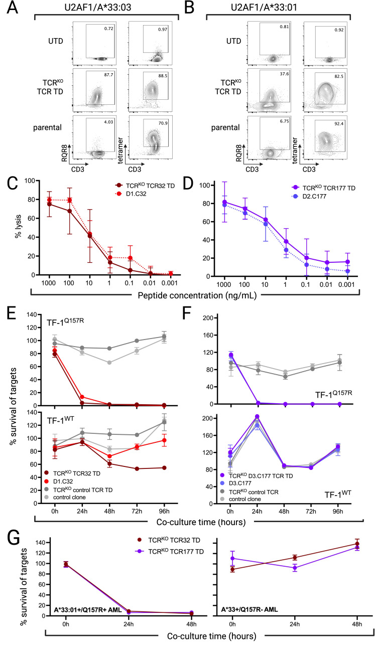
Methods: We identified candidate neoantigen epitopes from recurrent protein-coding mutations in the spliceosome genes SRSF2 and U2AF1 using a multistep in silico process. Candidate epitopes predicted to bind human leukocyte antigen (HLA) class I, be processed and presented from the parent protein, and not to be subject to tolerance then underwent in vitro immunogenicity screening. CD8+ T cells recognizing immunogenic neoantigen epitopes were evaluated in in vitro assays to assess functional avidity, confirm the predicted HLA restriction, the potential for recognition of similar peptides, and the ability to kill neoplastic cells in an antigen-specific manner. Neoantigen-specific TCR were sequenced, cloned into lentiviral vectors, and transduced into third-party T cells after knock-out of endogenous TCR, then tested in vitro for specificity and ability to kill neoplastic myeloid cells presenting the neoantigen. The efficacy of neoantigen-specific T cells was evaluated in vivo in a murine cell line-derived xenograft model.

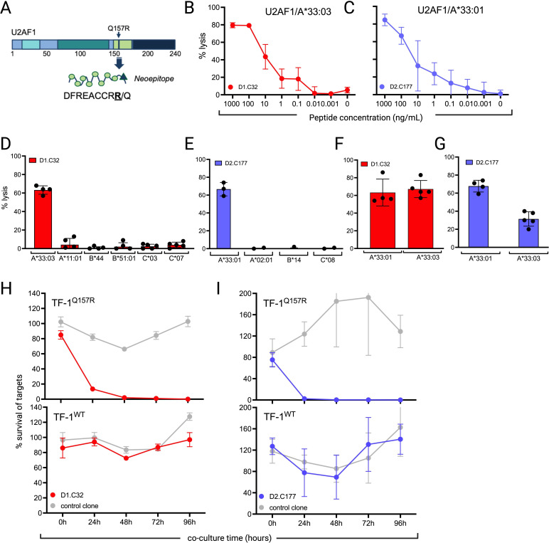
Results: We identified two neoantigens created from a recurrent mutation in U2AF1, isolated CD8+ T cells specific for the neoantigens, and demonstrated that transferring their TCR to third-party CD8+ T cells is feasible and confers specificity for the U2AF1 neoantigens. Finally, we showed that these neoantigen-specific TCR-T cells do not recognize normal hematopoietic cells but efficiently kill malignant myeloid cells bearing the specific U2AF1 mutation, including primary cells, in vitro and in vivo.

Conclusions: These data serve as proof-of-concept for developing precision medicine approaches that use neoantigen-directed T-cell receptor-transduced T cells to treat MDS and sAML.

Keywords: Antigens, Neoplasm; Hematologic Neoplasms; Immunotherapy, Adoptive.


Conflict of interest statement

Competing interests: MB is a Founder and Scientific Advisory Board member of HighPassBio, and a Scientific Advisory Board member of Orca Bio, and has also received compensation from Miltenyi Biotec for presentations at conferences and corporate symposia pertaining to research unrelated to the current manuscript. MB and MAB have filed a provisional patent application number 63/274,681 covering applications of T cell immunotherapy for U2AF1-mutated malignancies.

Figures

Figure 1
Figure 1
Two candidate neoantigen epitopes are predicted from recurrent myelodysplastic syndromes-associated and secondary acute myeloid leukemia-associated U2AF1 mutations. (A) Schematic representation of the workflow used for in silico identification of candidate neoantigens. wt, wild-type. (B) Predictions were made using IEDB Stabilized Matrix Method and Artificial Neural Network methods, netMHCpan 4.1, and netCTLpan and summarized graphically. Predicted HLA binders are shown in green. Predicted processed and presented peptides are shown in blue. Epitopes for which the corresponding wild-type peptide is not predicted to bind HLA are in bright green and denoted with an asterisk. Candidate epitopes selected for further study (defined as having predicted IC50 was <250 nM or rank <1 by at least two methods, processing/prediction rank <1%, and no predicted HLA binding of the equivalent wild-type peptide) are shown in bright blue with double asterisk. HLA, human leukocyte antigen; IEDB, Immune Epitope Database.
Figure 2
Figure 2
A U2AF1Q157R HLA-A*33-restricted neoantigen epitope is immunogenic and primes epitope-specific CD8+ T-cell clones. (A) Schematic of U2AF1Q157R protein and neoantigen epitope peptide. (B and C) One clone from an HLA-A*33:03-positive donor (B D1.C32) and one clone from an HLA-A*33:01-positive donor (C D2.C177) were each identified after primary in vitro stimulation of CD8+ T cells with DFREACCRR peptide, then tested for functional avidity in peptide titration CRA using autologous LCL pulsed with varying peptide concentrations (three technical replicate experiments). (D and E) HLA restriction of clone D1.C32 from HLA-A*33:03+ donor (D) and clone D2.C177 from HLA-A*33:01+ donor (E) were confirmed by testing in CRA against a panel of HLA-typed LCL with single HLA overlap with the original T-cell donor. LCL were pulsed with DFREACCRR peptide at 1000 ng/mL prior to co-culture. (F) Clone D1.C32 lyses DFREACCRR peptide-pulsed (1000 ng/mL) HLA-A*33:01+ as well as A*33:03+ LCL. (G) Clone D2.C177 lyses HLA-A*33:03+ as well as A*33:01+ LCL pulsed with 1000 ng/mL DFREACCRR peptide. (H and I) Per cent survival of TF-1 minigene-transduced cells co-cultured with either D1.C32 (H red), D2.C177 (I blue) or irrelevant neoantigen-specific clone (gray) in flow cytometry-based cytotoxicity assay. For (H and I) top plots, U2AF1Q157R minigene; bottom plots, U2AF1 minigene. Mean and SEM from >3 technical replicates shown. CRA, Cr-release cytotoxicity assays; HLA, human leukocyte antigen; LCL, lymphoblastoid cell lines.
Figure 3
Figure 3
U2AF1Q157R/A*33-specific clones do not recognize similar peptides. (A) Alanine scanning for U2AF1Q157R/A*33:03-specific clone D1.C32 was performed using autologous LCL pulsed with a panel of peptides (1000 ng/mL) with alanine residues substituted at each position, along with two peptides with either a glycine or valine substitution at position five, a natural alanine residue in the DFREACCRR peptide (three technical replicate experiments). These data were used to identify critical residues for HLA and TCR binding and define the core motifs DxxExCCRR. (B) Peptides derived from wild-type human proteins and sharing the core motifs for the HLA-A*33:03-restricted clones clone were identified using the ScanProsite tool. To evaluate for cross-recognition of these peptides by clones, autologous LCL were pulsed with each peptide (1000 ng/mL) and used as targets for clones in CRA (three technical replicate experiments). (C) Alanine scanning for U2AF1Q157R/A*33:01-specific clone D2.C177 identified critical residues for HLA and TCR binding identified, defining the core motif xxxExCCxR for this clone. (D) Human peptides sharing the core motif were identified as using ScanProsite and clone D2.C177 tested for their recognition in CRA using autologous LCL pulsed with each peptide (1000 ng/mL) as targets. For all experiments, mean and SEM are shown. CRA, Cr-release cytotoxicity assays; HLA, human leukocyte antigen; LCL, lymphoblastoid cell lines; TCR, T-cell receptor.
Figure 4
Figure 4
Transfer of U2AF1Q157R neoantigen-specific TCR confers specificity and function. (A) Representative flow plots demonstrating expression of D1.C32 U2AF1Q157R/A*33:03-specific TCR (TCR32) transduced (TD) into primary human CD8+ T cells after CRISPR/Cas9-mediated knock-out of endogenous TCR alpha and beta chains (TCRko). Staining for the RQR8 transduction marker (CD34), left, and U2AF1Q157R/A*33:03-pHLA tetramer, right. (B) Representative flow plots demonstrating expression of clone D2.C177 U2AF1Q157R/A*33:01-specific TCR (TCR177) TD into TCRko primary human CD8+ T cells. CD34 staining, left, and U2AF1Q157R/A*33:03-pHLA tetramer, right. For A and B top, untransduced (UTD); middle, TCR TD; bottom, parental clone. (C) Testing of TCR32 TD T cells (dark red, solid) with parental clone control (light red, dashed) and (D) testing of TCR177 TD T cells (dark purple, solid) and parental clone control (light purple, dashed), for specific lytic activity in peptide titration CRA. For both C and D technical triplicates shown. (E) Per cent survival of TF-1 minigene-transduced cells co-cultured with TCR32 TD T cells (dark red) or parental clone control (light red) in flow cytometry-based cytotoxicity assay. (F) Per cent survival of TF-1 minigene-transduced cells co-cultured with TCR177 TD T cells (dark purple) or parental clone control (light purple) in flow cytometry-based cytotoxicity assay. For both E and F >3 technical replicates; top plot, U2AF1Q157R minigene TD TF-1; bottom plots, wild-type (WT) U2AF1 minigene TD TF-1. (G) Per cent survival of U2AF1Q157R/ HLA-A*33:01+ primary AML (left) or wild-type U2AF1 (Q157R-)/HLA-A*33:03+ primary AML (right) co-cultured with either TCR32 TD T cells (red) or TCR177 TD T cells (purple) in flow cytometry-based cytotoxicity assay. Data from >3 technical replicates. Mean and SEM are shown. AML, acute myeloid leukemia; HLA, human leukocyte antigen; pHLA, peptide-HLA; TCR, T-cell receptor.
Figure 5
Figure 5
U2AF1Q157 neoantigen-specific TCR-T cells control myeloid neoplasms in vivo. (A) Experimental schematic. After sublethal irradiation, immunodeficient NSG-SGM3 mice were injected intravenous with naturally HLA-A*33:03-positive TF-1 cells transduced with a U2AF1Q157 minigene, allowed to engraft for 5 days, then injected with either U2AF1Q157R/A*33:03-specific TCR-T cells (TCR32), irrelevant neoantigen-specific control TCR-T cells, or vehicle. A fourth group was engrafted with TF-1 cells transduced with a wild-type U2AF1 minigene, then treated with U2AF1Q157R/A*33:03-specific TCR-T cells (n=7 per group). (B) Serial bioluminescence imaging from mice engrafted with U2AF1 minigene-transduced TF-1 cells before and at time points after T-cell injection. (C) Quantitation of bioluminescence flux (p/s) in animals in each group at serial time points. Pairwise comparisons (t-tests) were performed on the ranked data between groups. (D) Kaplan-Meier survival curve (Mantel-Cox log-rank test). (E) Summary of minigene expression in retrieved TF-1 cells in terminal bone marrow as indicated by total absolute numbers of CD20 staining in treatment groups. (Mann-Whitney test, **, p value<0.005; ***, p value<0.001). In B–E animals engrafted with U2AF1Q157R minigene-transduced TF-1 cells and treated with TCR32 T cells, solid red; engrafted with U2AF1Q157R TD TF-1 and treated with control TCR-T cells, gray; engrafted with U2AF1Q157R TD TF-1 and treated with vehicle, black; engrafted with wild-type U2AF1 minigene-transduced TF-1 cells and treated with U2AF1Q157R/A*33:03-specific TCR-T cells, dashed/open red. TCR-T, T-cell receptor-transduced T cell; WT, wild-type.

References

    1. Ogawa S. Genetics of MDS. Blood 2019;133:1049–59. 10.1182/blood-2018-10-844621 - DOI - PMC - PubMed
    1. Sperling AS, Gibson CJ, Ebert BL. The Genetics of myelodysplastic syndrome: from clonal haematopoiesis to secondary leukaemia. Nat Rev Cancer 2017;17:5–19. 10.1038/nrc.2016.112 - DOI - PMC - PubMed
    1. Estey EH. Acute myeloid leukemia: 2021 update on risk-stratification and management. Am J Hematol 2020;95:1368–98. 10.1002/ajh.25975 - DOI - PubMed
    1. Kolb HJ. Graft-versus-leukemia effects of transplantation and donor lymphocytes. Blood 2008;112:4371–83. 10.1182/blood-2008-03-077974 - DOI - PubMed
    1. Biernacki MA, Brault M, Bleakley M. T-cell receptor-based immunotherapy for hematologic malignancies. Cancer J 2019;25:179–90. 10.1097/PPO.0000000000000378 - DOI - PMC - PubMed

Publication types

MeSH terms