. 2024 Apr 12;52(6):3310-3326.
doi: 10.1093/nar/gkad1244.

Dissolution of ribonucleoprotein condensates by the embryonic stem cell protein L1TD1

Affiliations

Dissolution of ribonucleoprotein condensates by the embryonic stem cell protein L1TD1

Sang Woo Jin et al. Nucleic Acids Res. .

Abstract

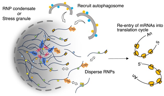
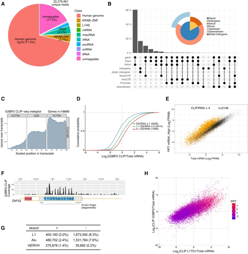
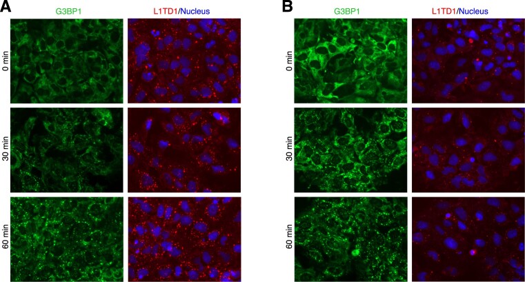
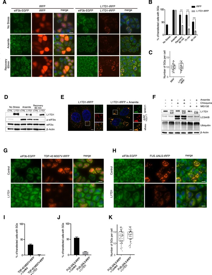
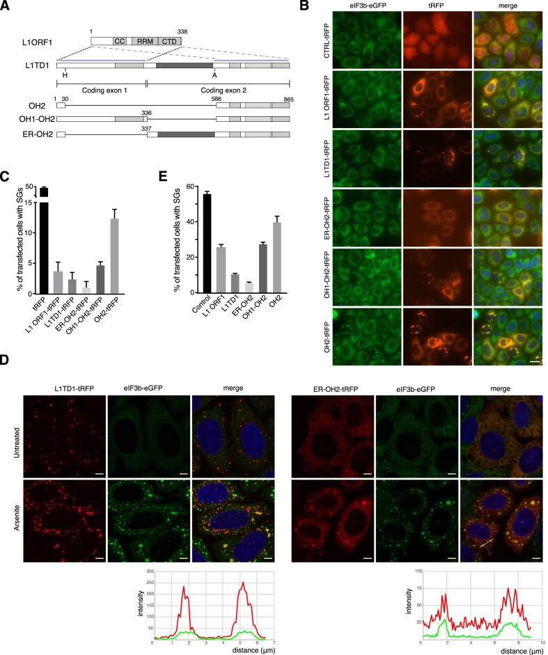
L1TD1 is a cytoplasmic RNA-binding protein specifically expressed in pluripotent stem cells and, unlike its mouse ortholog, is essential for the maintenance of stemness in human cells. Although L1TD1 is the only known protein-coding gene domesticated from a LINE-1 (L1) retroelement, the functional legacy of its ancestral protein, ORF1p of L1, and how it is manifested in L1TD1 are still unknown. Here, we determined RNAs associated with L1TD1 and found that, like ORF1p, L1TD1 binds L1 RNAs and localizes to high-density ribonucleoprotein (RNP) condensates. Unexpectedly, L1TD1 enhanced the translation of a subset of mRNAs enriched in the condensates. L1TD1 depletion promoted the formation of stress granules in embryonic stem cells. In HeLa cells, ectopically expressed L1TD1 facilitated the dissolution of stress granules and granules formed by pathological mutations of TDP-43 and FUS. The glutamate-rich domain and the ORF1-homology domain of L1TD1 facilitated dispersal of the RNPs and induced autophagy, respectively. These results provide insights into how L1TD1 regulates gene expression in pluripotent stem cells. We propose that the ability of L1TD1 to dissolve stress granules may provide novel opportunities for treatment of neurodegenerative diseases caused by disturbed stress granule dynamics.


Figures

Graphical Abstract
Graphical Abstract
Figure 1.
Figure 1.
L1TD1 binds L1 RNAs and mRNAs in H1 hESCs. (A) Distribution of L1TD1-CLIP-seq reads on the genome (UCSC reference genome hg19). L1TD1 rarely binds abundant noncoding RNAs, such as rRNAs, tRNAs, snRNAs, snoRNAs and miRNAs. L1HS denotes sense strand transcripts of the L1 human specific. (B) L1TD1 binds transcripts from genic regions and noncoding RNAs from intergenic regions. The UpSet plot and the venn pie diagram show fractions of reads mapping on the annotated genomic regions. (C) L1TD1 interacted with sense strand RNAs of L1 elements. The number of L1TD1 CLIP-seq reads for each retroelement is shown. (D) L1TD1-CLIP-seq reads are mapped on broad regions of L1HS RNAs encoding ORF1p and ORF2p. Alignments on repetitive sequences do not represent the actual binding of L1TD1. (E) Reads from L1TD1 CLIP libraries aligned to ZNF92 mRNA sequence allowing unique, first-match, or multiple mapping. No mismatches were allowed. Interactions between L1TD1 and KRAB-ZNF mRNAs are apparently confined to regions encoding zinc finger motifs that are not degenerate (blue rectangles). (F) Metagene profile analysis of CLIP-seq reads. L1TD1 preferentially binds 3′UTRs of mRNAs.
Figure 2.
Figure 2.
L1TD1 affects association of mRNAs with ribosomes in hESCs. (A) Expression levels of mRNAs are not affected by L1TD1 depletion. The scatter plot shows FPKMs of total mRNAs in control (siCtrl) and L1TD1-depleted H1 cells (siL1TD1). (B) The association of some mRNAs with ribosomes is reduced by L1TD1 depletion. The scatter plot shows FPKMs of ribosome-associated mRNAs of siCtrl- and siL1TD1-treated cells that were enriched by immunoprecipitation using an anti-5.8S rRNA antibody. Red dots indicate mRNAs for which L1TD1 increased association with ribosomes by at least 4-fold. (C) Gene ontology analysis of mRNAs, the association of which with ribosomes was enhanced by L1TD1. Top 20 significantly enriched KEGG pathways, ranked by p-values in -log10 scale were depicted in a bar plot. The numbers of enriched genes were denoted beside the bars. (D) L1TD1 binding and reduction of ribosome association by L1TD1 depletion are correlated. The cumulative distribution function plot shows the mRNAs enriched by L1TD1-CLIP tend to be less associated with ribosomes in L1TD1-depleted cells. The log2 fold changes of ribosome IP were grouped by the enrichment in L1TD1-CLIP. (E, F) mRNAs with L1TD1-dependent ribosome association are related to mRNAs in the RNA granules of heterologous cells. Cumulative distributions of L1TD1-dependent enrichment of ribosome-associated mRNAs were plotted by the enrichment in P-bodies (PB) of HEK293 (E) and stress granules (SGs) of arsenite-treated U2OS (F). Analyses of HEK293 mRNAs and U2OS mRNAs were from the previous reports of Hubstenberger et al. and Khong et al. (42,43). P-values in (D–F) were calculated using the Mann–Whitney U test.
Figure 3.
Figure 3.
Disproportionate partitioning of mRNAs in hESCs. (A, B) SGs (A) or P-bodies (B) are not apparent in unstressed H1 cells. Arsenite treatment significantly induced the formation of L1TD1 foci that co-localize with an SG marker, G3BP1, and a P-body marker, DDX6. The cells were treated with 125 μM arsenite for 60 min. (C) Localization of RNA-binding proteins in RNP condensates. The total cytoplasmic extracts (Cyt) prepared with buffers containing either 50 or 200 mM of potassium chloride were fractionated by centrifugation. RNA binding proteins in the cytoplasm (Cyt), the supernatant (SN) and the precipitate (PPT) were analyzed by western blot. PPT was dissolved in one fourth of the initial volume of Cyt. L22 and S6 are proteins in the large and small ribosomal subunit, respectively. (D) mRNAs are disproportionately localized in the PPT. FPKMs of mRNAs in the total cytoplasmic extract are correlated with FPKMs of mRNAs in the high-salt precipitate. (E) Similar mRNA species are enriched in the precipitates prepared with different salt concentration. (F, G) Enrichment in the precipitate is correlated with the U2OS mRNAs enriched in SGs (F) and the HEK293T mRNAs enriched in P-bodies (G). (H) Gene length affects the partitioning of mRNAs. Longer mRNAs are enriched in the precipitates, as are SG mRNAs. (I) L1TD1-bound RNAs are enriched in RNP condensates. mRNAs enriched more than 10-fold (RPKM of L1TD1 CLIP-seq reads/FPKM of mRNA-seq reads) are depicted in orange in the scatter plots of expression levels of Cyt and PPT mRNAs. (J) L1TD1 prefers longer mRNAs. p-values calculated using the Mann–Whitney U test in all sets in (F–H, J) are less than 2.2 × 10−16. (K) Cumulative distributions of L1TD1-dependent enrichment of ribosome-associated mRNAs were plotted by the enrichment in PPT.
Figure 4.
Figure 4.
G3BP2 binding sites are different from L1TD1 binding sites. (A) Distribution of G3BP2-CLIP-seq reads on the genome. G3BP2 barely binds L1HS RNA and coding regions of KRAB-ZNF mRNAs. The reads mapping in the mRNAs encoding KRAB-ZNF are mostly located in the 3′UTRs. (B) G3BP2 binds transcripts from genic regions and noncoding RNAs from intergenic regions. The UpSet plot and the Venn pie diagram show read numbers mapping on the annotated genomic regions. (C) Metagene profile analysis of CLIP-seq reads. Metagene plots show that G3BP2 preferentially binds 3′UTRs of mRNAs. (D) Target mRNAs of G3BP2 in hESCs were enriched in SGs of U2OS cells. P-values calculated using the Mann–Whitney U test were less than 2.2 × 10−16. (E) G3BP2-bound RNAs are enriched in RNP condensates. mRNAs enriched by CLIP-seq were depicted Colored dots in the scatter plot of the total and the PPT mRNAs. (F) Reads from G3BP2-CLIP libraries aligned to the ZNF92 mRNA sequence with multiple mapping allowed. G3BP2 barely binds regions encoding zinc finger motifs (blue rectangles) of KRAB-ZNF proteins. (G) The preferred retroelements of G3BP2 are different from those of L1TD1. (H) L1TD1 and G3BP2 share targets. The enrichment in the precipitate (log2 FPKM ratio of PPT mRNA/ Cyt mRNA) was color-coded.
Figure 5.
Figure 5.
L1TD1 affects the dynamics of SG formation in H1 hESCs. Forty-eight hours after siRNA transfection, control (A) and L1TD1-depleted (B) cells were treated with 75 μM arsenite for the indicated times. The proportions of cells without discrete G3BP1 granules at 30 min were 16.23 ± 1.93% (A) and 4.93 ± 0.69% (B), respectively (P = 0.0007).
Figure 6.
Figure 6.
L1TD1 facilitates dissolution of SGs in HeLa. (A) HeLa eIF3B-GFP cells were transfected with plasmids expressing tRFP or L1TD1-tRFP. After 24 h, the cells were treated with 0.5 mM sodium arsenite for 15 mins, then allowed to recover for 60 min before fixation. (B) The percentage of cells with SGs (eGFP-foci) among tRFP-positive cells were counted. 100 tRFP-positive cells were counted from each triplicate. (C) The number of SGs per cell was counted after 60 min recovery from arsenite treatment. Fifty cells from each set were examined. (D) Phosphorylation of eIF2-α is reduced in cells expressing L1TD1. Note that L1TD1 decreased after arsenite treatment. (E) L1TD1 foci colocalize with an autophagy marker LC3. (F) Degradation of L1TD1 by arsenite treatment is dependent on autophagy. L1TD1-transfected cells were treated with 50μM chloroquine for 12 h or 10μM MG-132 for 4 h before treatment with arsenite. Numbers are average relative intensities of L1TD in arsenite-treated cells compared to control cells. The intensities in each lane were normalized by those of actin. The mitigated decrease by chloroquine-treatment is statistically significant (P= 0.038, Student's t-test). (G-J) L1TD1 facilitates dissolution of granules induced by the mutants of TDP-43 (G, I) and FUS (H, J) in HeLa cells. Scale bars indicate 5 μm in (D) and 25 μm, elsewhere. (K) The number of SGs per cell in (H) was counted. Fifty cells from each set were examined.
Figure 7.
Figure 7.
Requirement of the non-conserved extra domain of L1TD1 for efficient dissolution of SGs. (A) Schematic diagrams of L1 ORF1p and L1TD1. CC, RRM and CTD stand for the coiled-coil motifs, RNA-recognition motifs and the C-terminal domain, respectively. Dotted lines and pale blue lines show the first and the second ORF-homology domains (OH1 and OH2). Dark grey rectangles indicate the glutamate-rich domain (ER). H and A indicate the restriction enzymes HpaI and AflII, which were used to generate the deletion mutants. (B) Intracellular distribution of L1 ORF1p and L1TD1 deletion mutants fused to tRFP in HeLa eIF3B-GFP cells. (C) Cells with SGs among transfected cells in (B) were counted. (D) Dispersed distribution of the deletion mutant ER-OH2-tRFP. Intensity profiles across the dotted lines are shown at the bottom. Scale bars indicate 25 μm in (B) and 5 μm in (D). (E) The FUS-ΔNLS-tRFP plasmid was co-transfected with a plasmid expressing L1 ORF1p or one of the L1TD1 deletion mutants into HeLa eIF3B-GFP cells (Supplementary Figure S12). Cells with SGs among transfected cells were counted.

Similar articles

Cited by

References

    1. Mitsui K., Tokuzawa Y., Itoh H., Segawa K., Murakami M., Takahashi K., Maruyama M., Maeda M., Yamanaka S.. The homeoprotein nanog is required for maintenance of pluripotency in mouse epiblast and ES cells. Cell. 2003; 113:631–642. - PubMed
    1. Iwabuchi K.A., Yamakawa T., Sato Y., Ichisaka T., Takahashi K., Okita K., Yamanaka S.. ECAT11/L1td1 Is enriched in ESCs and rapidly activated during iPSCGeneration, but it Is dispensable for the maintenance and induction of pluripotency. PLoS One. 2011; 6:e20461. - PMC - PubMed
    1. Wong R.C.-B., Ibrahim A., Fong H., Thompson N., Lock L.F., Donovan P.J.. L1TD1 Is a marker for undifferentiated Human embryonic stem cells. PLoS One. 2011; 6:e19355. - PMC - PubMed
    1. Närvä E., Rahkonen N., Emani M.R., Lund R., Pursiheimo J.-P., Nästi J., Autio R., Rasool O., Denessiouk K., Lähdesmäki H.et al. .. RNA-binding protein L1TD1 interacts with LIN28 via RNA and is required for Human embryonic stem cell self-renewal and cancer cell proliferation. Stem Cells. 2012; 30:452–460. - PMC - PubMed
    1. Santos M.C.T., Silva P.B.G., Rodini C.O., Furukawa G., Marco Antonio D.S., Zanotto-Filho A., Moreira J.C.F., Okamoto O.K.. Embryonic stem cell-related protein L1TD1 is required for cell viability, neurosphere formation, and chemoresistance in medulloblastoma. Stem Cells Dev. 2015; 24:2700–2708. - PubMed