. 2024 Mar;99(3):336-349.
doi: 10.1002/ajh.27184. Epub 2024 Jan 2.

Newly identified roles for PIEZO1 mechanosensor in controlling normal megakaryocyte development and in primary myelofibrosis

Affiliations

Newly identified roles for PIEZO1 mechanosensor in controlling normal megakaryocyte development and in primary myelofibrosis

Vittorio Abbonante et al. Am J Hematol. 2024 Mar.

Abstract

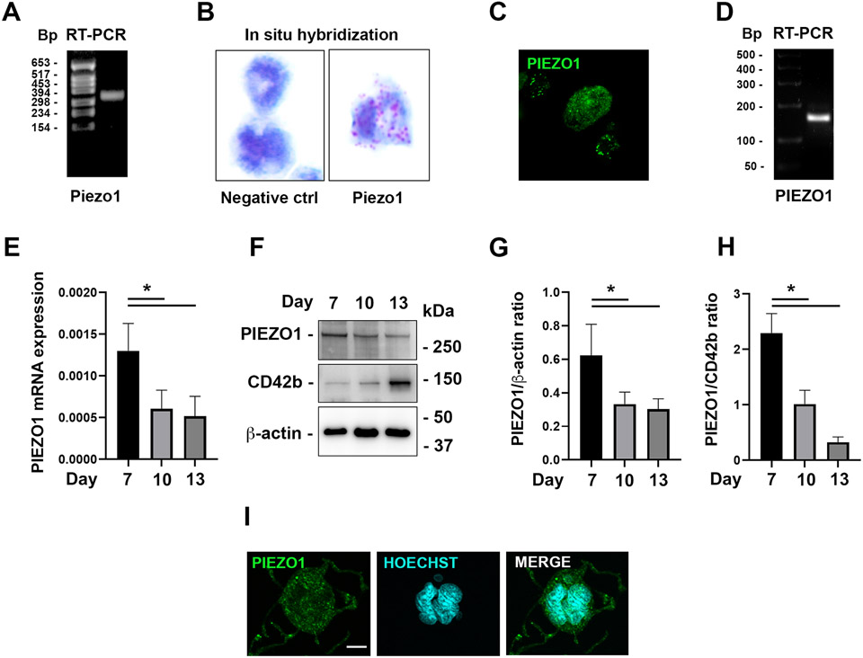
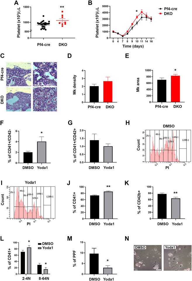
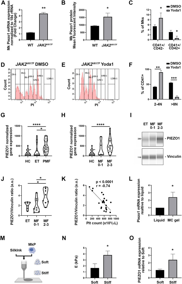
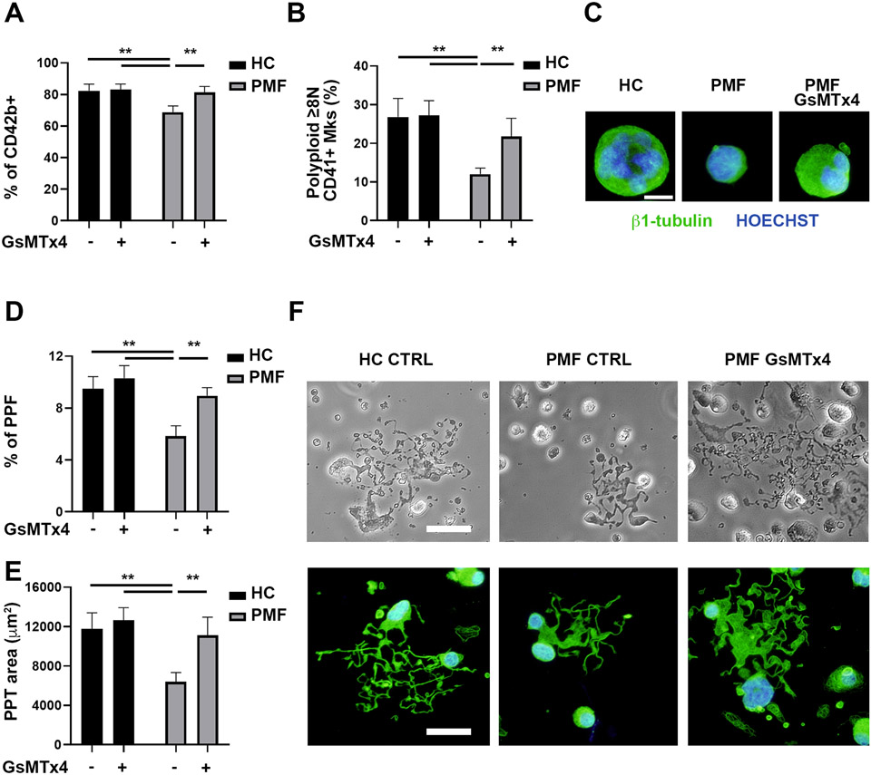
Mechanisms through which mature megakaryocytes (Mks) and their progenitors sense the bone marrow extracellular matrix to promote lineage differentiation in health and disease are still partially understood. We found PIEZO1, a mechanosensitive cation channel, to be expressed in mouse and human Mks. Human mutations in PIEZO1 have been described to be associated with blood cell disorders. Yet, a role for PIEZO1 in megakaryopoiesis and proplatelet formation has never been investigated. Here, we show that activation of PIEZO1 increases the number of immature Mks in mice, while the number of mature Mks and Mk ploidy level are reduced. Piezo1/2 knockout mice show an increase in Mk size and platelet count, both at basal state and upon marrow regeneration. Similarly, in human samples, PIEZO1 is expressed during megakaryopoiesis. Its activation reduces Mk size, ploidy, maturation, and proplatelet extension. Resulting effects of PIEZO1 activation on Mks resemble the profile in Primary Myelofibrosis (PMF). Intriguingly, Mks derived from Jak2V617F PMF mice show significantly elevated PIEZO1 expression, compared to wild-type controls. Accordingly, Mks isolated from bone marrow aspirates of JAK2V617F PMF patients show increased PIEZO1 expression compared to Essential Thrombocythemia. Most importantly, PIEZO1 expression in bone marrow Mks is inversely correlated with patient platelet count. The ploidy, maturation, and proplatelet formation of Mks from JAK2V617F PMF patients are rescued upon PIEZO1 inhibition. Together, our data suggest that PIEZO1 places a brake on Mk maturation and platelet formation in physiology, and its upregulation in PMF Mks might contribute to aggravating some hallmarks of the disease.


Conflict of interest statement

Conflict of interest disclosure

All authors have no conflicts of interest to declare that are relevant to the content of this article.

Figures

Figure 1.
Figure 1.. PIEZO1 is expressed in mouse and human megakaryocytes.
A) RT-PCR analysis of Piezo1 gene expression performed on mRNA from the mouse Mk culture. B) PIEZO1 in situ hybridization showing its expression in fully mature MKs, revealed by red dots. C) Representative immuno-staining image (40x) of gradient-purified cultured Mks (see methods) derived from 12 weeks old wild type mice and stained with anti-Piezo1, showing punctate peripheral staining in medium-size Mks, and diffused more dim staining in large ones. D) RT-PCR analysis of PIEZO1 gene expression in human Mks. E) qRT-PCR analysis of PIEZO1 gene expression by human Mks during differentiation, n=3. F-H) Western blot analysis of PIEZO1 protein expression by human Mks during differentiation, n=3. I) Immunofluorescence staining of PIEZO1 protein (green) in human Mks. Nuclei were counterstained with HOECHST. Scale bar is 10 μm. Data are expressed as mean ± standard deviation. * p < 0.05.
Figure 2.
Figure 2.. PIEZO1 activation impairs megakaryocyte maturation and proplatelet formation.
A) Mouse platelet count in DKO mice compared to Pf4-cre control mice; data are expressed as mean ± standard error of the mean (sem), n= 28 for Pf4-cre control mice, 8 for DKO mice. B) Platelet kinetic recovery following 5-fluorouracil (5-FU) intra peritoneal administration at day 0 (150 mg/kg); data are expressed as mean ± sem, n=8 mice. C) Representative Hematoxylin and eosin staining of bone marrow sections from Pf4-cre and DKO mice. Scale bar is 200 μm. D) Number of Mks per square surface unit (5,000 μm2) of bone marrow. Data are expressed as mean ± standard deviation (SD), n=3. E) Area of Mks expressed in μm2, Data are expressed as mean ± SD, n=3. F-G) Percentages of CD41+CD42− and CD41+CD42+ mouse Mks on day 4 of Dimethyl sulfoxide (DMSO; vehicle) or 4 μM Yoda1-treated bone marrow cultures were determined by flow cytometry. Cells were derived from 8-16 weeks old female C57BL/6J control mice. At least 4 experiments were performed, and 3 mice were used per group in each experiment. Data are expressed as mean ± SD. H-I) Representative ploidy profiles of Mks derived as in panels F-G and treated with vehicle (DMSO) or 4 μM Yoda1 (see also Supplementary Fig. 2). J-N) Human CB-derived CD34+ cells were differentiated to Mks in presence or not of 3 μM of the PIEZO1 pharmacological agonist Yoda1. DMSO was used as vehicle control. Data are expressed as mean ± SD, n=4 DMSO and n=4 Yoda1. J-K) Flow cytometry analysis of the early Mk marker CD41 and the late Mk marker CD42b at the end of the culture. L) Flow cytometry analysis of low ploidy (2-4N) and high ploidy (8-64N) CD41+ Mks. M) Proplatelet formation assay. Mks extending proplatelets were counted and expressed as the percentage of adhered Mks with or without proplatelet extensions. A minimum of 40 Mks per sample was analyzed. N) Representative image of Mks extending proplatelets. Scale bar is 100 μm. * p<0.05; ** p<0.01.
Figure 3.
Figure 3.. PIEZO1 expression is increased in a PMF mouse model and in patients affected by JAK2V617F PMF.
A) Mk Piezo1 mRNA was determined by qRT-PCR in isolated Mks from cultured bone marrow cells derived from 12-16 weeks old male and female JAK2V617F (n=8) and matching controls (n=9). Data are expressed as mean ± standard deviation (SD). B) Piezo1 cell surface expression was determined by staining bone marrow cultures derived from matching control and JAK2V617F male mice with CD41+ and Piezo1 antibody. Data shown are the mean fluorescent intensity ± SD, n=3. C) Percentages of CD41+CD42− and CD41+CD42+ Mks on day 4 of DMSO- (vehicle) or 4 μM Yoda1-treated bone marrow cultures were determined by flow cytometry. Cells were derived from 8-16 weeks old female or male JAK2V617F mice. At least 4 experiments were performed, and 3 mice were used per group in each experiment. Data are expressed as mean ± SD. D-E) Representative ploidy profiles of Mks derived as in panels C-D and treated with vehicle (DMSO; left panel) or 4 μM Yoda (right panel). F) Quantification of the percentage of CD41+ mouse Mks with lower (2-4N) and higher (>8N) ploidy derived as in panels C-D and treated with vehicle (DMSO) or 4 μM Yoda1. G) PIEZO1 normalized platelet gene expression in healthy controls (HC), ET and PMF patients. H) PIEZO1 normalized platelet gene expression in HC, early PMF (fibrosis grade 0-1) and overt PMF (fibrosis grade 2-3) patients. I) Mks were isolated from bone marrow aspirates derived from patients affected by ET and PMF. PIEZO1 protein expression was analyzed by quantitative capillary-based electrophoresis with anti-PIEZO1 antibody. Anti-vinculin antibody was used for loading control. Representative data are shown as blots. J) Peak areas were quantified and expressed as the ratio of PIEZO1/vinculin. K) Correlation (r) between PIEZO1 protein expression in bone marrow Mks and peripheral blood platelet count. L) qRT-PCR analysis of Piezo1 gene expression in mouse Mks grown in 3D gel compared to liquid culture. Data are expressed as mean ± standard error of the mean normalized to liquid culture, n=3. M) Human Mks were cultured in 3D bone marrow-like tissues having different mechanical features. N) Elastic modulus of fibroin silk-based 3D bone marrow-like tissues. Data are expressed as mean ± SD, n=4. O) qRT-PCR analysis of PIEZO1 gene expression in human Mks cultured in the 3D bone marrow-like tissues having different mechanical features. Data are expressed as mean ± SD, n=3. * p<0.05; ** p<0.01.
Figure 4.
Figure 4.. PIEZO1 inhibition increases megakaryocyte maturation and proplatelet formation of JAK2V617F PMF megakaryocytes.
CD34+ progenitors derived from healthy controls (HC, n=5) and patients affected by JAK2V617F Primary Myelofibrosis (PMF, n=5) were differentiated in Mks in presence or not of 5 μM of the PIEZO1 inhibitor GsMTx4. A) Flow cytometry analysis of CD42b+ Mks at the end of the culture. B) Flow cytometry analysis of the ploidy of CD41+ Mks. C) Representative immunofluorescence images of HC and PMF Mks stained with anti-β1-tubulin (green). Nuclei were counterstained with HOECHST. Scale bar is 20 μm. Where indicated PMF Mks were differentiated in presence of 5 μM GsMTx4. D) Proplatelet formation (PPF) assay. Mks extending proplatelets were counted and expressed as the percentage of adhered Mks. A minimum of 40 Mks per sample was analyzed. E) The proplatelet (PPT) area was calculated. F) Representative phase contrast and immunofluorescence microscopy images of proplatelet extending Mks. Scale bar is 100 μm. Data are expressed as mean ± SD * p<0.05; ** p<0.01.

References

    1. Shin JW, Swift J, Spinler KR, Discher DE. Myosin-II inhibition and soft 2D matrix maximize multinucleation and cellular projections typical of platelet-producing megakaryocytes. Proc Natl Acad Sci U S A. 2011;108(28):11458–11463. - PMC - PubMed
    1. Pallotta I, Lovett M, Rice W, Kaplan DL, Balduini A. Bone marrow osteoblastic niche: a new model to study physiological regulation of megakaryopoiesis. PLoS One. 2009;4(12):e8359. - PMC - PubMed
    1. Malara A, Gruppi C, Pallotta I, et al. Extracellular matrix structure and nano-mechanics determine megakaryocyte function. Blood. 2011;118(16):4449–4453. - PMC - PubMed
    1. Aguilar A, Pertuy F, Eckly A, et al. Importance of environmental stiffness for megakaryocyte differentiation and proplatelet formation. Blood. 2016. - PubMed
    1. Abbonante V, Di Buduo CA, Gruppi C, et al. A new path to platelet production through matrix sensing. Haematologica. 2017;102(7):1150–1160. - PMC - PubMed

Publication types