. 2024 Feb 13;102(3):e207966.
doi: 10.1212/WNL.0000000000207966. Epub 2023 Dec 28.

Characteristics, Prevalence, and Clinical Relevance of Juxtacortical Paramagnetic Rims in Patients With Multiple Sclerosis

Affiliations

Characteristics, Prevalence, and Clinical Relevance of Juxtacortical Paramagnetic Rims in Patients With Multiple Sclerosis

Riccardo Galbusera et al. Neurology. .

Erratum in

Abstract

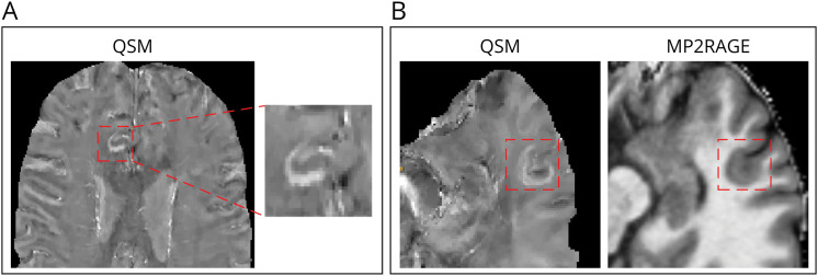
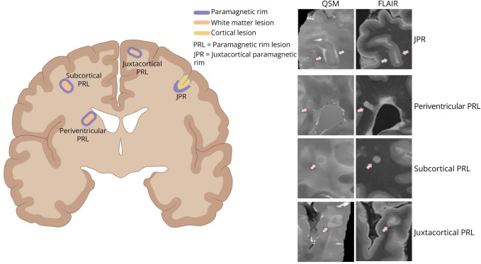
Background and objectives: A subgroup of patients with multiple sclerosis (MS) presents focal paramagnetic rims at the border between cortex and white matter (juxtacortical paramagnetic rims [JPRs]). We investigated the presence of this finding in our in vivo MS cohort and explored its potential clinical relevance. Moreover, we exploited postmortem MRI of fixed whole MS brains to (1) detect those rims and (2) investigate their histologic correlation.

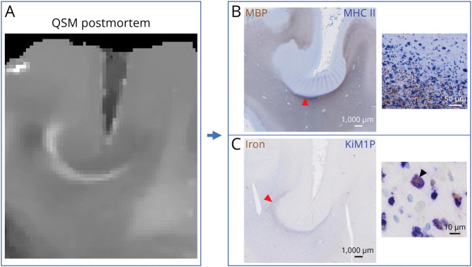
Methods: Quantitative susceptibility mapping (QSM) and magnetization-prepared 2 rapid acquisition gradient-echo (MP2RAGE) images at 3T-MRI of 165 patients with MS from the in vivo cohort were screened for JPRs and the presence of cortical lesions. Five postmortem brains from patients with MS were imaged with 3T-MRI to obtain QSM and MP2RAGE sequences. Tissue blocks containing JPRs were excised and paraffin-embedded slices stained by immunohistochemistry for myelin basic protein (for myelin) and anti-CR3/43 (for major histocompatibility complex II-positive microglia/macrophages). DAB-Turnbull stain was performed to detect iron.

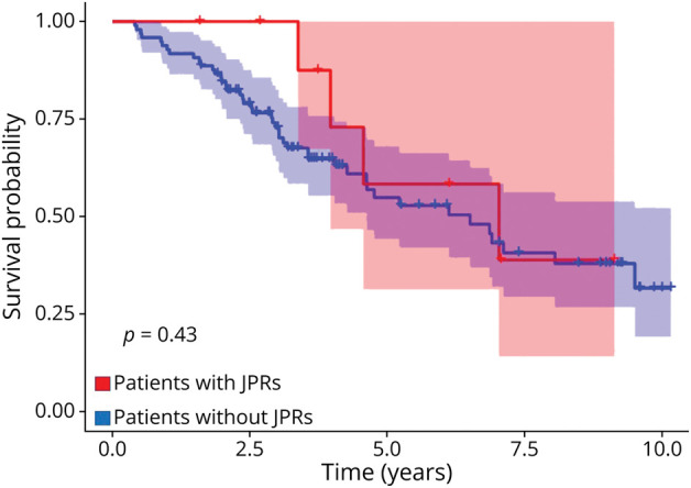
Results: JPRs are present in approximately 10% of in vivo patients and are associated with increased cortical lesion load. One of the 5 postmortem brains showed JPRs. Histologically, JPRs correspond to an accumulation of activated iron-laden phagocytes and are associated with demyelination of the whole overlying cortical ribbon.

Discussion: JPRs are a novel potential MRI biomarker of focal cortical demyelination, which seems related to global cortical pathology and might be useful for diagnostic and stratification purposes in a clinical setting.


Conflict of interest statement

The authors report no relevant disclosures. Go to Neurology.org/N for full disclosures.

Figures

Figure 1
Figure 1. Examples of JPRs in Patients With MS In Vivo
In vivo QSM of a patient with multiple sclerosis included in our cohort showing a JPR (A) and in vivo QSM of another patient showing a JPR and the corresponding MP2RAGE revealing an underlying focal cortical/juxtacortical hypointensity (B). JPR = juxtacortical paramagnetic rim; MP2RAGE = magnetization-prepared 2 rapid acquisition gradient-echo; MS = multiple sclerosis; QSM = quantitative susceptibility mapping.
Figure 2
Figure 2. Estimation of the Disease Progression Rate of Patients With and Without JPRs
Kaplan-Meier estimate for time to first clinical worsening (EDSS progression). Number of patients with JPRs = 10, number of patients without JPRs = 98. This is a retrospective analysis, and the group (JPR) was not defined at the beginning of the observation time. EDSS = Expanded Disability Status Scale; JPR = juxtacortical paramagnetic rim; MS = multiple sclerosis.
Figure 3
Figure 3. Histologic Characterization of JPRs
Postmortem QSM showing a juxtacortical rim (A) and its histologic correlate showing accumulation of activated phagocytes (B) and iron-laden macrophages (C). Cortical demyelination is reaching the cortical layer VI. JPR = juxtacortical paramagnetic rim; QSM = quantitative susceptibility mapping.
Figure 4
Figure 4. Different Types and Locations of Paramagnetic Rims in MS
PRLs are lesions located in the periventricular, subcortical, and juxtacortical white matter, which are surrounded by a rim of increased susceptibility. They represent the imaging correlation of chronic active white matter lesions. JPRs are areas of local increase in susceptibility that are located in the juxtacortical white matter and that surround a cortical lesion involving the whole cortical ribbon (not a white matter lesion). FLAIR = fluid-attenuated inversion recovery; JPR = juxtacortical paramagnetic rim; MS = multiple sclerosis; PRL = paramagnetic rim lesion; QSM = quantitative susceptibility mapping.

References

    1. Reich DS, Lucchinetti CF, Calabresi PA. Multiple sclerosis. N Engl J Med. 2018;378(2):169-180. doi:10.1056/NEJMra1401483 - DOI - PMC - PubMed
    1. Lucchinetti CF, Popescu BFG, Bunyan RF, et al. . Inflammatory cortical demyelination in early multiple sclerosis. N Engl J Med. 2011;365(23):2188-2197. doi:10.1056/NEJMoa1100648 - DOI - PMC - PubMed
    1. Calabrese M, Rocca MA, Atzori M, et al. . A 3-year magnetic resonance imaging study of cortical lesions in relapse-onset multiple sclerosis. Ann Neurol. 2010;67(3):376-383. doi:10.1002/ana.21906 - DOI - PubMed
    1. Junker A, Wozniak J, Voigt D, et al. . Extensive subpial cortical demyelination is specific to multiple sclerosis. Brain Pathol. 2020;30(3):641-652. doi:10.1111/bpa.12813 - DOI - PMC - PubMed
    1. Harrison DM, Roy S, Oh J, et al. . Association of cortical lesion burden on 7-T magnetic resonance imaging with cognition and disability in multiple sclerosis. JAMA Neurol. 2015;72(9):1004-1012. doi:10.1001/jamaneurol.2015.1241 - DOI - PMC - PubMed