

A simple and rapid preparation of smooth muscle myosin 2 for the electron microscopic analysis
- PMID: 38165512
- PMCID: PMC10761634
- DOI: 10.1186/s42649-023-00094-5
A simple and rapid preparation of smooth muscle myosin 2 for the electron microscopic analysis
Abstract
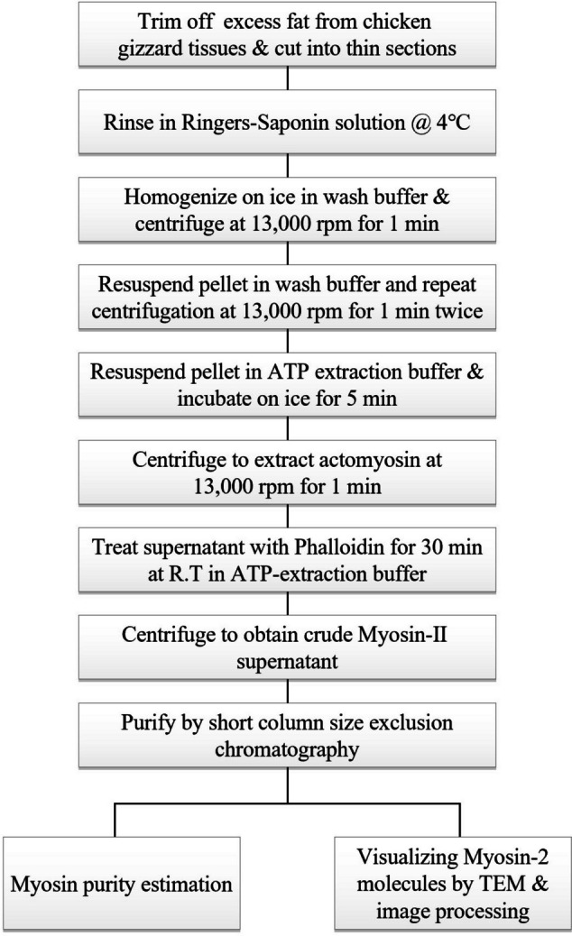
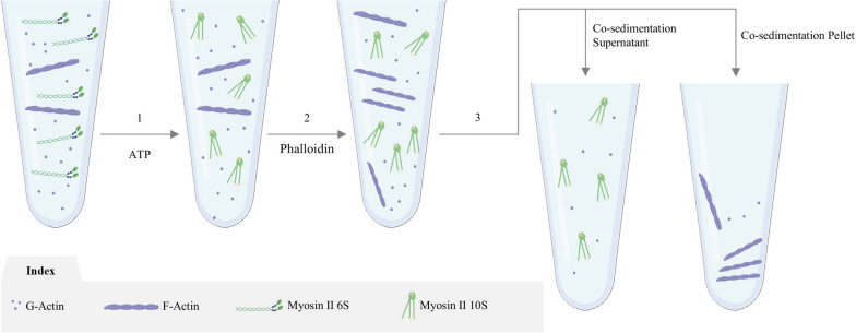
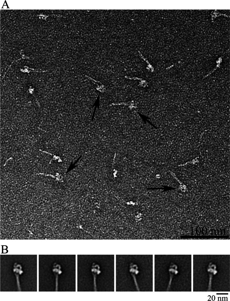
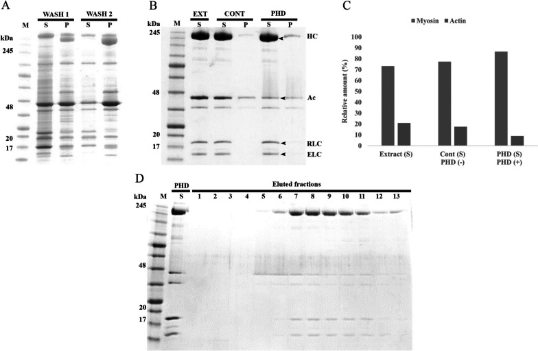
There has been an increase in the demand for purified protein as a result of recent developments in the structural biology of myosin 2. Although promising, current practices in myosin purification are usually time-consuming and cumbersome. The reported increased actin to myosin ratio in smooth muscles adds to the complexity of the purification process. Present study outlines a streamlined approach to isolate smooth muscle myosin 2 molecules from actomyosin suspension of chicken gizzard tissues. The procedure entails treating actomyosin for a brief period with actin-binding peptide phalloidin, followed by co-sedimentation and short column size exclusion chromatography. Typical myosin molecule with heavy and light chains and approximately 95% purity was examined using gel electrophoresis. Negative staining electron microscopy and image processing showed intact 10S myosin 2 molecules, proving that phalloidin is effective at eliminating majority of actin in the form of F-actin without dramatic alteration in the structure of myosin. The entire purification discussed here can be completed in a few hours, and further analysis can be done the same day. Thus, by offering quick and fresh supplies of native myosin molecules suited for structural research, specially cryo-electron microscopy, this innovative approach can be adapted to get around the drawbacks of time-intensive myosin purifying processes.
Keywords: 10S myosin; Phalloidin; Protein purification; Smooth muscle myosin 2; Transmission electron microscopy.
© 2023. The Author(s).
Conflict of interest statement
The authors declare that they have no competing interests.
Figures
Similar articles
-
Actin-facilitated assembly of smooth muscle myosin induces formation of actomyosin fibrils.J Cell Biol. 1992 Jun;117(6):1223-30. doi: 10.1083/jcb.117.6.1223. J Cell Biol. 1992. PMID: 1607384 Free PMC article.
-
Purification of native myosin filaments from muscle.Biophys J. 2001 Nov;81(5):2817-26. doi: 10.1016/S0006-3495(01)75923-1. Biophys J. 2001. PMID: 11606293 Free PMC article.
-
Vascular smooth muscle caldesmon.J Biol Chem. 1986 Jun 15;261(17):8028-35. J Biol Chem. 1986. PMID: 2940249
-
Regulation of actomyosin and contraction in smooth muscle.World J Urol. 1994;12(5):292-7. doi: 10.1007/BF00191210. World J Urol. 1994. PMID: 7866427 Review.
-
Purification of Myosin from Bovine Tracheal Smooth Muscle, Filament Formation and Endogenous Association of Its Regulatory Complex.Cells. 2023 Feb 3;12(3):514. doi: 10.3390/cells12030514. Cells. 2023. PMID: 36766856 Free PMC article. Review.
References
LinkOut - more resources
Full Text Sources