. 2024 Jan 2;65(1):4.
doi: 10.1167/iovs.65.1.4.

Transmembrane Protein CMTM6 Alleviates Ocular Inflammatory Response and Improves Corneal Epithelial Barrier Function in Experimental Dry Eye

Affiliations

Transmembrane Protein CMTM6 Alleviates Ocular Inflammatory Response and Improves Corneal Epithelial Barrier Function in Experimental Dry Eye

Yifan Zhou et al. Invest Ophthalmol Vis Sci. .

Abstract

Purpose: To investigate the impact of transmembrane protein CMTM6 on the pathogenesis of dry eye disease (DED) and elucidate its potential mechanisms.

Methods: CMTM6 expression was confirmed by database analysis, real-time polymerase chain reaction (RT-PCR), western blot, and immunohistochemistry. Tear secretion was measured using the phenol red thread test. Immune cell infiltration was assessed through flow cytometry. Barrier function was evaluated by fluorescein sodium staining, immunofluorescence staining of zonula occludens 1 (ZO-1), and electric cell-substrate impedance sensing (ECIS) assessment. For silencing CMTM6 expression, siRNA and shRNA were employed, along with lentiviral vector-mediated overexpression of CMTM6. Proinflammatory cytokine levels were analyzed by RT-PCR and cytometric bead array (CBA) analysis.

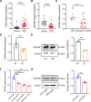
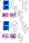
Results: CMTM6 showed high expression in healthy human and mouse corneal and conjunctival epithelium but was notably reduced in DED. Notably, this downregulation was correlated with disease severity. Cmtm6-/- dry eye (DE) mice displayed reduced tear secretion, severe corneal epithelial defects, decreased conjunctival goblet cell density, and upregulated inflammatory response. Additionally, Cmtm6-/- DE mice and CMTM6 knockdown human corneal epithelial cell-transformed (HCE-T) cells showed more severe barrier disruption and reduced expression of ZO-1. Knockdown of CMTM6 in HCE-T cells increased inflammatory responses induced by hyperosmotic stress, which was significantly mitigated by CMTM6 overexpression. Moreover, the level of phospho-p65 in hyperosmolarity-stimulated HCE-T cells increased after silencing CMTM6. Nuclear factor kappa B (NF-κB) p65 inhibition (JSH-23) reversed the excessive inflammatory responses caused by hyperosmolarity in CMTM6 knockdown HCE-T cells.

Conclusions: The reduction in CMTM6 expression on the ocular surface contributes to the pathogenesis of DED. The CMTM6-NF-κB p65 signaling pathway may serve as a promising therapeutic target for DED.


Conflict of interest statement

Disclosure: Y. Zhou, None; B. Ma, None; Q. Liu, None; H. Duan, None; Y. Huo, None; L. Zhao, None; J. Chen, None; W. Han, None; H. Qi, None

Figures

Figure 1.
Figure 1.
CMTM6 is expressed at a high level in corneal and conjunctival epithelium. (A) The tissue and organ distributions of Cmtm6 were analyzed within the BioGPS database. (B) The detection of CMTM6 expression in adult human corneas and adjacent conjunctivas was carried out utilizing GSE182583 (n = 4). (C) The expression of Cmtm6 in corneal epithelium, corneal stroma, and conjunctiva of healthy mice was assessed by RT-PCR (n = 5 in each group). (D, E) The protein level of CMTM6 was evaluated by IHC. Representative images of cornea (D) and conjunctiva (E) are shown. Scale bars: 20 µm. The results of a one-way ANOVA and Dunnett's multiple comparisons test are displayed as mean ± SD; “n” represents both eyes from a single mouse (C). **P < 0.01, ****P < 0.0001. CS_Epi, corneal superficial epithelial cells; CB_Epi, corneal basal epithelial cells; CSKs, corneal stroma keratocytes; C_Endo, corneal endothelial cells; FC_Endo, fibroblastic corneal endothelial cells; Conj_Epi, conjunctival epithelial cells.
Figure 2.
Figure 2.
CMTM6 is significantly decreased in patients with DED and DE models in vivo and in vitro. (A, B) Conjunctival epithelial cells from patients with DED (n = 34) and healthy individuals (n = 20) were collected, and the expression of Tnfa (A) and Cmtm6 (B) was detected. (C) Patients with DED were divided into two groups according to CFS scores: mild DED (n = 8) and moderate to severe DED (n = 26). The expression of Cmtm6 was examined by RT-PCR. (D, E) A mouse model of DE was established using scopolamine hydrobromide injection. At day 14, all of the mice were sacrificed and corneas were collected (n = 6 in each group). (D) CMTM6 mRNA expression was detected by RT-PCR. (E) CMTM6 protein expression was measured by western blot analysis. (F, G) An in vitro DE model was established by culturing HCE-T cells in a hypertonic environment (500 mOsM). (F) Cmtm6 mRNA expression levels were measured at 3, 8, and 12 hours following hyperosmotic stimulation. (G) CMTM6 protein expression level was measured at 24 hours. Results are shown as mean ± SD. Two-group statistical analysis was conducted using the unpaired Student's t-test (AE, G). Four groups were compared using one-way ANOVA followed by Dunnett's multiple comparisons test (F); “n” represents both eyes from a single mouse (D, E). **P < 0.01, ***P < 0.001, ****P < 0.0001.
Figure 3.
Figure 3.
CMTM6 alleviates damage in DE mice. (A, D) CFS was used to evaluate corneal epithelial defects. (B, E) Tear production was examined using the phenol red thread test. (C, F) The conjunctival goblet cells were stained with PAS, and the positive cells were counted. Scale bars: 20 µm. Results are shown as mean ± SD (n = 6–8 in each group). A two-group statistical analysis was conducted using the unpaired Student's t-test; “n” represents one eye per mouse (AF). **P < 0.01, ***P < 0.001; ns, non-significant.
Figure 4.
Figure 4.
CMTM6 reduces DLNs Th1 and Th17 cell infiltration and proinflammatory cytokine production in DE mice. (A) Representative flow cytometry plots and quantitative summary of Th1, Th17, and Treg percentages among the DLNs. (BD) RT-PCR was used to examine the production of proinflammatory cytokines Tnfa (B), Il1b (C), and Il6 (D) in corneas. (E) The expression of TNF-α in tears was measured using CBA. Results are shown as mean ± SD (n = 3–4 in each group). A two-group statistical analysis was conducted using the unpaired Student's t-test; “n” represents bilateral draining lymph nodes (A) or both eyes (BE) from a single mouse. *P < 0.05, **P < 0.01, ***P < 0.001.
Figure 5.
Figure 5.
CMTM6 reduces barrier disruption in vivo and in vitro. (A) Immunofluorescence analysis of ZO-1 in wholemount corneal tissues with quantification by ImageJ. Scale bars: 20 µm. (BE) Changes in the barrier resistance of HCE-T cells were examined using ECIS. (B) Resistance measurements generated at 4000 Hz from HCE-T cells grown in 310- and 500-mOsM culture media. (C) The endpoint resistance values of HCE-T cells. (D) Rb parameters were measured to reflect the paracellular barrier strength after the stabilization of resistance. (E) The endpoint Rb values of HCE-T cells. (F, G) After Cmtm6 knockdown in HCE-T cells using siRNA (F) or shRNA (G), protein levels of ZO-1 were detected using western blot analysis. Results are shown as mean ± SD. Four groups were compared using one-way ANOVA followed by Tukey's multiple comparisons test (C, EG). *P < 0.05, **P < 0.01, ***P < 0.001, ****P < 0.0001.
Figure 6.
Figure 6.
CMTM6 attenuates inflammation stimulated by hyperosmolarity in HCE-T cells. A TG006-CMTM6-IRES-EGFP lentiviral vector was used to overexpress CMTM6. The TG006 lentiviral vector was used as a negative control. (A, B) Overexpression of CMTM6 in HCE-T cells was validated using RT-PCR (A) and western blot analyses (B). (CE) RT-PCR was used to detect the mRNA levels of TNFA (C), IL1B (D), and IL6 (E). (F) The protein level of IL-6 in the culture supernatant was measured using CBA. Two-group statistical analysis was conducted using the unpaired Student's t-test (A). Four groups were compared using one-way ANOVA followed by Tukey's multiple comparisons test (BF). *P < 0.05, **P < 0.01, ***P < 0.001.
Figure 7.
Figure 7.
Knockdown of CMTM6 enhances inflammation stimulated by hyperosmolarity in HCE-T cells. Knockdown of CMTM6 using shRNA promotes inflammation stimulated by hyperosmolarity in HCE-T cells. A lentivirus encoding a CMTM6 shRNA was used to silence CMTM6 expression. A lentivirus expressing control shRNA (shNC) was used as a negative control. (A) RT-PCR was used to detect the mRNA level of CMTM6 in HCE-T cells. (B) Western blot analysis was used to examine the protein level of CMTM6 in HCE-T cells. (CE) Cultured cells were collected to assess the mRNA expression of TNFA (C), IL1B (D), and IL6 (E). (F) IL-6 release was quantified in the culture supernatant using CBA. Six groups were compared using one-way ANOVA followed by Tukey's multiple comparisons test. *P < 0.05, **P < 0.01, ***P < 0.001, ****P < 0.0001.
Figure 8.
Figure 8.
CMTM6 suppresses inflammation through the NF-κB/p65 pathway in HCE-T cells under hyperosmotic stress. (A, B) The effect of CMTM6 on the activation of p38 (A), Erk (A), and p65 (B) was examined using western blot analysis. (CG) For the treatment of p65 inhibition, 50 µM JSH-23 was added to the cell culture medium, followed by incubation with 500-mOsM media for 8 or 24 hours. DMSO was used as a control. (C) The effect of JSH-23 was detected using western blot analysis. (DF) The mRNA levels of TNFA (D), IL1B (E), and IL6 (F) were examined using RT-PCR. (G) The protein level of IL-6 in cell culture supernatants was detected using CBA. Six groups were compared using one-way ANOVA followed by Tukey's multiple comparisons test. *P < 0.05, **P < 0.01, ***P < 0.001.
Figure 9.
Figure 9.
Graph abstract of how CMTM6 regulates inflammatory response and barrier function in the pathogenesis of DED. Under normal conditions, CMTM6 exhibits high expression levels in the corneal epithelium. However, following the onset of dry eye, CMTM6 expression is notably downregulated. CMTM6 plays a pivotal role in preventing corneal epithelial cells from generating an excessive inflammatory response by inhibiting NF-κB p65, thereby mitigating inflammatory response and ameliorating epithelial barrier disruption in DED. Figure was created using BioRender (http://biorender.com/).

Similar articles

Cited by

References

    1. Craig JP, Nichols KK, Akpek EK, et al. .. TFOS DEWS II Definition and Classification Report. Ocul Surf. 2017; 15(3): 276–283. - PubMed
    1. Chen Y, Dana R.. Autoimmunity in dry eye disease – an updated review of evidence on effector and memory Th17 cells in disease pathogenicity. Autoimmun Rev. 2021; 20(11): 102933. - PMC - PubMed
    1. Stapleton F, Alves M, Bunya VY, et al. .. TFOS DEWS II Epidemiology Report. Ocul Surf. 2017; 15(3): 334–365. - PubMed
    1. Morthen MK, Magno MS, Utheim TP, Hammond CJ, Vehof J.. The work-related burden of dry eye. Ocul Surf. 2023; 28: 30–36. - PubMed
    1. Yang W, Luo Y, Wu S, et al. .. Estimated annual economic burden of dry eye disease based on a multi-center analysis in China: a retrospective study. Front Med (Lausanne). 2021; 8: 771352. - PMC - PubMed