. 2024 Jan 2;19(1):e0295504.
doi: 10.1371/journal.pone.0295504. eCollection 2024.

Neuroprotective effect of the RNS60 in a mouse model of transient focal cerebral ischemia

Affiliations

Neuroprotective effect of the RNS60 in a mouse model of transient focal cerebral ischemia

Gloria Patricia Baena-Caldas et al. PLoS One. .

Abstract

Background: Stroke is a major cause of death, disability, and public health problems. Its intervention is limited to early treatment with thrombolytics and/or endovascular clot removal with mechanical thrombectomy without any available subacute or chronic neuroprotective treatments. RNS60 has reduced neuroinflammation and increased neuronal survival in several animal models of neurodegeneration and trauma. The aim here was to evaluate whether RNS60 protects the brain and cognitive function in a mouse stroke model.

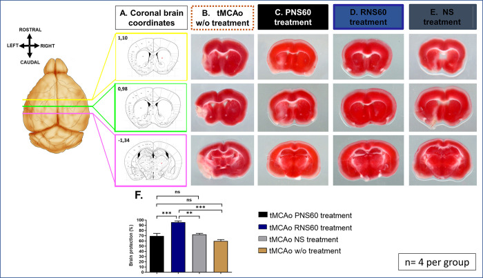
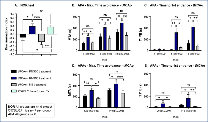
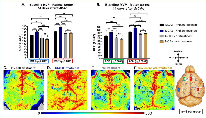
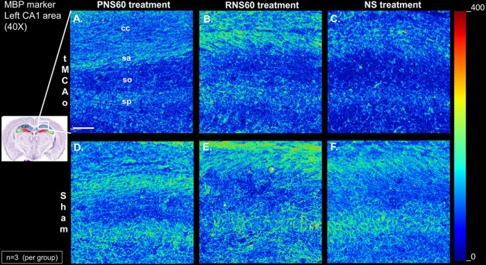
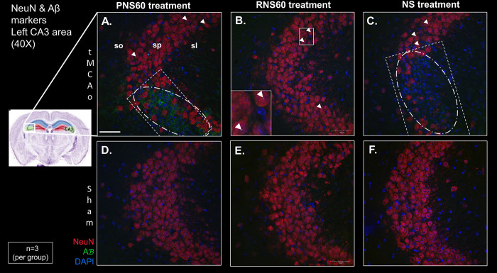
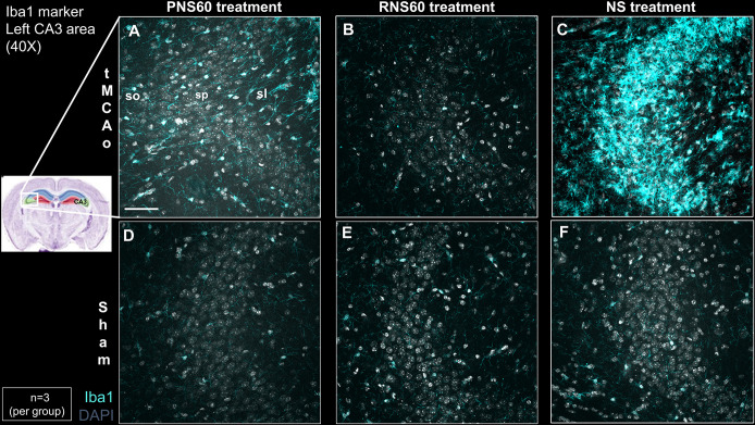
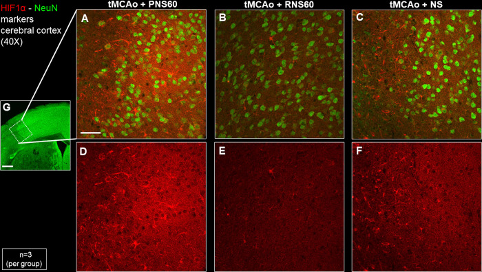
Methods: Male C57BL/6J mice were subjected to sham or ischemic stroke surgery using 60-minute transient middle cerebral artery occlusion (tMCAo). In each group, mice received blinded daily administrations of RNS60 or control fluids (PNS60 or normal saline [NS]), beginning 2 hours after surgery over 13 days. Multiple neurobehavioral tests were conducted (Neurological Severity Score [mNSS], Novel Object Recognition [NOR], Active Place Avoidance [APA], and the Conflict Variant of APA [APAc]). On day 14, cortical microvascular perfusion (MVP) was measured, then brains were removed and infarct volume, immunofluorescence of amyloid beta (Aβ), neuronal density, microglial activation, and white matter damage/myelination were measured. SPSS was used for analysis (e.g., ANOVA for parametric data; Kruskal Wallis for non-parametric data; with post-hoc analysis).

Results: Thirteen days of treatment with RNS60 reduced brain infarction, amyloid pathology, neuronal death, microglial activation, white matter damage, and increased MVP. RNS60 reduced brain pathology and resulted in behavioral improvements in stroke compared to sham surgery mice (increased memory-learning in NOR and APA, improved cognitive flexibility in APAc).

Conclusion: RNS60-treated mice exhibit significant protection of brain tissue and improved neurobehavioral functioning after tMCAo-stroke. Additional work is required to determine mechanisms, time-window of dosing, and multiple dosing volumes durations to support clinical stroke research.


Conflict of interest statement

The authors have declared that no competing interests exist. Drs. SG and AK are full-time employees of the Revalesio Corporation but have not conflict of interest or any disclosures. They are authors because they participated in the experimental design, visualization, and manuscript preparation but did not participate in any data collection or analysis.

Figures

Fig 1
Fig 1. RNS60 protected against hemispheric loss in mice subjected to left tMCAo.
A) The cerebral slices of the coordinates 1.10, 0.98 and -1.34 from the Paxinos mouse atlas were used for analysis with ImageJ software. B-E) Representative TTC images of tMCAo brain slices (coordinates shown in Fig A) of untreated (B) mice or mice treated with PNS60 (C), RNS60 (D) or NS (E). F) Quantification of the TTC staining shows increased ipsilateral protection of brain loss in tMCAo mice treated with RNS60 (blue bar) compared to those treated with PNS60 (black bar) or NS (gray bar) as well as mice without any treatment (brown bar). The percentage of protection is shown as mean ± SEM. **p < 0.01, ***p < 0.001 and ns = no statistical significance.
Fig 2
Fig 2. RNS60 treatment improved performance in cognitive evaluation tests.
A) NOR task showed that mice subjected to left tMCAo and treated with RNS60 (blue bar) performed similarly to healthy C57BL/6J mice without surgery and treatment (w/o Sx and Tx, green bar). In contrast, tMCAo mice treated with PNS60 (black bar) showed no preference for the new object (discrimination index near 0) suggesting that memory is impaired, and mice with left tMCAo and treated with NS (grey bar) responded with an aversion to the new object (negative discrimination index). B-E) Active place avoidance memory task shows that animals treated with RNS60 have better memory and cognitive flexibility as assessed by APA (B, C) and APA conflict (D, E) tests using two parameters of learning and memory: Maximum Time Avoidance (MTA) (B, D), and time to the 1st entrance (T1stE) (C, E). *p<0.05, **p<0.01, ***p<0.001, ns = non-significant.
Fig 3
Fig 3. RNS60 treatment increased the cortical cerebral blood flow in mice subjected to tMCAo (60-min) followed by 14 days treatment.
Plotted are means ± SEM, showing a significant increase of cerebral cortical microvascular perfusion (MVP) in parietal and motor cortex of both brain sides during baseline period in mice subjected to left transient middle cerebral artery occlusion (tMCAo) treated with PNS60 (black bar), RNS60 (blue bar), NS (grey bar) and without treatment (brown bar) (A and B). Microcirculation (pseudo-colored) images show the cerebral basal blood flow (baseline) of mice subjected to tMCAo followed by 14 days treatment with PNS60 (C), RNS60 (D), NS (E) and without treatment (F). Each image represents an independent mouse. *p<0.05, **p<0.01, ***p<0.001, ns = non-significant.
Fig 4
Fig 4. RNS60 treatment of tMCAo mice reduced alterations of myelin basic protein (MBP) in corpus callosum contiguous to the CA1 of the hippocampus.
MBP staining in tMCAo mice with RNS60 treatment (B) shows a similar pattern and intensity as in Sham mice (D-F) whereas PNS60- (A) and NS-treated (C) mice showed an altered pattern and lower intensity. A representative staining from each treatment group is shown with colormap intensity scale where red is the highest intensity and blue the lowest. cc, corpus callosum; sa, stratum alveus; so, stratum oriens; sp, stratum pyramidale. Each panel represents an independent mouse (A-C n = 3 and D-F n = 3). Scale bar = 50μm.
Fig 5
Fig 5. RNS60 treatment decreased Aß aggregation and neurodegeneration in the stratum pyramidale in the CA3 region of the hippocampus of tMCAo animals 14 days after stroke.
After 14 days of treatment, mice (n = 3) were sacrificed, and the brains were processed to quantitate Aß and NeuN levels by immunofluorescence. Representative microphotographs of Aß (green), NeuN (red) and DAPI (blue) in the stratum pyramidale (sp) in the CA3 region of the hippocampus are shown. tMCAo (A—C) and Sham (D–F) animals treated with PNS60 (A, D), RNS60 (B, E) and NS (C, F). Rectangle = area of neuronal cell loss; oval = area of extracellular Aß; arrowheads = intracellular Aß. so, stratum oriens in CA3; sp, stratum pyramidale in CA3; sl, stratum lucidum in CA3; sr, stratum radiatum in CA3; Scale bar = 50μm.
Fig 6
Fig 6. RNS60 treatment of tMCAo mice decreased microglial activation in the CA3 region of the ipsilateral hippocampus.
Photomicrographs of Iba1 (light blue) and DAPI (grey) show that tMCAo mice treated with RNS60 (B) exhibit reduced Iba1 signal intensity compared to tMCAo mice treated with PNS60 (A) or NS (C). Iba1 staining in tMCAo mice with RNS60 treatment (B) resembles Sham (D-F). so, stratum oriens; sp, pyramidale stratum; sl, stratum lucidum. Scale bar = 50 μm.
Fig 7
Fig 7. RNS60 treatment decreased expression of hypoxia inducible factor 1α (HIF1α) in the peri infarct cortical area of tMCAo mice 4 days after onset of daily dose of RNS60.
Representative photomicrographs of HIF1α (red) and NeuN (green) staining show that tMCAo mice treated with RNS60 (B, E) exhibit reduced HIF1α signal intensity compared to tMCAo mice treated with PNS60 (A, D) or NS (C, F). Inset in G, shows the peri-infarct-penumbra in the cortical area of the brain selected to image A-F. The samples are coronal cerebral slices of the coordinates between 0.62 mm and 0.50 mm anterior to Bregma. Scale bar A-F = 50 μm; and G = 500 μm (n = 3 per group).

Similar articles

References

    1. Kalaria RN, Akinyemi R, Ihara M. Stroke injury, cognitive impairment and vascular dementia. Biochim Biophys Acta. 2016;1862(5):915–25. doi: 10.1016/j.bbadis.2016.01.015 - DOI - PMC - PubMed
    1. Johnson W, Onuma O, Owolabi M, Sachdev S. Stroke: a global response is needed. Bull World Health Organ. 2016;94(9):634–A. doi: 10.2471/BLT.16.181636 - DOI - PMC - PubMed
    1. Feigin VL, Stark BA, Johnson CO, Roth GA, Bisignano C, Abady GG, et al.. Global, regional, and national burden of stroke and its risk factors, 1990–2019: A systematic analysis for the Global Burden of Disease Study 2019. The Lancet Neurology. 2021;20(10):795–820. doi: 10.1016/S1474-4422(21)00252-0 - DOI - PMC - PubMed
    1. Guzik A, Bushnell C. Stroke Epidemiology and Risk Factor Management. Continuum (Minneap Minn). 2017;23(1):15–39. doi: 10.1212/CON.0000000000000416 - DOI - PubMed
    1. Chugh C. Acute Ischemic Stroke: Management Approach. Indian J Crit Care Med. 2019;23(Suppl 2):S140–S6. doi: 10.5005/jp-journals-10071-23192 - DOI - PMC - PubMed