. 2024 Jan 2;20(1):e1011913.
doi: 10.1371/journal.ppat.1011913. eCollection 2024 Jan.

Overproduction of mycotoxin biosynthetic enzymes triggers Fusarium toxisome-shaped structure formation via endoplasmic reticulum remodeling

Affiliations

Overproduction of mycotoxin biosynthetic enzymes triggers Fusarium toxisome-shaped structure formation via endoplasmic reticulum remodeling

Minhui Wang et al. PLoS Pathog. .

Abstract

Mycotoxin deoxynivalenol (DON) produced by the Fusarium graminearum complex is highly toxic to animal and human health. During DON synthesis, the endoplasmic reticulum (ER) of F. graminearum is intensively reorganized, from thin reticular structure to thickened spherical and crescent structure, which was referred to as "DON toxisome". However, the underlying mechanism of how the ER is reorganized into toxisome remains unknown. In this study, we discovered that overproduction of ER-localized DON biosynthetic enzyme Tri4 or Tri1, or intrinsic ER-resident membrane proteins FgHmr1 and FgCnx was sufficient to induce toxisome-shaped structure (TSS) formation under non-toxin-inducing conditions. Moreover, heterologous overexpression of Tri1 and Tri4 proteins in non-DON-producing fungi F. oxysporum f. sp. lycopersici and F. fujikuroi also led to TSS formation. In addition, we found that the high osmolarity glycerol (HOG), but not the unfolded protein response (UPR) signaling pathway was involved in the assembly of ER into TSS. By using toxisome as a biomarker, we screened and identified a novel chemical which exhibited high inhibitory activity against toxisome formation and DON biosynthesis, and inhibited Fusarium growth species-specifically. Taken together, this study demonstrated that the essence of ER remodeling into toxisome structure is a response to the overproduction of ER-localized DON biosynthetic enzymes, providing a novel pathway for management of mycotoxin contamination.


Conflict of interest statement

The authors have declared that no competing interests exist.

Figures

Fig 1
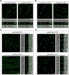
Fig 1. Overproduction of Tri4 and Tri1 induced toxisome-shaped structure (TSS) formation under DON non-inducing conditions.
(A-B) Localization of Tri4-GFP (A) and Tri1-GFP (B) under native promoter (left panels) and gpda promoter (right panels) in YEPD medium or TBI medium. RFP-HDEL is used as the ER marker. Images of each strain were taken after incubation at 28°C for 48 h. DIC indicates differential interference contrast. Bar = 10 μm. (C-D) The protein abundance of Tri4-GFP (C) and Tri1-GFP (D) isolated from the same set of samples used in A and B was determined by western blot assay with the anti-GFP antibody. The protein abundance of H3 of each sample served as a loading control. The intensities of the western blot bands were quantified using the Image J software, and numbers below the bands represent relative intensity of GFP normalized to H3. Native and gpda represent Tri proteins that are expressed under native and gpda promoters, respectively.
Fig 2
Fig 2. An essential role of the transmembrane domain of Tri4 in the formation of Tri4-marked TSS.
(A) Domain architecture of Tri4 and Tri1. The number at the bottom indicates the deduced amino acid positions of corresponding domains. (B) The TSS formation patterns in truncated Tri4 proteins. Each strain was grown in TBI medium for 48 h. Bar = 10 μm. (C) The average number of toxisome-shaped structures in an examination field of 135 μm × 135 μm. (D) DON production of the truncated Tri4 complemented strains. After growth in TBI for 7 d, each strain was determined for DON production. Data represent the mean ± s.d. from three independent experiments. Different letters indicate a significant difference (P < 0.05) based on one-way ANOVA followed by Tukey’s multiple comparison test.
Fig 3
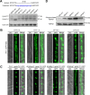
Fig 3. Overexpression of ER-resident proteins FgHmr1 and FgCnx triggers the ER remodeling into TSS.
(A-B) Localization of FgHmr1-GFP (A) and FgCnx-GFP (B) under native promoter (upper panels) and gpda promoter (lower panels) in YEPD. Each strain was grown in YEPD medium at 28°C for 48 h. Bar = 10 μm. (C-D) The protein abundance of FgHmr1-GFP (C) and FgCnx-GFP (D) under native and gpda promoters was determined by western blot assay using the anti-GFP antibody. The protein H3 was used as a reference. The intensities of the western blot bands were quantified using the Image J software, and numbers below the bands represent relative intensity of GFP normalized to H3.
Fig 4
Fig 4. Remodeling of the ER in other fungi that do not produce DON.
(A-B) Heterologous overexpression of Tri4-GFP (A) and Tri1-GFP (B) in F. oxysporum f. sp. lycopersici (Fol) (left panels) and F. fujikuroi (Ff) (right panels) induced the formation of TSS. Each strain was incubated in YEPD at 28°C for 48 h. Bar = 10 μm. (C) Localization of ER-resident protein Hmr1 under its native promoter in Fol (upper panel) and Ff (lower panel). Each strain was incubated in YEPD medium at 28°C for 48 h. Bar = 10 μm. (D) Upon overexpression of the endogenous Hmr1 in Fol (upper panel) and Ff (lower panel) by gpda promoter, the ER was reorganized into TSS. Images of each strain were taken after incubation in YEPD medium at 28°C for 48 h. Bar = 10 μm.
Fig 5
Fig 5. Regulation of the Tri4-marked TSS formation by the TRI cluster.
(A) The effect of gene deletion in the TRI cluster on the formation of TSS under DON inducing conditions. The wild-type strain and TRI gene deletion mutants were grown in TBI medium at 28°C for 48 h. RFP-HDEL is used as the ER marker. Bar = 10 μm. (B) Accumulation of Tri4-GFP proteins in TRI gene deletion mutants was determined by western blot with the anti-GFP antibody (left panel). The protein abundance of H3 in each sample served as a loading control. The relative intensity of Tri4-GFP in each strain was shown in right panel, and was calculated by determining the intensity of Tri4-GFP band normalized to the intensity of H3 band. The relative intensity of the wild-type strain PH-1 was normalized to 1. Data represent the mean ± s.d. from three independent experiments.
Fig 6
Fig 6. The HOG, but not the UPR signaling pathway, is involved in TSS assembly in F. graminearum.
(A) The FgHAC1 mRNA splicing pattern in TBI and YEPD medium. DNA sequence alignment of the spliced and unspliced FgHAC1 (upper panel). The 20 bp non-canonical intron is indicated with lowercase blue letters (upper panel). The FgHAC1 mRNA splicing pattern was analyzed by RT-PCR using the total RNA of PH-1 after incubation for the indicated times in TBI and YEPD medium (lower panel). PH-1 cultured in YEPD for 12 h and then treated with the ER stress agent DTT (10 mM) for 2 h was used as the positive control. Unspliced and spliced FgHAC1 transcripts yielded 211 and 191-bp products, respectively. The transcripts of FgACTIN gene (FGRAMPH1_01G24551) served as the loading control. (B) DTT induces an aggregation of ER membrane. The strain PH-1::GFP-HDEL was used to visualize the ER and cultured in YEPD medium for 12 h followed by treatment with DTT for the indicated times. Bar = 10 μm. (C) The FgHog1 deletion mutant failed to induce the TSS formation under DON non-inducing conditions (YEPD medium). Images of each strain were taken after incubation in YEPD at 28°C for 48 h. Bar = 10 μm. (D) Time course analysis of the phosphorylation of FgHog1 in TBI. After growing in TBI medium at 28°C for the indicated times, mycelia of PH-1 were harvested for protein extraction in western blot analysis. FgHog1 and phosphorylated FgHog1 were detected using anti-Hog1 antibody and Anti-phospho-p38 antibody, respectively.
Fig 7
Fig 7. The novel compound ZJU212 effectively blocks the ER remodeling into TSS and subsequent DON biosynthesis.
(A) Inhibition of each compound at the indicated concentration against mycelial growth of F. graminearum on PDA. DMSO was used as a control. Photos were taken after 2 days of incubation at 25°C on PDA amended without or with relevant compounds. (B) Toxisome formation in the mycelia of ΔTri1::np-Tri1-GFP (under native promoter) treated with each antifungal compound at indicated concentrations. After growth in TBI for 24 h, ΔTri1::np-Tri1-GFP was treated with antifungal compounds at indicated concentrations for another 24 h before examination. Bar = 10 μm. (C) DON production was assayed for the wild-type PH-1 grown in TBI supplemented with each antifungal compound at indicated concentrations. DON was extracted from mycelia of each strain cultured in TBI for 7 days and each compound was added after incubation of PH-1 in TBI medium for 24 h. Data represent the mean ± s.d. from three independent experiments. Statistical differences were analyzed by two-sided, unpaired Student’s t-test (ns: not significant, *P < 0.05, **P < 0.01, ****P < 0.0001). (D) TSS formation in the mycelia of ΔTri1::gpda-Tri1-GFP (under gpda promoter) treated with each antifungal compound at indicated concentrations. After growth in YEPD medium for 12 h, ΔTri1::gpda-Tri1-GFP was treated with antifungal compounds at indicated concentrations for another 36 h before examination. Bar = 10 μm.

Similar articles

Cited by

References

    1. Xu X, Nicholson P. Community ecology of fungal pathogens causing wheat head blight. Annu Rev Phytopathol. 2009; 47:83–103. doi: 10.1146/annurev-phyto-080508-081737 - DOI - PubMed
    1. Dean R, Van Kan JA, Pretorius ZA, Hammond-Kosack KE, Di Pietro A, Spanu PD, et al.. The Top 10 fungal pathogens in molecular plant pathology. Mol Plant Pathol. 2012; 13(4):414–30. doi: 10.1111/j.1364-3703.2011.00783.x - DOI - PMC - PubMed
    1. Figueroa M, Hammond-Kosack KE, Solomon PS. A review of wheat diseases-a field perspective. Mol Plant Pathol. 2018; 19(6):1523–1536. doi: 10.1111/mpp.12618 - DOI - PMC - PubMed
    1. Chen Y, Kistler HC, Ma Z. Fusarium graminearum Trichothecene Mycotoxins: Biosynthesis, Regulation, and Management. Annu Rev Phytopathol. 2019; 57:15–39. doi: 10.1146/annurev-phyto-082718-100318 - DOI - PubMed
    1. Ma Z, Xie Q, Li G, Jia H, Zhou J, Kong Z, et al.. Germplasms, genetics and genomics for better control of disastrous wheat Fusarium head blight. Theor Appl Genet. 2020; 133(5):1541–1568. doi: 10.1007/s00122-019-03525-8 - DOI - PubMed