Case Reports
. 2023 Dec;5(6):e230165.
doi: 10.1148/ryct.230165.

Filamin C-associated Cardiomyopathy on Cardiac MR Images

Affiliations
Case Reports

Filamin C-associated Cardiomyopathy on Cardiac MR Images

Segolene Weller et al. Radiol Cardiothorac Imaging. 2023 Dec.
No abstract available

Keywords: Cardiac MRI; Cardiomyopathy; Filamin C; MR Imaging.


Conflict of interest statement

Disclosures of conflicts of interest: S.W. No relevant relationships. C.B.O. No relevant relationships. C.F. Grant from the National Institutes of Health; royalties from Elsevier and Siemens Healthineers; payment for the Society of Cardiovascular Computed Tomography Board Review; travel support from the Society of Cardiovascular Computed Tomography; member of the Society of Cardiovascular Computed Tomography Board of Directors; spouse owns stock in ViewRay; associate editor for Radiology: Cardiothoracic Imaging. E.H.W. No relevant relationships.

Figures

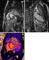
Cardiac MR images in a 21-year-old male patient with filamin C
variant–associated cardiomyopathy. Postcontrast T1-weighted images acquired
with a 1.5-T MRI scanner in (A) short-axis view and (B) vertical long-axis view
demonstrate marked concentric (or “ringlike”) midmyocardial late
gadolinium enhancement of the left ventricle (yellow arrows). (C) Native T1 mapping
image in short-axis view demonstrates an elevated T1 value (1120 msec).
Cardiac MR images in a 21-year-old male patient with filamin C variant–associated cardiomyopathy. Postcontrast T1-weighted images acquired with a 1.5-T MRI scanner in (A) short-axis view and (B) vertical long-axis view demonstrate marked concentric (or “ringlike”) midmyocardial late gadolinium enhancement of the left ventricle (yellow arrows). (C) Native T1 mapping image in short-axis view demonstrates an elevated T1 value (1120 msec).

References

    1. Ortiz-Genga MF , Cuenca S , Dal Ferro M , et al. . Truncating FLNC Mutations Are Associated With High-Risk Dilated and Arrhythmogenic Cardiomyopathies . J Am Coll Cardiol 2016. ; 68 ( 22 ): 2440 – 2451 . - PubMed
    1. Augusto JB , Eiros R , Nakou E , et al. . Dilated cardiomyopathy and arrhythmogenic left ventricular cardiomyopathy: a comprehensive genotype-imaging phenotype study . Eur Heart J Cardiovasc Imaging 2020. ; 21 ( 3 ): 326 – 336 . - PubMed
    1. Wang H , Bo K , Gao Y , Zhou Z , Xu L . Prognosis evaluation of chronic inflammatory cardiomyopathy with ring-like late gadolinium enhancement . ESC Heart Fail 2023. ; 10 ( 3 ): 1735 – 1744 . - PMC - PubMed