. 2023 Dec;5(6):e230155.
doi: 10.1148/ryct.230155.

Cardiac MRI and Clinical Outcomes in TMEM43 Arrhythmogenic Cardiomyopathy

Affiliations

Cardiac MRI and Clinical Outcomes in TMEM43 Arrhythmogenic Cardiomyopathy

João Matos et al. Radiol Cardiothorac Imaging. 2023 Dec.

Abstract

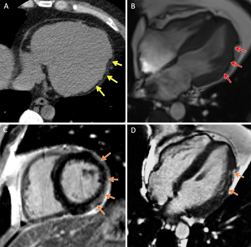
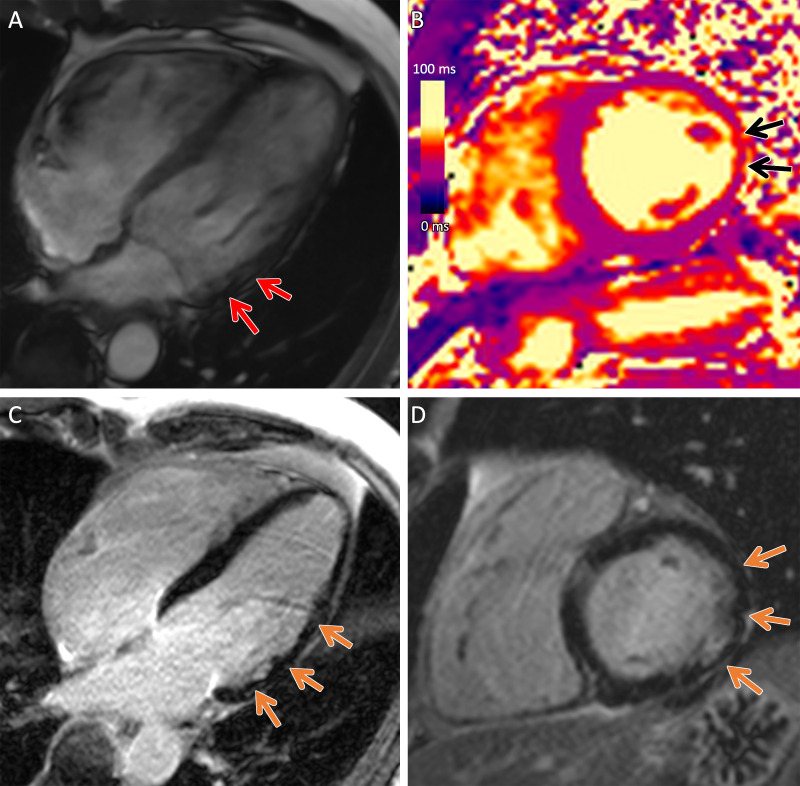
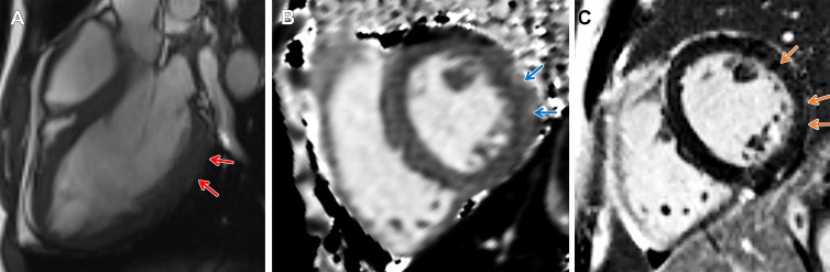
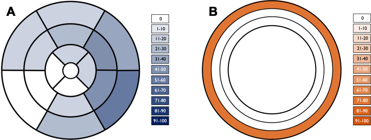
Arrhythmogenic cardiomyopathy is an inherited cardiomyopathy that can involve both ventricles. Several genes have been identified as pathogenic in arrhythmogenic cardiomyopathy, including TMEM43. However, there are limited data on cardiac MRI findings in patients with TMEM43 variants to date. In this case series, cardiac MRI findings and clinical outcomes are described in 14 patients with TMEM43 variants, including eight (57%) with the pathogenic p.Ser358Leu variant (six female patients; mean age, 33 years ± 15 [SD]) and six (43%) with a TMEM43 variant of unknown significance (three female patients; mean age, 38 years ± 11). MRI findings demonstrated left ventricular systolic dysfunction in eight (57%) patients and right ventricular dysfunction in four (29%) patients. Among the nine patients with late gadolinium enhancement imaging, left ventricular late gadolinium enhancement was present in seven (78%; all subepicardial) patients. In summary, TMEM43 variants are associated with high prevalence of subepicardial late gadolinium enhancement and left ventricular dysfunction. Keywords: Arrhythmogenic Cardiomyopathy, Arrhythmogenic Right Ventricular Cardiomyopathy, TMEM43, Cardiac MRI, Genetic Variants Supplemental material is available for this article.

Keywords: Arrhythmogenic Cardiomyopathy; Arrhythmogenic Right Ventricular Cardiomyopathy; Cardiac MRI; Genetic Variants; TMEM43.


Conflict of interest statement

Disclosures of conflicts of interest: J.M. No relevant relationships. E.H. No relevant relationships. M.C. Speaker honoraria from Blueprint Genetics. Y.M. No relevant relationships. M.H.G. No relevant relationships. P.T. Consultation fees from GE. D.S. No relevant relationships. K.H. Payment from Sanofi; associate editor for Radiology and Radiology: Cardiothoracic Imaging.

Figures

TMEM43 arrhythmogenic cardiomyopathy. Cardiac CT and MR images in a
male patient between 30 and 39 years of age (exact age not provided due to
potential reidentification risk) with a TMEM43 variant of unknown
significance (p.Gly280Glu) with palpitations and premature ventricular beats
at Holter monitoring. (A) Axial noncontrast cardiac CT image demonstrates
extensive subepicardial fat along the lateral left ventricular wall (yellow
arrows). (B) Four-chamber 1.5-T steady-state free precession MR image
demonstrates subepicardial chemical shift artifact along the lateral left
ventricular wall (red arrows). (C) Short-axis and (D) four-chamber late
gadolinium enhancement images demonstrate subepicardial late gadolinium
enhancement involving the midventricular anterolateral wall, inferolateral
wall, and inferior wall (orange arrows).
Figure 1:
TMEM43 arrhythmogenic cardiomyopathy. Cardiac CT and MR images in a male patient between 30 and 39 years of age (exact age not provided due to potential reidentification risk) with a TMEM43 variant of unknown significance (p.Gly280Glu) with palpitations and premature ventricular beats at Holter monitoring. (A) Axial noncontrast cardiac CT image demonstrates extensive subepicardial fat along the lateral left ventricular wall (yellow arrows). (B) Four-chamber 1.5-T steady-state free precession MR image demonstrates subepicardial chemical shift artifact along the lateral left ventricular wall (red arrows). (C) Short-axis and (D) four-chamber late gadolinium enhancement images demonstrate subepicardial late gadolinium enhancement involving the midventricular anterolateral wall, inferolateral wall, and inferior wall (orange arrows).
TMEM43 arrhythmogenic cardiomyopathy. Cardiac CT and MR images in a
female patient between 50 and 59 years of age (exact age not provided due to
potential reidentification risk) with a TMEM43 variant of unknown
significance (p.Glu142Gly) with premature ventricular beats at Holter
monitoring. (A, B) Short-axis late gadolinium enhancement images at the (A)
base and (B) midventricle demonstrate subepicardial late gadolinium
enhancement involving the basal to mid inferolateral wall (orange arrows).
(C) Axial and (D) short-axis noncontrast cardiac CT images demonstrate
subepicardial fat along the lateral basal left ventricular wall (yellow
arrows) and right ventricular implantable cardioverter defibrillator lead
(blue arrows).
Figure 2:
TMEM43 arrhythmogenic cardiomyopathy. Cardiac CT and MR images in a female patient between 50 and 59 years of age (exact age not provided due to potential reidentification risk) with a TMEM43 variant of unknown significance (p.Glu142Gly) with premature ventricular beats at Holter monitoring. (A, B) Short-axis late gadolinium enhancement images at the (A) base and (B) midventricle demonstrate subepicardial late gadolinium enhancement involving the basal to mid inferolateral wall (orange arrows). (C) Axial and (D) short-axis noncontrast cardiac CT images demonstrate subepicardial fat along the lateral basal left ventricular wall (yellow arrows) and right ventricular implantable cardioverter defibrillator lead (blue arrows).
Arrhythmogenic cardiomyopathy with TMEM43 pathogenic variant
(p.Ser358Leu). Cardiac 3-T MR images in a male patient between 50 and 59
years of age (exact age not provided due to potential reidentification risk)
with chest pain and premature ventricular beats at Holter monitoring. The
patient had a family history of arrhythmogenic cardiomyopathy. (A)
Four-chamber steady-state free precession MR image demonstrates
subepicardial chemical shift artifact along the basal to midventricular
lateral left ventricular wall (red arrows). (B) Native T2 map demonstrates
regional high T2 values at the midventricular inferolateral wall (black
arrows). (C) Four-chamber and (D) short-axis late gadolinium enhancement
images demonstrate subepicardial late gadolinium enhancement involving the
basal to midventricular anterolateral and inferolateral wall (orange
arrows).
Figure 3:
Arrhythmogenic cardiomyopathy with TMEM43 pathogenic variant (p.Ser358Leu). Cardiac 3-T MR images in a male patient between 50 and 59 years of age (exact age not provided due to potential reidentification risk) with chest pain and premature ventricular beats at Holter monitoring. The patient had a family history of arrhythmogenic cardiomyopathy. (A) Four-chamber steady-state free precession MR image demonstrates subepicardial chemical shift artifact along the basal to midventricular lateral left ventricular wall (red arrows). (B) Native T2 map demonstrates regional high T2 values at the midventricular inferolateral wall (black arrows). (C) Four-chamber and (D) short-axis late gadolinium enhancement images demonstrate subepicardial late gadolinium enhancement involving the basal to midventricular anterolateral and inferolateral wall (orange arrows).
Arrhythmogenic cardiomyopathy with TMEM43 pathogenic variant
(p.Ser358Leu). Short-axis 3-T MR images in a male patient between 18 and 19
years of age (exact age not provided due to potential reidentification risk)
with palpitations and premature ventricular beats at Holter monitoring. He
had a family history of arrhythmogenic cardiomyopathy in his grandfather,
father, and brother. (A) Three-chamber steady-state free precession MR image
demonstrates subepicardial chemical shift artifact along the basal to mid
left ventricular inferolateral wall (red arrows). (B) Native T1 map
demonstrates regional high T1 values at the subepicardial midventricular
inferolateral wall (blue arrows). (C) There is subepicardial late gadolinium
enhancement involving the midventricular anterior wall, anterolateral wall,
and inferolateral wall (orange arrows).
Figure 4:
Arrhythmogenic cardiomyopathy with TMEM43 pathogenic variant (p.Ser358Leu). Short-axis 3-T MR images in a male patient between 18 and 19 years of age (exact age not provided due to potential reidentification risk) with palpitations and premature ventricular beats at Holter monitoring. He had a family history of arrhythmogenic cardiomyopathy in his grandfather, father, and brother. (A) Three-chamber steady-state free precession MR image demonstrates subepicardial chemical shift artifact along the basal to mid left ventricular inferolateral wall (red arrows). (B) Native T1 map demonstrates regional high T1 values at the subepicardial midventricular inferolateral wall (blue arrows). (C) There is subepicardial late gadolinium enhancement involving the midventricular anterior wall, anterolateral wall, and inferolateral wall (orange arrows).
Segmental distribution of late gadolinium enhancement. (A)
Color-shaded polar plot represents the percentage of patients with late
gadolinium enhancement for each myocardial segment according to a
standardized 17-segment model. (B) Color-shaded plot of the ventricular
myocardium represents the percentage of patients with late gadolinium
enhancement in each myocardial layer (from outer to inner: subepicardial,
midwall, and subendocardial).
Figure 5:
Segmental distribution of late gadolinium enhancement. (A) Color-shaded polar plot represents the percentage of patients with late gadolinium enhancement for each myocardial segment according to a standardized 17-segment model. (B) Color-shaded plot of the ventricular myocardium represents the percentage of patients with late gadolinium enhancement in each myocardial layer (from outer to inner: subepicardial, midwall, and subendocardial).

Similar articles

Cited by

References

    1. Corrado D , Basso C , Judge DP . Arrhythmogenic Cardiomyopathy . Circ Res 2017. ; 121 ( 7 ): 784 – 802 . - PubMed
    1. Marcus FI , Fontaine GH , Guiraudon G , et al. . Right ventricular dysplasia: a report of 24 adult cases . Circulation 1982. ; 65 ( 2 ): 384 – 398 . - PubMed
    1. Corrado D , Perazzolo Marra M , Zorzi A , et al. . Diagnosis of arrhythmogenic cardiomyopathy: The Padua criteria . Int J Cardiol 2020. ; 319 : 106 – 114 . - PubMed
    1. Fressart V , Duthoit G , Donal E , et al. . Desmosomal gene analysis in arrhythmogenic right ventricular dysplasia/cardiomyopathy: spectrum of mutations and clinical impact in practice . Europace 2010. ; 12 ( 6 ): 861 – 868 . - PubMed
    1. Merner ND , Hodgkinson KA , Haywood AFM , et al. . Arrhythmogenic right ventricular cardiomyopathy type 5 is a fully penetrant, lethal arrhythmic disorder caused by a missense mutation in the TMEM43 gene . Am J Hum Genet 2008. ; 82 ( 4 ): 809 – 821 . - PMC - PubMed