Review
. 2023 Nov;248(22):2001-2019.
doi: 10.1177/15353702231215897. Epub 2024 Jan 2.

Modeling immunity in microphysiological systems

Affiliations
Review

Modeling immunity in microphysiological systems

Brian J Kwee et al. Exp Biol Med (Maywood). 2023 Nov.

Abstract

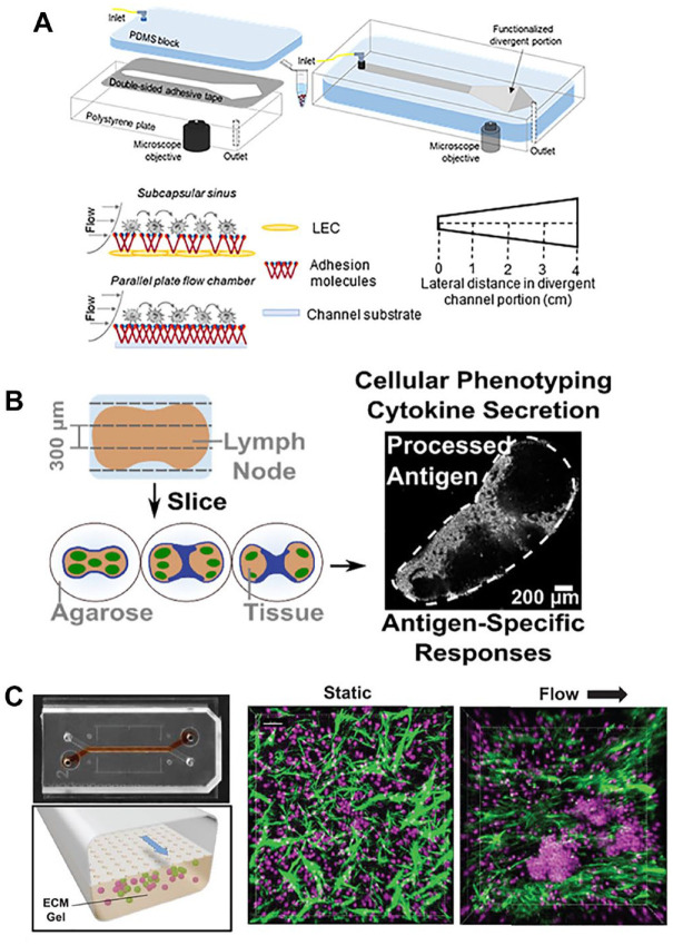
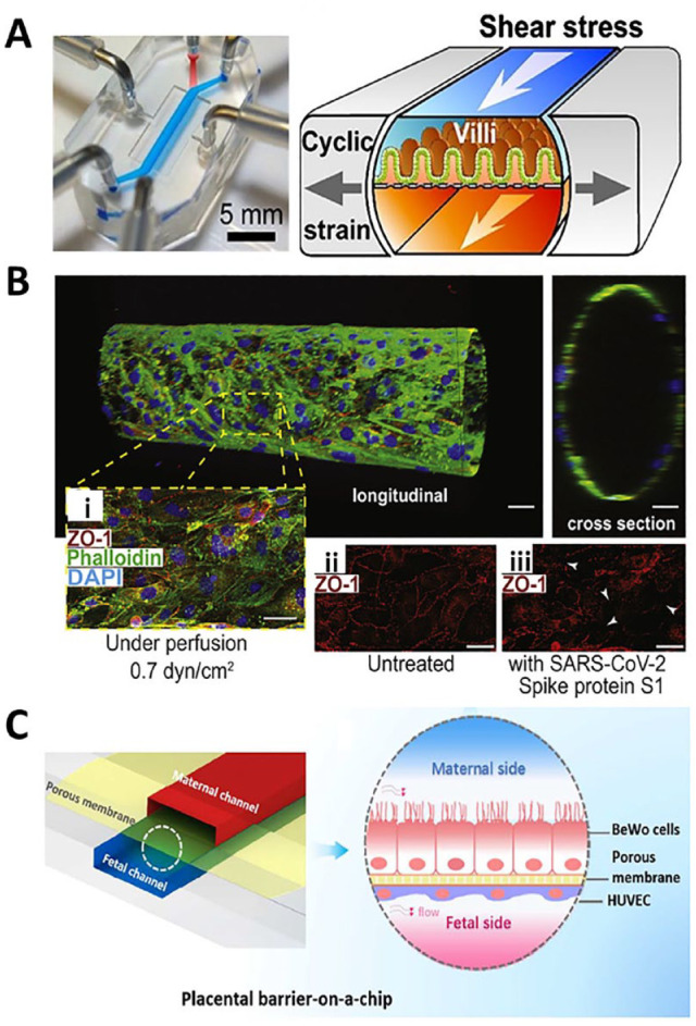
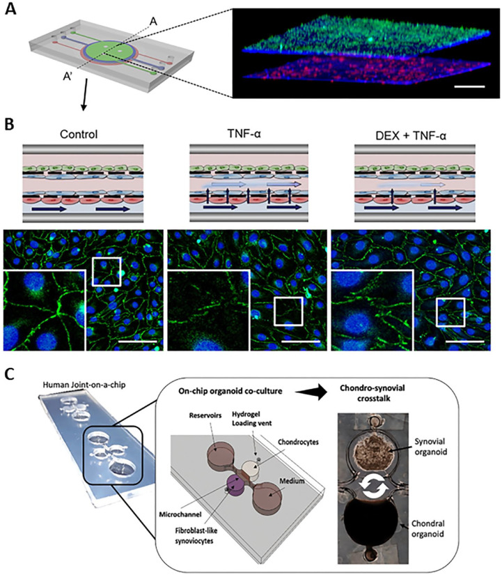
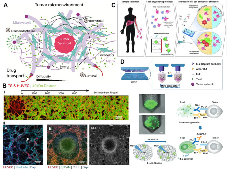
There is a need for better predictive models of the human immune system to evaluate safety and efficacy of immunomodulatory drugs and biologics for successful product development and regulatory approvals. Current in vitro models, which are often tested in two-dimensional (2D) tissue culture polystyrene, and preclinical animal models fail to fully recapitulate the function and physiology of the human immune system. Microphysiological systems (MPSs) that can model key microenvironment cues of the human immune system, as well as of specific organs and tissues, may be able to recapitulate specific features of the in vivo inflammatory response. This minireview provides an overview of MPS for modeling lymphatic tissues, immunity at tissue interfaces, inflammatory diseases, and the inflammatory tumor microenvironment in vitro and ex vivo. Broadly, these systems have utility in modeling how certain immunotherapies function in vivo, how dysfunctional immune responses can propagate diseases, and how our immune system can combat pathogens.

Keywords: Microphysiological systems; immunity; inflammatory diseases; lymphatics; tissue interfaces; tumor microenvironment.


Conflict of interest statement

Declaration Of Conflicting InterestsThe author(s) declared no potential conflicts of interest with respect to the research, authorship, and/or publication of this article.

Figures

Figure 1.
Figure 1.
Examples of lymphatic microphysiological systems that may be used to evaluate vaccines. (A) A divergent channel perfusion system is used to evaluate the effect of lymph node subcapsular sinus microenvironment-mimicking flow on cancer and immune cell adhesion in vitro. (Source: Reproduced with permission, Copyright 2021 Elsevier.) (B) Murine lymph node slices cultured ex vivo can model cellular adaptive immune responses and antigen-specific responses. (Source: Reproduced with permission, Copyright 2021 American Chemical Society.) (C) A lymph node follicle on a chip can induce ectopic lymph node follicle formation from primary B- and T-cells under flow to model lymph node germinal centers. In right confocal images, green denotes extracellular matrix fibers by second harmonic imaging and magenta denotes Hoechst staining of culture lymphocytes. (Source: Reproduced with permission, an open-access journal printed by 2022 Wiley-VCH GmbH.)
Figure 2.
Figure 2.
Examples of microphysiological systems to model tissue interface. (A) A GI-on-a-chip displays mechanical strain (gray arrows) and shear force (white arrows) contribute to the microvillous formation. (Source: Reproduced with permission, Copyright 2015 National Academy of Sciences (NAS).) (B) A 3D human BBB model shows an endothelial monolayer at longitudinal and cross-sectional view, and that SARS-CoV-2 spike protein S1 disrupt tight junctions (white arrows). (Source: Reproduced with permission, an open-access journal printed by 2020 Elsevier.) Scale bar: 20 μm. (C) A placenta-on-a-chip model shows the trophoblasts and endothelial cells on the opposite of a microporous membrane, and the flow mimic the physiological environment. (Source: Reproduced with permission, Copyright 2018 American Chemical Society.)
Figure 3.
Figure 3.
Examples of microphysiological systems to model pathological inflammation. (A). A skin-on-a-chip microfluidic device presented a three-dimensional (3D) fluorescence image of the cross-section (A-A’). The 3D image showed four layers of three cell types that were uniformly stacked on two porous membranes. HaCaT cells, HS27 fibroblasts (Fbs), and HUVECs were stained in green, blue, and red, respectively. Scale bars: 300 μm. (B) Schematic of the skin edema model in a microfluidic device showed a control treatment, an inflammatory condition with TNF-α exposure, and a therapeutic treatment with TNF-α exposure after a dexamethasone (Dex) pretreatment on the top panel. On the bottom panel, immunofluorescence microscopy showed DAPI (blue) staining of HUVEC nuclei, and zonula occluden (ZO-1) (green) staining of HUVEC tight junctions. Gap junction was disrupted in the TNF-α-treat chip, and Dex prevented this TNF-α-induced disruption of the tight junctions. Scale bars: 100 μm. (Source: Reprinted from Figure 3(F) for Figure 3(A), Figure 7(B) for Figure 3(B), top panel, and Figure 6(G) to (I) for Figure 3(B), bottom panel from Wufuer et al. Distributed under a Creative Commons Attribution 4.0 International License (CC BY 4.0) http://creativecommons.org/licenses/by/4.0/.) (C) A human joint-on-a-chip model displayed a co-culture system comprising chondral and fibroblasts-like synovial compartments for reciprocal inflammatory cross talk studies in arthritis research. (Source: Reprinted from Figure 1(B) of Rothbauer et al. with permission from the Royal Society of Chemistry. Distributed under a Creative Commons Attribution 3.0 Unported License (CC BY 3.0) https://creativecommons.org/licenses/by/3.0/.)
Figure 4.
Figure 4.
How microphysical tumor models are being used to understand the tumor microenvironment and possible cancer therapeutics. (A) Illustration of the complexity of the tumor microenvironment and some of its main components and how drug transport interacts with this environment. (Source: Reproduced with permission, Copyright 2021 WILEY – VCH VERLAG GMBH & CO. KGAA). (B) The top image illustrates HUVEC-derived vasculature created in a microfluidic device used to assess permeability and shows how a tumor spheroid influences vascular network density within the in vitro model. The bottom three images show different cancer types modeled and vascularized within a microfluidic device. The image on the bottom left illustrates HUVEC-derived vasculature within ovarian carcinoma, where the white arrow indicating a ring of fibroblast taken at day 7. The middle and right most images show lung adenocarcinoma fixed at day 7. (Scale bar 200 microns). (Source: Reproduced with permission, Copyright 2021 WILEY – VCH VERLAG GMBH & CO. KGAA.) (C) Illustrates the process by which T-cell therapeutics are developed and used in a microfluidics to assess their anticancer ability. (Source: Reproduced with permission, Copyright 2021 American Society for Clinical Investigation.) (D) Illustrates the immunotherapeutic high-throughput observation chamber (iHOC) system which uses tumor spheroids as a microphysical model for the exploration of PDL-1 in T-cell infiltration of the tumor environment. Also included is an illustration showing PD-1’s role in T-cell immunosuppression and how this effects tumor infiltration within the tumor microenvironment. (Source: Edited and reproduced with permission, Copyright 2020 WILEY VCH.)

Similar articles

Cited by

References

    1. Descotes JJ. Methods of evaluating immunotoxicity. Expert Opin Drug Metab Toxicol 2006;2:249–59 - PubMed
    1. Pineda CC, Castañeda Hernández G, Jacobs IA, Alvarez DF, Carini C. Assessing the immunogenicity of biopharmaceuticals. Biodrugs 2016;30:195–206 - PMC - PubMed
    1. Cerf-Bensussan N, Gaboriau-Routhiau V. The immune system and the gut microbiota: friends or foes? Nat Rev Immunol 2010;10:735–44 - PubMed
    1. Park JE, Barbul A. Understanding the role of immune regulation in wound healing. Am J Surg 2004;187:11S–6 - PubMed
    1. Zschaler JJ, Schlorke D, Arnhold J. Differences in innate immune response between man and mouse. Crit Rev Immunol 2014;34:433–54 - PubMed