

Leukocyte differential gene expression prognostic value for high versus low seizure frequency in temporal lobe epilepsy
- PMID: 38166692
- PMCID: PMC10759702
- DOI: 10.1186/s12883-023-03459-1
Leukocyte differential gene expression prognostic value for high versus low seizure frequency in temporal lobe epilepsy
Abstract
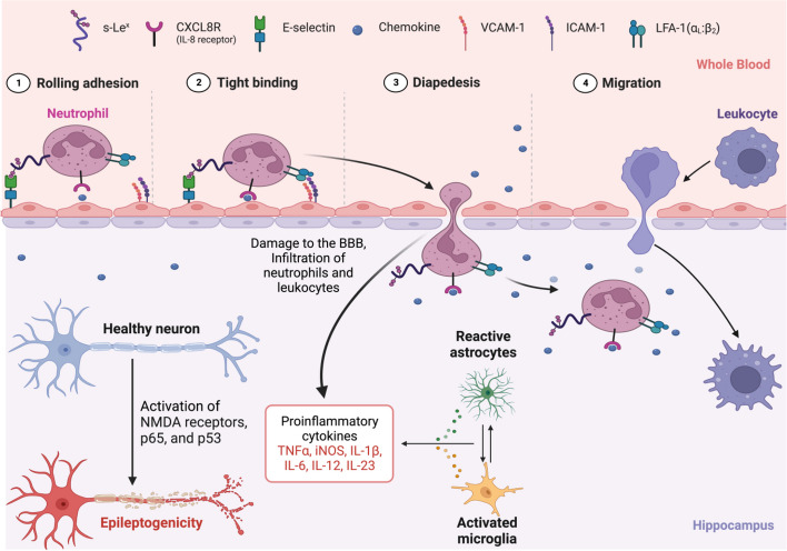
Background: This study was performed to test the hypothesis that systemic leukocyte gene expression has prognostic value differentiating low from high seizure frequency refractory temporal lobe epilepsy (TLE).
Methods: A consecutive series of patients with refractory temporal lobe epilepsy was studied. Based on a median baseline seizure frequency of 2.0 seizures per month, low versus high seizure frequency was defined as ≤ 2 seizures/month and > 2 seizures/month, respectively. Systemic leukocyte gene expression was analyzed for prognostic value for TLE seizure frequency. All differentially expressed genes were analyzed, with Ingenuity® Pathway Analysis (IPA®) and Reactome, to identify leukocyte gene expression and biological pathways with prognostic value for seizure frequency.
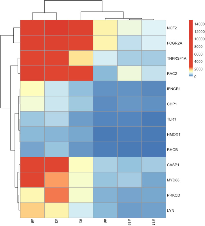
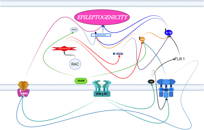
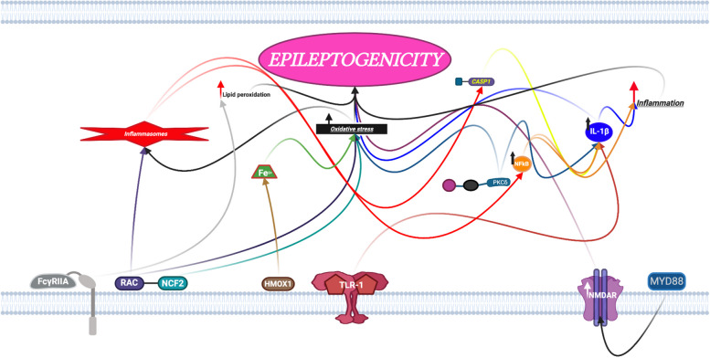
Results: There were ten males and six females with a mean age of 39.4 years (range: 16 to 62 years, standard error of mean: 3.6 years). There were five patients in the high and eleven patients in the low seizure frequency cohorts, respectively. Based on a threshold of twofold change (p < 0.001, FC > 2.0, FDR < 0.05) and expression within at least two pathways from both Reactome and Ingenuity® Pathway Analysis (IPA®), 13 differentially expressed leukocyte genes were identified which were all over-expressed in the low when compared to the high seizure frequency groups, including NCF2, HMOX1, RHOB, FCGR2A, PRKCD, RAC2, TLR1, CHP1, TNFRSF1A, IFNGR1, LYN, MYD88, and CASP1. Similar analysis identified four differentially expressed genes which were all over-expressed in the high when compared to the low seizure frequency groups, including AK1, F2R, GNB5, and TYMS.
Conclusions: Low and high seizure frequency TLE are predicted by the respective upregulation and downregulation of specific leukocyte genes involved in canonical pathways of neuroinflammation, oxidative stress and lipid peroxidation, GABA (γ-aminobutyric acid) inhibition, and AMPA and NMDA receptor signaling. Furthermore, high seizure frequency-TLE is distinguished prognostically from low seizure frequency-TLE by differentially increased specific leukocyte gene expression involved in GABA inhibition and NMDA receptor signaling. High and low seizure frequency patients appear to represent two mechanistically different forms of temporal lobe epilepsy based on leukocyte gene expression.
Keywords: Gene expression; Leukocyte; Seizure frequency; Temporal lobe epilepsy.
© 2023. The Author(s).
Conflict of interest statement
The authors declare that they have no competing interests. Author Dr. Weinand has been assigned the following patent application by the University of Arizona (patent ID# UA23-142) titled, “METHODS FOR THE PROGNOSIS AND TREATMENT OF TEMPORAL LOBE EPILEPSY.” for work relating to the research described in this manuscript. Dr. Weinand has received no financial compensation for the above assigned patent.
Figures


Similar articles
-
Altered expression of signaling pathways regulating neuronal excitability in hippocampal tissue of temporal lobe epilepsy patients with low and high seizure frequency.Epilepsy Res. 2019 Sep;155:106145. doi: 10.1016/j.eplepsyres.2019.05.013. Epub 2019 May 24. Epilepsy Res. 2019. PMID: 31195185
-
Leukocyte expression profiles reveal gene sets with prognostic value for seizure-free outcome following stereotactic laser amygdalohippocampotomy.Sci Rep. 2019 Jan 31;9(1):1086. doi: 10.1038/s41598-018-37763-5. Sci Rep. 2019. PMID: 30705324 Free PMC article.
-
Nocturnal temporal lobe epilepsy.Neurology. 1998 Jun;50(6):1772-7. doi: 10.1212/wnl.50.6.1772. Neurology. 1998. PMID: 9633726
-
Myeloid differentiation factor 88 is up-regulated in epileptic brain and contributes to experimental seizures in rats.Exp Neurol. 2017 Sep;295:23-35. doi: 10.1016/j.expneurol.2017.05.008. Epub 2017 May 18. Exp Neurol. 2017. PMID: 28529112
-
Deep brain stimulation for refractory temporal lobe epilepsy: a systematic review and meta-analysis with an emphasis on alleviation of seizure frequency outcome.Childs Nerv Syst. 2018 Feb;34(2):321-327. doi: 10.1007/s00381-017-3596-6. Epub 2017 Sep 18. Childs Nerv Syst. 2018. PMID: 28921161
Cited by
-
Increased Immunoglobulin and Proteoglycan Synthesis in Resected Hippocampal Tissue Predicts Post-Surgical Seizure Recurrence in Human Temporal Lobe Epilepsy.Pathophysiology. 2025 Apr 14;32(2):15. doi: 10.3390/pathophysiology32020015. Pathophysiology. 2025. PMID: 40265440 Free PMC article.
References
-
- de Barros Lourenço FH, Marques LHN, de Araujo Filho GM. Electroencephalogram alterations associated with psychiatric disorders in temporal lobe epilepsy with mesial sclerosis: a systematic review. Epilepsy Behav. 2020;108:107100. 10.1016/j.yebeh.2020.107100. - PubMed
MeSH terms
Substances
Grants and funding
LinkOut - more resources
Full Text Sources
Molecular Biology Databases
Research Materials
Miscellaneous
