

Application of contrast-enhanced ultrasound in the diagnosis of tuberous vas deferens tuberculosis
- PMID: 38166757
- PMCID: PMC10763423
- DOI: 10.1186/s12879-023-08886-6
Application of contrast-enhanced ultrasound in the diagnosis of tuberous vas deferens tuberculosis
Abstract
Background: To assess the value of contrast-enhanced ultrasound (CEUS) in the diagnosis of tuberous vas deferens tuberculosis (VD TB) and improve the positive diagnostic rate of VD TB.
Methods: CEUS and routine ultrasound (US) images of 17 patients with tuberous VD TB confirmed by surgery, pathology, or laboratory semen examination were retrospectively analyzed and summarized, and the positive rates of both imaging techniques were compared.
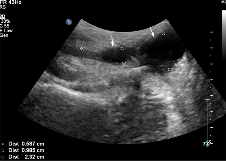
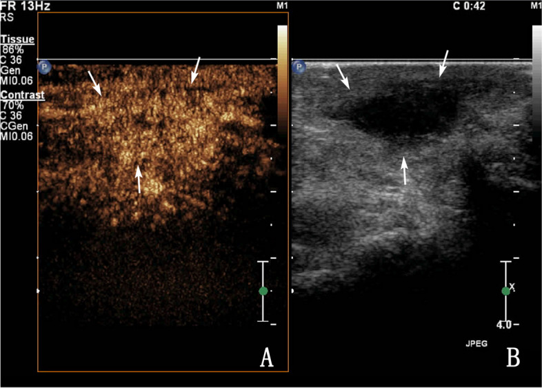
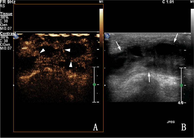
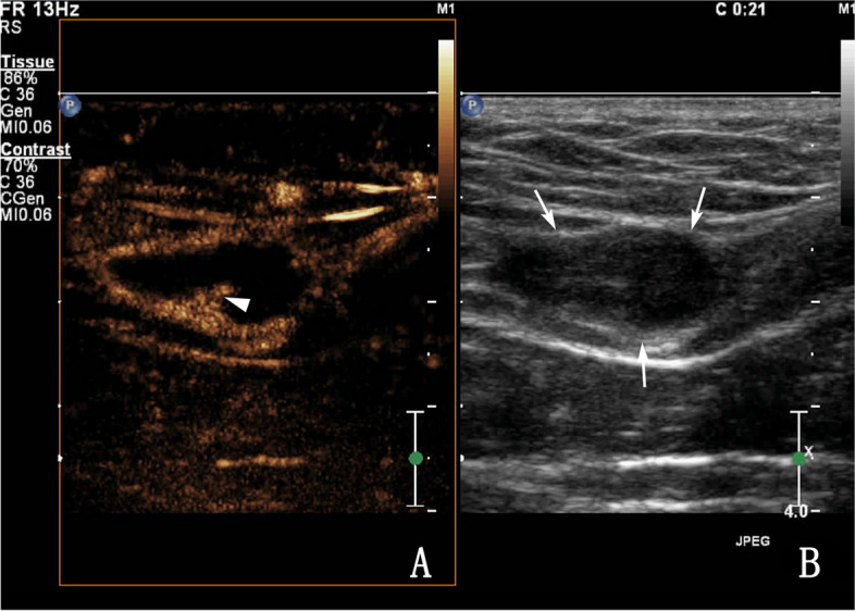
Results: The 19 VD lesions of the 17 patients were divided into two types according to the CEUS findings: Type I and Type II, and type II was divided into Types IIa, IIb, and IIc. Of the nodules with transverse diameters > 1 cm, 100% presented as type II. Of the nodules with transverse diameters < 1 cm, 37.5% (3/8) presented as type I and 62.5% (5/8) presented as type II. The sonographic manifestations of tuberous VD TB were hypoechoic and mixed echoic. The positive diagnostic rate was 89.5% for CEUS and 68.4% for US, but the difference was not significant (χ2 = 2.533; P = 0.111).
Conclusions: CEUS was able to show the blood supply characteristics of tuberous VD TB, the internal necrosis of nodules was more easily observed by CEUS than by routine US, which is helpful for the diagnosis of tuberous VD TB.
Keywords: Contrast-enhanced ultrasound; Male infertility; Reproductive system; Tuberculosis; Vas deferens.
© 2023. The Author(s).
Conflict of interest statement
The authors declare no competing interests.
Figures
Similar articles
-
The Role of Contrast-Enhanced Ultrasound in the Differential Diagnosis of Tuberous Vas Deferens Tuberculosis and Metastatic Inguinal Lymph Nodes.Diagnostics (Basel). 2023 May 17;13(10):1762. doi: 10.3390/diagnostics13101762. Diagnostics (Basel). 2023. PMID: 37238246 Free PMC article.
-
Vas deferens sonographic appearances of tuberculosis lesions of 19 cases of male genital systemic tuberculosis.Medicine (Baltimore). 2019 Mar;98(11):e14843. doi: 10.1097/MD.0000000000014843. Medicine (Baltimore). 2019. PMID: 30882677 Free PMC article.
-
Contrast-enhanced Ultrasound for Diagnosis of Renal Cystic Mass.Curr Med Imaging. 2022;18(3):292-298. doi: 10.2174/1573405617666210719141831. Curr Med Imaging. 2022. PMID: 34825641
-
[Application value of elastic ultrasound and contrast-enhanced ultrasonography in evaluating testicular blood supply after blockage of the vas deferens artery].Zhonghua Nan Ke Xue. 2019 Nov;25(11):990-995. Zhonghua Nan Ke Xue. 2019. PMID: 32233232 Chinese.
-
Contrast-enhanced ultrasound of malignant liver lesions.Abdom Radiol (NY). 2018 Apr;43(4):819-847. doi: 10.1007/s00261-017-1360-8. Abdom Radiol (NY). 2018. PMID: 29094174 Review.
References
-
- Kulchavenya E. Male genital tuberculosis. Cham: Springer International Publishing; 2014.
MeSH terms
Substances
Grants and funding
- 20190101A09/Hangzhou Agriculture and Social Development Plan
- 20190101A09/Hangzhou Agriculture and Social Development Plan
- 20190101A09/Hangzhou Agriculture and Social Development Plan
- 20190101A09/Hangzhou Agriculture and Social Development Plan
- 20190101A09/Hangzhou Agriculture and Social Development Plan
LinkOut - more resources
Full Text Sources
