

Evaluation of extravascular lung water and cardiac function in normal vaginal delivery by intrapartum bedside ultrasound
- PMID: 38166871
- PMCID: PMC10759567
- DOI: 10.1186/s12884-023-06201-4
Evaluation of extravascular lung water and cardiac function in normal vaginal delivery by intrapartum bedside ultrasound
Abstract
Background: Healthy parturients may experience pulmonary edema and disturbed cardiac function during labor. We aimed to evaluate the extravascular lung water (EVLW), intravascular volume, and cardiac function of normal parturients during spontaneous vaginal delivery by bedside ultrasound. And to explore the correlation between EVLW and intravascular volume, cardiac function.
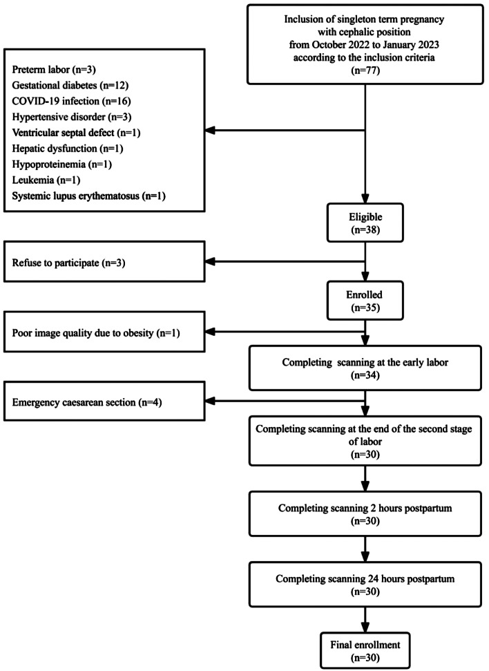
Methods: This was a prospective observational study including 30 singleton-term pregnant women undergoing spontaneous vaginal delivery. Bedside ultrasound was performed at the early labor, the end of the second stage of labor, 2 and 24 h postpartum, and 120 scanning results were recorded. EVLW was evaluated by the echo comet score (ECS) obtained by the 28-rib interspaces technique. Inferior vena cava collapsibility index (IVC-CI), left ventricle ejection fraction, right ventricle fractional area change, left and right ventricular E/A ratio, and left and right ventricular index of myocardial performance (LIMP and RIMP) were measured. Measurements among different time points were compared, and the correlations between ECS and other measurements were analyzed.
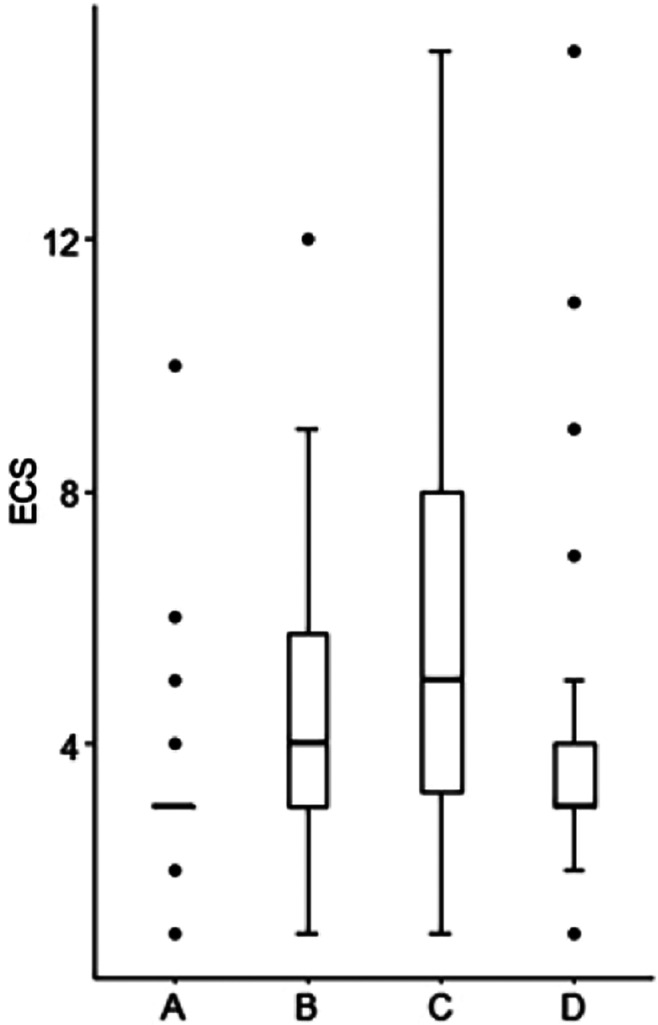
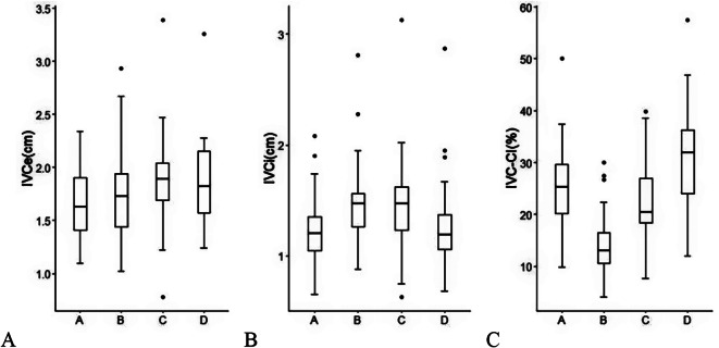
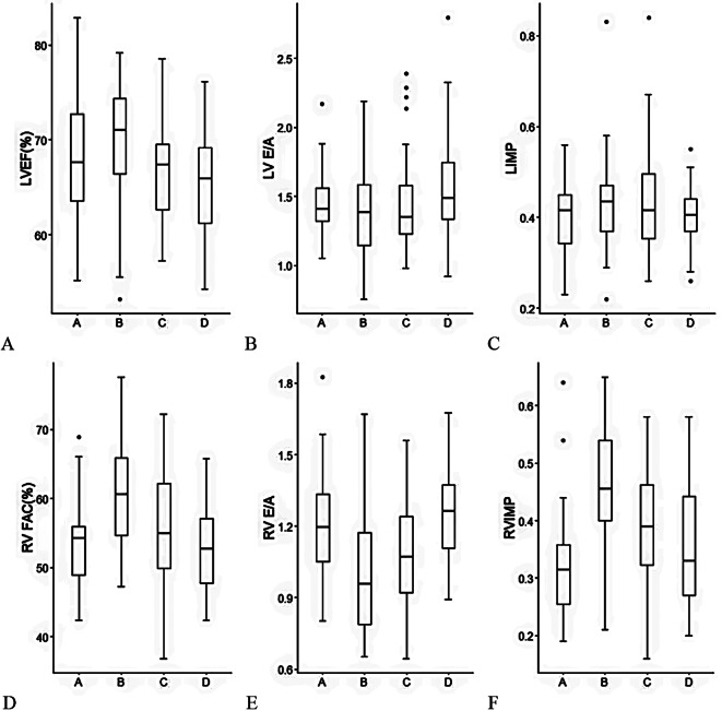
Results: During the spontaneous vaginal delivery of healthy pregnant women, 2 had a mild EVLW increase at the early labor, 8 at the end of the second stage of labor, 13 at 2 h postpartum, and 4 at 24 h postpartum (P < 0.001). From the early labor to 24 h postpartum, ECS first increased and then decreased, reaching its peak at 2 h postpartum (P < 0.001). IVC-CI first decreased and then increased, reaching its minimum at the end of the second stage of labor (P < 0.001). RIMP exceeded the cut-off value of 0.43 at the end of the second stage of labor. ECS was weakly correlated with IVC-CI (r=-0.373, P < 0.001), LIMP (r = 0.298, P = 0.022) and RIMP (r = 0.211, P = 0.021).
Conclusions: During spontaneous vaginal delivery, the most vital period of perinatal care is between the end of the second stage of labor and 2 h postpartum, because the risk of pulmonary edema is higher and the right ventricle function may decline. IVC-CI can be used to evaluate maternal intravascular volume. The increase in EVLW may be related to the increase in intravascular volume and the decrease in ventricular function.
Keywords: Cardiac function; Echocardiography; Labor; Lung ultrasound; Pulmonary edema.
© 2023. The Author(s).
Conflict of interest statement
The authors declare no competing interests.
Figures
Similar articles
-
Point-of-Care Lung Ultrasound Pattern in Healthy Parturients: Prevalence of Pulmonary Interstitial Syndrome Following Vaginal Delivery, Elective and Unplanned Intrapartum Cesarean Delivery.Anesth Analg. 2021 Sep 1;133(3):739-746. doi: 10.1213/ANE.0000000000005464. Anesth Analg. 2021. PMID: 33721873
-
Assessment of stress-induced pulmonary interstitial edema by chest ultrasound during exercise echocardiography and its correlation with left ventricular function.J Am Soc Echocardiogr. 2006 Apr;19(4):457-63. doi: 10.1016/j.echo.2005.11.013. J Am Soc Echocardiogr. 2006. PMID: 16581487 Clinical Trial.
-
Lung ultrasound to quantitatively evaluate extravascular lung water content and its clinical significance.J Matern Fetal Neonatal Med. 2022 Aug;35(15):2904-2914. doi: 10.1080/14767058.2020.1812057. Epub 2020 Sep 16. J Matern Fetal Neonatal Med. 2022. PMID: 32938256
-
Intrapartum ultrasound measurement of angle of progression at the onset of the second stage of labor for prediction of spontaneous vaginal delivery in term singleton pregnancies: a systematic review and meta-analysis.Am J Obstet Gynecol. 2022 Feb;226(2):205-214.e2. doi: 10.1016/j.ajog.2021.07.031. Epub 2021 Aug 9. Am J Obstet Gynecol. 2022. PMID: 34384775
-
Ultrasound lung comets: a clinically useful sign of extravascular lung water.J Am Soc Echocardiogr. 2006 Mar;19(3):356-63. doi: 10.1016/j.echo.2005.05.019. J Am Soc Echocardiogr. 2006. PMID: 16500505 Review.
References
Publication types
MeSH terms
LinkOut - more resources
Full Text Sources
