. 2024 Jan 3;22(1):13.
doi: 10.1186/s12967-023-04825-w.

AIM2 promotes irradiation resistance, migration ability and PD-L1 expression through STAT1/NF-κB activation in oral squamous cell carcinoma

Affiliations

AIM2 promotes irradiation resistance, migration ability and PD-L1 expression through STAT1/NF-κB activation in oral squamous cell carcinoma

Hui-Wen Chiu et al. J Transl Med. .

Abstract

Background: Radioresistance and lymph node metastasis are common phenotypes of refractory oral squamous cell carcinoma (OSCC). As a result, understanding the mechanism for radioresistance and metastatic progression is urgently needed for the precise management of refractory OSCC. Recently, immunotherapies, e.g. immune checkpoint inhibitors (ICIs), were employed to treat refractory OSCC; however, the lack of predictive biomarkers still limited their therapeutic effectiveness.

Methods: The Cancer Genome Atlas (TCGA)/Gene Expression Omnibus (GEO) databases and RT-PCR analysis were used to determine absent in melanoma 2 (AIM2) expression in OSCC samples. Colony-forming assay and trans-well cultivation was established for estimating AIM2 function in modulating the irradiation resistance and migration ability of OSCC cells, respectively. RT-PCR, Western blot and flow-cytometric analyses were performed to examine AIM2 effects on the expression of programmed death-ligand 1 (PD-L1) expression. Luciferase-based reporter assay and site-directed mutagenesis were employed to determine the transcriptional regulatory activity of Signal Transducer and Activator of Transcription 1 (STAT1) and NF-κB towards the AIM2-triggered PD-L1 expression.

Results: Here, we found that AIM2 is extensively upregulated in primary tumors compared to the normal adjacent tissues and acts as a poor prognostic marker in OSCC. AIM2 knockdown mitigated, but overexpression promoted, radioresistance, migration and PD-L1 expression via modulating the activity of STAT1/NF-κB in OSCC cell variants. AIM2 upregulation significantly predicted a favorable response in patients receiving ICI treatments.

Conclusions: Our data unveil AIM2 as a critical factor for promoting radioresistance, metastasis and PD-L1 expression and as a potential biomarker for predicting ICI effectiveness on the refractory OSCC.

Keywords: AIM2; Immune checkpoint inhibitors; Metastasis; Oral squamous cell carcinoma; PD-L1; Radioresistance.


Conflict of interest statement

The authors declare that they have no competing interests.

Figures

Fig. 1
Fig. 1
AIM2 upregulation is highly detected in primary tumors compared to normal adjacent tissues and correlates with a poor response to radiotherapy in OSCC. A and B Boxplots and dot plots with line respectively present AIM2 mRNA levels in the unpaired normal tissues (N)/primary tumors (T) and paired normal adjacent tissues (NAT)/primary tumors derived from TCGA HNSCC (A) and GSE4273 OSCC (B) samples. C and D RT-PCR (C) and dot plots with lines (D) show the AIM2 gene expression in the paired primary tumors/normal adjacent tissues derived from OSCC samples in Taipei Medical University (TMU) Biobank. E Kaplan–Meier analyses for AIM2 mRNA levels using overall survival (OS) probability under a maximal risk condition against TCGA HNSCC (upper) and GSE42743 OSCC (lower) patients who received radiotherapy. F Scatchard plots for the correlation between AIM2 mRNA levels in the respective primary tumors and overall survival time of TCGA HNSCC and GSE42743 OSCC patients receiving radiotherapy (RT) with the overall survival endpoint as alive or dead
Fig. 2
Fig. 2
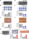
AIM2 expression is associated with cellular irradiation resistance and migration ability in OSCC cells. AI RT-PCR for the endogenous mRNA levels of AIM2 and GAPDH, crystal violet staining (upper)/histogram (lower) for colony formation post-treatment without or with irradiation at 4 Gy and Giemsa staining (upper)/histogram (lower) for the migrated cells in the tested OSCC cell lines (AC), HSC4 cell variants (DF) and SAS cell variants (GI). in A, D and G, GAPDH was used as an internal control of the experiment. The abbreviations of PT, NS, VC and OE are parental, non-silencing, vector control and overexpression, respectively. ***p < 0.001
Fig. 3
Fig. 3
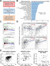
AIM2 upregulation highly correlates with the activation of interferon and inflammation-related pathways and the expression of PD-L1 in the primary tumors derived from OSCC patients with a poorer responsiveness to radiotherapy. A Experimental flowchart for generating AIM2 gene signature and performing GSEA simulation. B Histogram for Hallmark genesets with positive NES and statistical significance (p and q < 0.05) in GSEA simulation against AIM2 gene signature. C The plot of enrichment score for IFNγ-responsive and TNFα/NF-κB signaling axis-regulated genesets in GSEA simulation against AIM2 gene signature. D Scatchard plot derived from TIMER2.0 for the correlation of AIM2 mRNA levels with tumor purity and infiltration levels of macrophages, CD8 + T cells and NK cells against TCGA HNSCC. E Scatchard plot for the co-expression of AIM2 and PD-L1 in TCGA HNSCC primary tumors (left) and GSE42743 OSCC samples derived from patients without (middle) or with (right) the indicated records. In D and E, the solid and dashed lines shown in red represent the 95% confidence bands (dashed) of the best-fit line (solid) in Simple Linear Regression
Fig. 4
Fig. 4
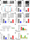
AIM2 upregulation promotes PD-L1 expression via activating STAT1 and NF-κB signaling pathways in OSCC cells. A and B RT-PCR (left)/Western blot analyses (right) for the endogenous mRNA/protein levels of PD-L1 and GAPDH (A) and histograms for the mean of fluorescent intensity (MFI) of isotype control (cyan) and PD-L1 (pink) antibodies in Flow-cytometric analysis (B) against SAS and HSC4 cells. C and D RT-PCR (upper)/Western blot analysis(middle) for the endogenous mRNA/protein levels of PD-L1 and GAPDH and histograms for the MFI of PD-L1 antibody normalized to control groups (NS and VC) in Flow-cytometric analysis against HSC4 (C) and SAS (D) cell variants. EH Western blot analyses for phosphorylated STAT1/NF-κB, total STAT1/NF-κB and GAPDH protein levels (upper) and histograms for the DNA-binding activity of STAT1/NF-κB normalized to control groups (NS and VC) in luciferase reporter assay (lower) against HSC4 (E and G) and SAS (F and H) cell variants. I Illustration for the locations and mutated nucleotides of NF-κB binding site/IFNγ activated site in the PD-L1 promoter region subcloned into the upstream of the firefly luciferase gene. JH Histograms for the normalized luciferase activity detected from HSC4/SAS cell variants (J), AIM2-overexpressing SAS cells (K) and HSC3/HSC4 cells (H) cells transfected with the PD-L1 promoter-driven firefly luciferase reporter vector without or with mutations at NF-κB binding site and IFNγ activated site. In A, C, D, E, F and G, GAPDH was used as an internal control of experiments. The symbol “***” and different alphabets denote p < 0.001 and 0.05, respectively
Fig. 5
Fig. 5
The AIM2-triggered IL-1β secretion positively regulates STAT1/NF-κB activation, irradiation resistance, cellular migration ability and PD-L1 expression in OSCC cells. A Dot blot (upper) of three independent experiments (Exp) and histograms (lower) of ELISA results for the IL-1β protein levels in the culture media from HSC4 (left) and SAS (right) cell variants. B Western blot analyses for phosphorylated STAT1/NF-κB, total STAT1/NF-κB and GAPDH protein levels (upper) and histograms for DNA-binding activity of STAT1/NF-κB in luciferase reporter assays (lower) against the AIM2-overexpressing SAS cells treated without or with IL-1β-neutralizing or isotype antibody (Ab, 10 μg/ml of each). CH Crystal violet staining (upper)/histogram (lower) for colony formation post-treatment without or with irradiation at 4 Gy, Giemsa staining (upper)/histogram (lower) for the migrated cells and RT-PCR/Western blot analyses for the mRNA/protein levels of PD-L1 and GAPDH (upper)/histograms for PD-L1 transcriptional activity in luciferase assays (lower) against the AIM2-overexpressing SAS cells cultivated in the absence or presence of IL-1β-neutralizing/isotype antibody (CE) and NF-κB inhibitor SN50 at the indicated concentrations (FH). In B, E and H, GAPDH was used as an internal control of experiments. The symbol “***” and different alphabets represent p < 0.001 and p < 0.05, respectively
Fig. 6
Fig. 6
AIM2 upregulation correlates with a favorable response in cancer patients receiving immune checkpoint inhibitors. A Box plots for AIM2 mRNA levels in the non-responder (non-R) and responder (R) and receiver operating characteristic (ROC) curve (right) for the predictive sensitivity of AIM2 expression in ROC Plotter cancer patients receiving the indicated ICIs. B Kaplan–Meier analyses for AIM2 mRNA levels using progression-free survival probability under a maximal risk condition against K-M Plotter cancer patients receiving the indicated ICIs. C A proposed model for the AIM2-promoted radioresistance, metastasis and PD-L1 expression in refractory OSCC

References

    1. Mortazavi H, Baharvand M, Mehdipour M. Oral potentially malignant disorders: an overview of more than 20 entities. J Dent Res Dent Clin Dent Prospects. 2014;8(1):6–14. - PMC - PubMed
    1. Dhanuthai K, Rojanawatsirivej S, Thosaporn W, et al. Oral cancer: a multicenter study. Med Oral Patol Oral Cir Bucal. 2018;23(1):e23–e29. - PMC - PubMed
    1. Liu C, Wang M, Zhang H, et al. Tumor microenvironment and immunotherapy of oral cancer. Eur J Med Res. 2022;27(1):198. doi: 10.1186/s40001-022-00835-4. - DOI - PMC - PubMed
    1. Zhang Y, Xu X, Cheng H, et al. AIM2 and psoriasis. Front Immunol. 2023;14:1085448. doi: 10.3389/fimmu.2023.1085448. - DOI - PMC - PubMed
    1. Wang B, Tian Y, Yin Q. AIM2 inflammasome assembly and signaling. Adv Exp Med Biol. 2019;1172:143–155. doi: 10.1007/978-981-13-9367-9_7. - DOI - PubMed

Publication types

MeSH terms