

The effects of postoperative malrotation alignment on outcomes of Gartland type III/IV paediatric supracondylar humeral fractures treated by close reduction and percutaneous K-wire fixation
- PMID: 38167111
- PMCID: PMC10763312
- DOI: 10.1186/s13018-023-04505-x
The effects of postoperative malrotation alignment on outcomes of Gartland type III/IV paediatric supracondylar humeral fractures treated by close reduction and percutaneous K-wire fixation
Abstract
Purpose: In this study, we aimed to investigate the effects of postoperative malrotation alignment on the outcomes of Gartland type III/IV paediatric supracondylar humeral fracture (SCHF) treated by close reduction and percutaneous K-wire fixation.
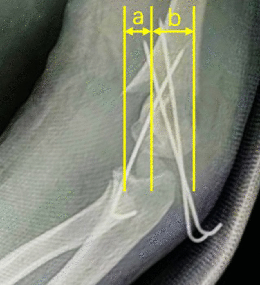
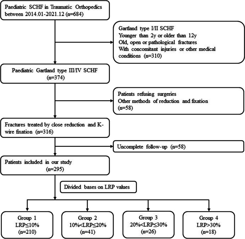
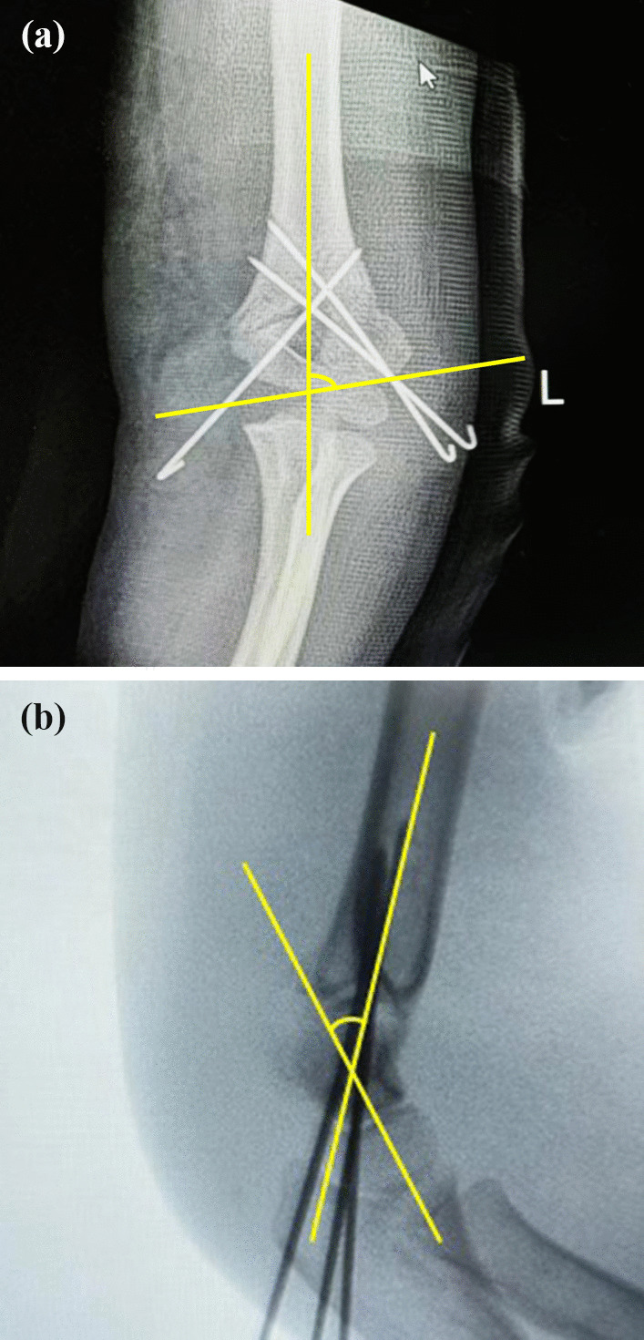
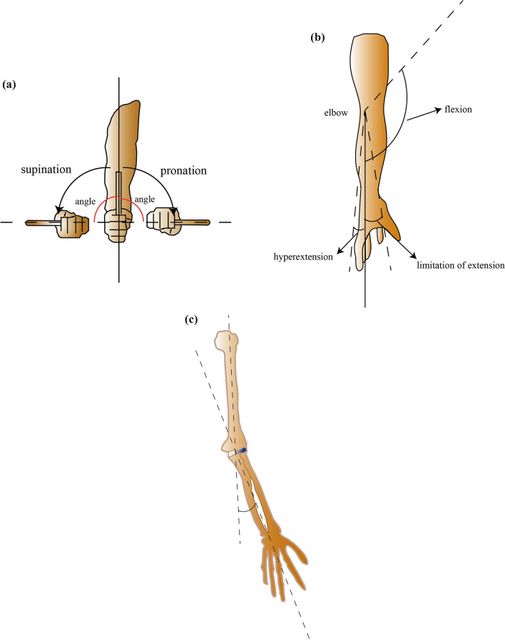
Methods: Between January 2014 and December 2021, 295 Gartland type III/IV paediatric SCHFs treated by close reduction and percutaneous K-wire fixation were selected for this retrospective study. The demographic, clinical and radiographic parameters of all cases were collected. The lateral rotation percentage (LRP) was measured on X-rays to evaluate postoperative malrotation alignment of the fracture. All cases were categorized into 4 groups according to LRP: LRP ≤ 10% (210, 71.2%), 10% < LRP ≤ 20% (41, 13.9%), 20% < LRP ≤ 30% (26, 8.8%) and LRP > 30% (18, 6.1%). The carrying angle, ranges of multidirectional motions, Mayo Elbow Performance Score (MEPS) and Flynn's Standard Score (FSS) of the injured elbow were assessed 6 months postoperation and compared among different groups. ROC analysis based on LRP and the excellent/good rate of FSS was performed to determine the acceptable maximum degree of postoperative malrotation alignment.
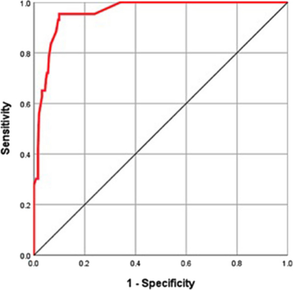
Results: There was no difference in the demographic characteristics (age, sex, injured side and fracture type), postoperative Baumann angle, carrying angle or range of forearm rotation among the 4 groups (P > 0.05). The operation time and time from operation to K-wire removal were longer in the 20% < LRP ≤ 30% and LRP > 30% groups than in the LRP < 10% and 10% < LRP ≤ 20% groups (P < 0.001). The shaft condylar angle, range of elbow flexion, MEPS and FSS of the injured elbow 6 months postoperatively were lower in the 20% < LRP ≤ 30% and LRP > 30% groups than in the LRP < 10% and 10% < LRP ≤ 20% groups (P < 0.001). ROC analysis based on LRP and the excellent/good rate of FSS showed an area under the curve of 0.959 (95% CI 0.936-0.983), with a cutoff value of 26.5%, sensitivity of 95.3% and specificity of 90.1%.
Conclusion: A certain degree of residual malrotation alignment deformity of the SCHF may reduce the shaft condylar angle and extend the time from operation to removing the K-wire and affect elbow function, especially the range of elbow flexion. The acceptable maximum degree of residual malrotation deformity expressed as the LRP value was 26.5%.
Keywords: Gartland type III/IV; Malrotation; Outcomes; Supracondylar humeral fracture.
© 2023. The Author(s).
Conflict of interest statement
The authors declare that they have no competing interests.
Figures
Similar articles
-
[Short-term effectiveness of transverse antecubital incision for failed closed reduction of Gartland type Ⅲ supracondylar humerus fractures in children].Zhongguo Xiu Fu Chong Jian Wai Ke Za Zhi. 2023 May 15;37(5):566-571. doi: 10.7507/1002-1892.202211033. Zhongguo Xiu Fu Chong Jian Wai Ke Za Zhi. 2023. PMID: 37190833 Free PMC article. Chinese.
-
The posterior intrafocal pin improves sagittal alignment in Gartland type III paediatric supracondylar humeral fractures.Injury. 2016 Apr;47(4):842-7. doi: 10.1016/j.injury.2015.12.031. Epub 2015 Dec 31. Injury. 2016. PMID: 26777466
-
[Zero-incision treatment of supracondylar humeral fractures in extremely unstable Gartland type Ⅳ children by percutaneous prying combined with modified rotary reduction with Kirschner wire].Zhongguo Gu Shang. 2025 Jan 25;38(1):92-6. doi: 10.12200/j.issn.1003-0034.20230481. Zhongguo Gu Shang. 2025. PMID: 39848941 Chinese.
-
Does posterior approach always lead to poor functional and cosmetic outcomes in displaced pediatric supracondylar humeral fractures?Ulus Travma Acil Cerrahi Derg. 2023 Apr;29(4):523-529. doi: 10.14744/tjtes.2022.29403. Ulus Travma Acil Cerrahi Derg. 2023. PMID: 36995201 Free PMC article. Review.
-
Examining Outcomes and Complications for Operative Versus Nonoperative Treatment of Pediatric Type II Supracondylar Humerus Fractures: A Systematic Review of Comparative Studies.J Pediatr Orthop. 2025 Jan 1;45(1):e1-e9. doi: 10.1097/BPO.0000000000002789. Epub 2024 Aug 22. J Pediatr Orthop. 2025. PMID: 39169804
Cited by
-
Efficacy of minimally invasive small-incision-assisted reduction with percutaneous Kirschner wire fixation in pediatric supracondylar humerus fractures.Am J Transl Res. 2025 Jul 15;17(7):5520-5529. doi: 10.62347/WMDY8791. eCollection 2025. Am J Transl Res. 2025. PMID: 40821044 Free PMC article.
References
MeSH terms
Grants and funding
- M2021063/Scientific Research Project of Jiangsu Commission of Health
- M2021063/Scientific Research Project of Jiangsu Commission of Health
- M2021063/Scientific Research Project of Jiangsu Commission of Health
- M2021063/Scientific Research Project of Jiangsu Commission of Health
- M2021063/Scientific Research Project of Jiangsu Commission of Health
LinkOut - more resources
Full Text Sources
Medical
Miscellaneous
