

Pan PPAR agonist stimulation of induced MSCs produces extracellular vesicles with enhanced renoprotective effect for acute kidney injury
- PMID: 38167146
- PMCID: PMC10763307
- DOI: 10.1186/s13287-023-03577-0
Pan PPAR agonist stimulation of induced MSCs produces extracellular vesicles with enhanced renoprotective effect for acute kidney injury
Abstract
Background: Acute kidney injury (AKI) has a complex pathophysiology and imposes serious health concerns worldwide. Extracellular vesicles (EVs) derived from induced mesenchymal stem cells (iMSCs) have been recognized as novel cell-free therapeutics for various inflammatory and degenerative disorders. In this study, we investigated whether iMSCs stimulated with a pan-peroxisome proliferator-activated receptor (PPAR) agonist could enhance the therapeutic efficacy of EVs against AKI.
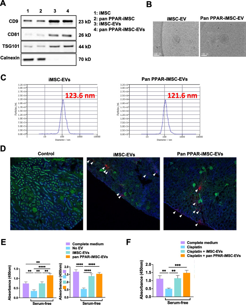
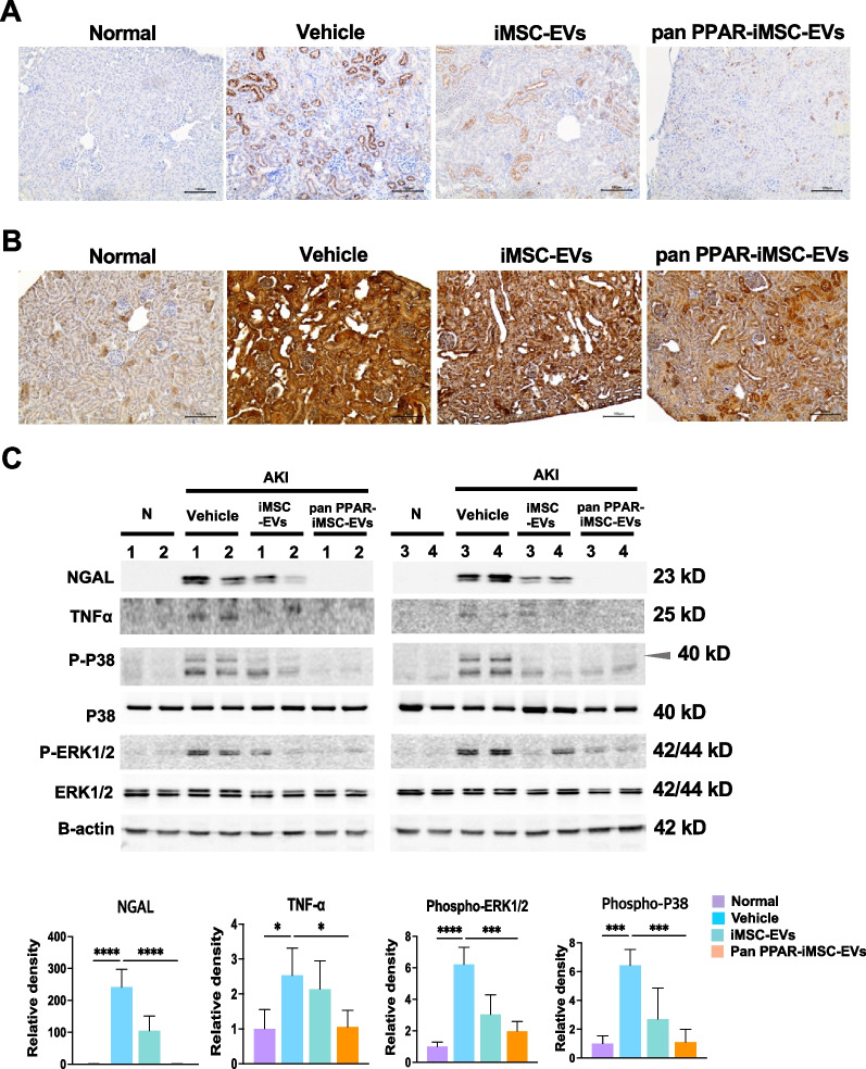
Methods: Human iMSCs were primed with or without lanifibranor, a PPAR agonist for 24 h, and EVs were collected after an additional 24 h. The basic characteristics of EVs were evaluated using cryo-transmission electron microscopy imaging, immunoblot detection of EV markers, nanoparticle tracking analysis, and localization in AKI kidneys. In vitro, the potential of the EVs to promote the growth and survival of HK-2 cells undergoing cisplatin-induced apoptosis and anti-inflammatory effects in M1-polarized THP-1 was compared. Subsequently, AKI was induced in BALB/c mice using cisplatin. After 8 and 24 h of cisplatin treatment, iMSC-EVs or pan-PPAR-iMSC-EVs were injected intravascularly. At 96 h after cisplatin administration, the renoprotective effects of iMSC-EVs or pan-PPAR-iMSC-EVs in inhibiting inflammation and apoptosis were compared using serum biochemistry, histology, immunohistochemistry, and gene expression analysis by qPCR.
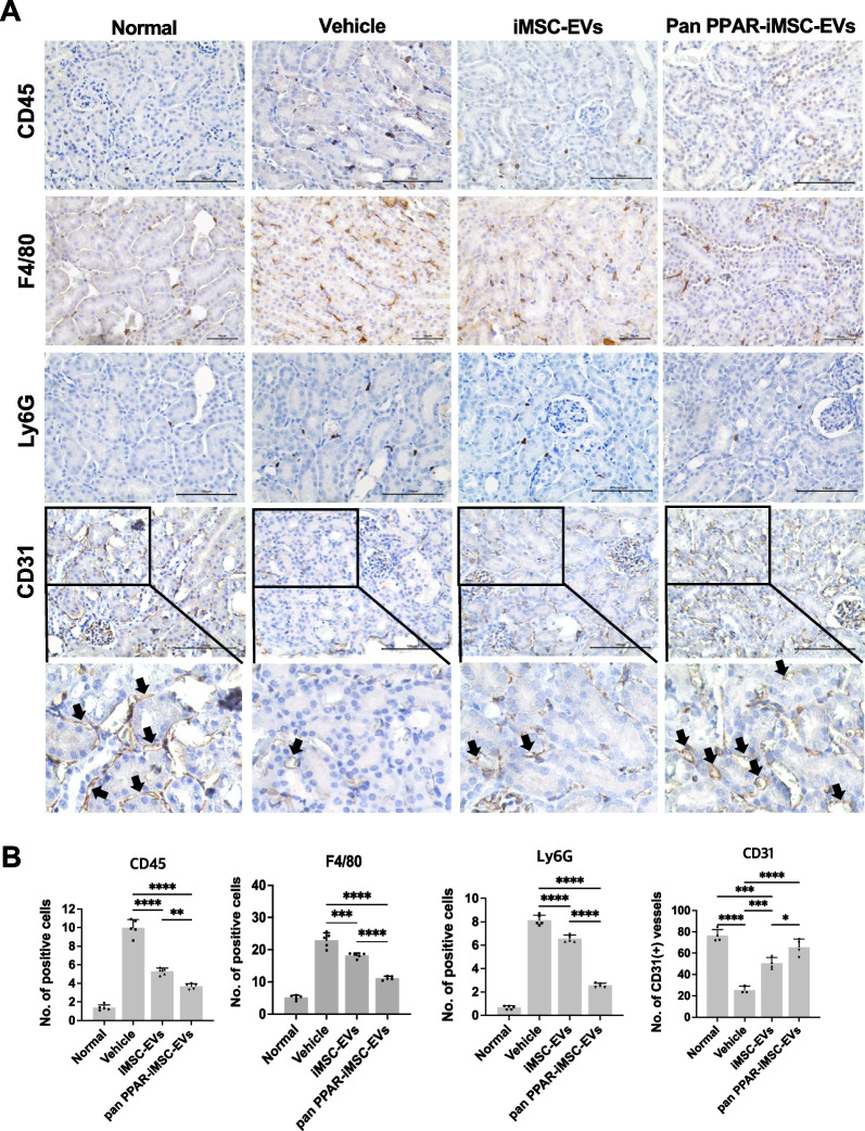
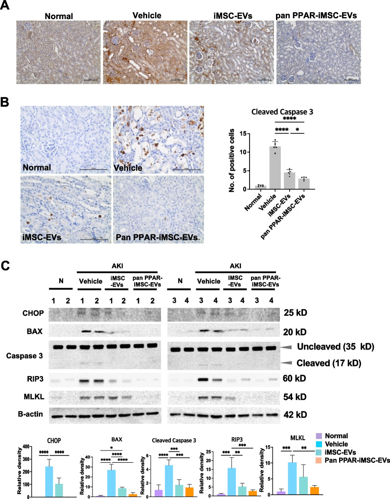
Results: Both EV types expressed EV markers and had typical EV morphology, and their localization in the renal tissue was confirmed. The proliferation and survival of HK-2 cells were higher in pan-PPAR-iMSC-EVs than those in iMSC-EVs. In M1-polarized THP-1 cells, the reduction in the mRNA expression of inflammatory cytokines was more significant in pan-PPAR-iMSC-EVs than that in iMSC-EVs. In the mouse model of cisplatin-induced AKI, pan-PPAR-iMSC-EVs markedly enhanced renoprotective effects compared to iMSC-EVs. Specifically, pan-PPAR-iMSC-EVs reduced tissue inflammation, immune cell infiltration, and apoptosis. Pan-PPAR-iMSC-EVs also increased renal capillary density.
Conclusion: Priming iMSCs with a PPAR agonist significantly improved the therapeutic potential of EVs by reducing inflammation and apoptosis. The reported strategy may contribute to the development of a novel cell-free option for AKI treatment.
Trial registration: Not applicable.
Keywords: Acute kidney injury; Extracellular vesicle; Induced mesenchymal stem cells; Priming.
© 2023. The Author(s).
Conflict of interest statement
S. K. is the chief executive officer of Brexogen Inc. Other authors declare no competing interests.
Figures

Similar articles
-
The application potential of iMSCs and iMSC-EVs in diseases.Front Bioeng Biotechnol. 2024 Jul 29;12:1434465. doi: 10.3389/fbioe.2024.1434465. eCollection 2024. Front Bioeng Biotechnol. 2024. PMID: 39135947 Free PMC article. Review.
-
Extracellular vesicles from induced pluripotent stem cell-derived mesenchymal stem cells enhance the recovery of acute kidney injury.Cytotherapy. 2024 Jan;26(1):51-62. doi: 10.1016/j.jcyt.2023.09.003. Epub 2023 Oct 16. Cytotherapy. 2024. PMID: 37843481
-
Cargo proteins in extracellular vesicles: potential for novel therapeutics in non-alcoholic steatohepatitis.J Nanobiotechnology. 2021 Nov 17;19(1):372. doi: 10.1186/s12951-021-01120-y. J Nanobiotechnology. 2021. PMID: 34789265 Free PMC article.
-
Improvement of androgenic alopecia by extracellular vesicles secreted from hyaluronic acid-stimulated induced mesenchymal stem cells.Stem Cell Res Ther. 2024 Sep 11;15(1):287. doi: 10.1186/s13287-024-03906-x. Stem Cell Res Ther. 2024. PMID: 39256806 Free PMC article.
-
Mesenchymal Stem Cell-Derived Extracellular Vesicles to the Rescue of Renal Injury.Int J Mol Sci. 2021 Jun 20;22(12):6596. doi: 10.3390/ijms22126596. Int J Mol Sci. 2021. PMID: 34202940 Free PMC article. Review.
Cited by
-
Extracellular Vesicles in Acute Kidney Injury: Mechanisms, Biomarkers, and Therapeutic Potential.Int J Nanomedicine. 2025 May 17;20:6271-6288. doi: 10.2147/IJN.S519345. eCollection 2025. Int J Nanomedicine. 2025. PMID: 40400780 Free PMC article. Review.
-
The application potential of iMSCs and iMSC-EVs in diseases.Front Bioeng Biotechnol. 2024 Jul 29;12:1434465. doi: 10.3389/fbioe.2024.1434465. eCollection 2024. Front Bioeng Biotechnol. 2024. PMID: 39135947 Free PMC article. Review.
-
Emerging Frontiers in acute kidney injury: The role of extracellular vesicles.Bioact Mater. 2025 Feb 18;48:149-170. doi: 10.1016/j.bioactmat.2025.02.018. eCollection 2025 Jun. Bioact Mater. 2025. PMID: 40046015 Free PMC article. Review.
References
-
- Hoste EAJ, Kellum JA, Selby NM, Zarbock A, Palevsky PM, Bagshaw SM, Goldstein SL, Cerda J, Chawla LS. Global epidemiology and outcomes of acute kidney injury. Nature. 2018;14(10):607–625. - PubMed
MeSH terms
Substances
Grants and funding
LinkOut - more resources
Full Text Sources
Molecular Biology Databases
