. 2024 Jan 2;15(1):33.
doi: 10.1038/s41467-023-44329-1.

Aspergillus fumigatus mitogen-activated protein kinase MpkA is involved in gliotoxin production and self-protection

Affiliations

Aspergillus fumigatus mitogen-activated protein kinase MpkA is involved in gliotoxin production and self-protection

Patrícia Alves de Castro et al. Nat Commun. .

Abstract

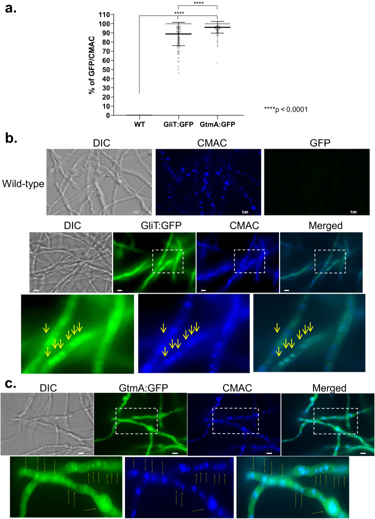
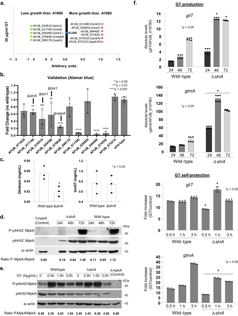
Aspergillus fumigatus is a saprophytic fungus that can cause a variety of human diseases known as aspergillosis. Mycotoxin gliotoxin (GT) production is important for its virulence and must be tightly regulated to avoid excess production and toxicity to the fungus. GT self-protection by GliT oxidoreductase and GtmA methyltransferase activities is related to the subcellular localization of these enzymes and how GT can be sequestered from the cytoplasm to avoid increased cell damage. Here, we show that GliT:GFP and GtmA:GFP are localized in the cytoplasm and in vacuoles during GT production. The Mitogen-Activated Protein kinase MpkA is essential for GT production and self-protection, interacts physically with GliT and GtmA and it is necessary for their regulation and subsequent presence in the vacuoles. The sensor histidine kinase SlnASln1 is important for modulation of MpkA phosphorylation. Our work emphasizes the importance of MpkA and compartmentalization of cellular events for GT production and self-defense.


Conflict of interest statement

The authors declare no competing interests.

Figures

Fig. 1
Fig. 1. GliT:GFP and GtmA:GFP have enriched vacuolar localization during GT production.
GliT:GFP and GtmA:GFP germlings were grown in liquid Czapek-dox medium for 24 h at 37 oC. Representative brightfield, differential interference contrast (DIC), and Cell tracker Blue CMAC (CellTracker Blue CMAC Dye (7-amino-4-chloromethylcoumarin) for vacuolar staining. a The number of GliT:GFP and GtmA:GFP germlings that co-localized with CMAC that was used for vacuolar staining were determined. We have counted three independent experiments with 45 germlings for each strain per experiment (N = 135 germlings) and the results were expressed as the mean values (%) of 3 independent experiments of GFP that co-localizes with CMAC. Yellow arrows indicate the localization of GliT:GFP and GtmA:GFP in magnified images of sub-cellular structures. The p-values were calculated using One-way ANOVA with Tukey’s multiple comparisons test, ****p < 0.0001. Germlings were assigned as positively present in the vacuoles if they have concomitant localization of GliT:GFP or GtmA:GFP in the same vacuole stained by CMC in the same germling. Representative images are shown in (b) and (c). Bars, 5 µm. Source data are provided as a Source Data file.
Fig. 2
Fig. 2. GliT:GFP and GtmA:GFP localize in the cytoplasm, endoplasmic reticulum, and small vesicle structures during GT self-protection.
GliT:GFP and GtmA:GFP germlings were grown in liquid MM for 24 h at 37 oC and exposed to GT 3 µg/ml for 2 h and co-stained with either CMAC, ER-Tracker Blue-White DPX, or FM4-64 a The number of GliT:GFP and GtmA:GFP germlings that co-localized with CMAC, ER-tracker, or FM4-64 staining were determined. We have counted two independent experiments with 45 germlings per experiment (N = 90 germlings) for each strain and the results were expressed as mean values (%) of the 2 independent experiments of GFP germlings that co-localizes with vacuoles (CMAC), Endoplasmic Reticulum, ER (ER-tracker) or vesicles (FM4-64). The percentage of germlings with punctuated structures of GliT:GFP and GtmA:GFP were also determined. Yellow arrows indicate the localization of GliT:GFP and GtmA:GFP in magnified images of sub-cellular structures. Germlings were assigned as positively present in the vacuoles, endoplasmic reticulum, vesicles, or endomembranes if at least one of these structures was co-localizing with GliT:GFP or GtmA:GFP in the same germling. Representative images are shown in (b), (c), (d), (e), (f), and (g). Bars, 5 µm. Source data are provided as a Source Data file.
Fig. 3
Fig. 3. Identification of proteins that interact with GliT:GFP and GtmA:GFP during GT production.
A. fumigatus was grown for 24, 48, and 72 h at 37 oC in liquid Czapek-dox medium. Proteins were extracted and immunoprecipitated (IP) with anti-GFP antibody. a Venn diagram showing the number of unique and shared IP proteins identified by mass spectrometry as interacting with GliT:GFP and/or GtmA:GFP. bd Fungifun categorization of proteins interacting with GliT:GFP, GtmA:GFP, and GliT:GFP and GtmA:GFP. (e and f) Co-IPs for GliT:GFP, GtmA:GFP, GliT:GFP MpkA:3xHA, GtmA:GFP MpkA:3xHA and MpkA:3xHA strains under GT production and self-protection. For GT production, A. fumigatus was grown under the same conditions described in (a) while for GT self-protection A. fumigatus was grown in liquid minimal medium for 24 h at 37 oC and exposed or not to GT 5 µg/mL for 30 min, 1 h and 3 h. The western blots are representative results from 2 independent experiments. Source data are provided as a Source Data file.
Fig. 4
Fig. 4. The mitogen-activated protein kinase (MAPK) MpkA is essential for GT production and self-protection.
a The wild-type, ΔmpkA and ΔmpkA::mpkA+ strains were grown for 5 days at 37 oC in solid minimal medium. b The wild-type and the MAPK mutants ΔsakA, ΔmpkC, ΔsakA ΔmpkC, ΔmpkA, and ΔmpkB were grown for 72 h at 37 oC in liquid Czapek-dox medium, secondary metabolites of the supernatant extracted and GT and bmGT identified. All the results are the mean of n = 6 biological replicates from 3 independent experiments ± SD, and p-values were calculated using One-way ANOVA with Tukey’s multiple comparisons test. ****p  <  0.0001. c Metabolic activity expressed by Alamar blue of A. fumigatus grown for 48 h in the absence or presence of different concentrations of GT. All the results are the mean of n = 6 biological replicates from 3 independent experiments ± SD; (****, p < 0.0001; One-way ANOVA with Tukey’s multiple comparisons test comparing the ΔmpkA, and ΔmpkA::mpkA+ mutants with the wild-type strain). d Western blot analysis for GT production and self-protection. GliT:GFP and GtmA:GFP were identified by anti-GFP antibody, MpkA-P and total MpkA were identified by using P-p44/42 and p44/42 antibodies while actin by anti-actin antibody. For GT production A. fumigatus was grown under the same conditions described in (b) while for GT self-protection A. fumigatus was grow in liquid minimal medium for 24 h at 37 oC and exposed or not to GT 5 µg/mL for 3 h. e, f RTqPCR for genes of the GT pathway and gtmA. and (g) RTqPCR for rglT and kojR, under GT production in liquid Czapek-dox medium for 24, 28 and 72 h, and GT self-protection conditions as described in (d). Results are the mean of 3 biologically independent experiments (with two technical replicates) ± SD; (*, p < 0.05, t-test). Source data are provided as a Source Data file.
Fig. 5
Fig. 5. Screening of the protein kinase null mutant library for GT susceptibility.
a Growth screening. b Metabolic activity expressed by Alamar blue of A. fumigatus grown for 48 h in the absence or presence of 30 µg/mL of GT (*, p < 0.05; **, p < 0.01, and ***, p < 0.001, One-way ANOVA with Dunnett’s multiple comparisons test). All the results are the average of three repetitions ± standard deviation. c GT and bmGT production in the A. fumigatus wild-type and ΔslnA strains. The strains were grown in liquid liquid Czapek-dox medium for 3 days at 37 oC. GT and bmGT were extracted from the supernatants and analyzed by LC-MS. Results are the mean of n = 6 biological replicates from 3 independent experiments ± SD; (*, p < 0.05, t-test). d, e Western blot analysis for GT production and self-protection. Western blot analysis for GT production and self-protection. GliT:GFP and GtmA:GFP were identified by anti-GFP antibody. MpkA-P and total MpkA were identified by using P-p44/42 and p44/42 antibodies while actin by anti-actin antibody. For GT production A. fumigatus was grown under the same conditions described in (c) while for GT self-protection A. fumigatus was grow in liquid minimal medium for 24 h at 37 oC and exposed or not to GT 5 µg/mL for 0.5 to 3 h. f RTqPCR for gliT and gtmA for wild-type and ΔslnA strains under GT production and self-protection conditions, as described in (d, e) of 3 biologically independent experiments (with two technical replicates). Results are the mean values ± SD; (*, p < 0.05, t-test). Source data are provided as a Source Data file.
Fig. 6
Fig. 6. Peroxisomes are required for GT production, self-protection, and virulence.
a The A. fumigatus wild-type, ΔpexE, and ΔpexG strains were grown for 5 days at 37 oC on MM and MM+biotin. b, c The A. fumigatus wild-type, ΔpexE, and ΔpexG strains were grown for 2 days at 37 oC on MM and MM + 30 µg/ml of GT. The radial growth results are the mean of 3 independent experiments ± SD, n = 6 biological replicates (*, p < 0.05, t-test) d The A. nidulans wild-type, ΔpexE, and ΔpexG strains were grown for 5 days at 37 oC on MM and MM+biotin. e, f The A. nidulans wild-type, ΔpexE, and ΔpexG strains were grown for 2 days at 37oC on YUU and YUU + 5 µg/ml of GT. Results are the mean of 3 independent experiments ± SD, n = 6 biological replicates (*, p < 0.05, t-test). g, h GT and bmGT production in the A. fumigatus wild-type, ΔpexE, and ΔpexG strains. The strains were grown in liquid Czapek-dox medium for 3 days at 37 oC. GT and bmGT were extracted from the supernatants and analyzed by mass spectrometry. Results are the mean of n = 6 biological replicates from 3 independent experiments ± SD; (*, p < 0.05, t-test). i, j Kaplan-Meier survival curves showing percentages of immunosuppressed mice infected intranasally with A. fumigatus strain (n = 10 mice/strain) and percentage of variation in the body weight. Phosphate buffered saline (PBS) was administered in a negative control group (n = 10). The indicated p-values determined with the use of the Logrank (Mantel-Cox test) and Gehan-Breslow-Wilcoxon test comparing the ΔpexE, and ΔpexG mutants with the wild-type strain, (*, p < 0.05 and **, p < 0.001). In the percentage of body weight variation, ΔpexE mutants are compared to the wild-type while ΔpexG mutants are compared to the PBS negative control (*, p < 0.001, One-way ANOVA with Tukey’s multiple comparisons test). Data represent the cumulative data of 2 separate experiments. Source data are provided as a Source Data file.

Update of

References

    1. Bignell E, Cairns TC, Throckmorton K, Nierman WC, Keller NP. Secondary metabolite arsenal of an opportunistic pathogenic fungus. Philos. Trans. R. Soc. B Biol. Sci. 2016;371:20160023. doi: 10.1098/rstb.2016.0023. - DOI - PMC - PubMed
    1. Blachowicz A, et al. Contributions of spore secondary metabolites to UV-C protection and virulence vary in different Aspergillus fumigatus strains. MBio. 2020;11:e03415-19. doi: 10.1128/mBio.03415-19. - DOI - PMC - PubMed
    1. Dolan SK, O’Keeffe G, Jones GW, Doyle S. Resistance is not futile: gliotoxin biosynthesis, functionality and utility. Trends Microbiol. 2015;23:419–428. doi: 10.1016/j.tim.2015.02.005. - DOI - PubMed
    1. Keller NP. Fungal secondary metabolism: regulation, function and drug discovery. Nat. Rev. Microbiol. 2019;17:167–180. doi: 10.1038/s41579-018-0121-1. - DOI - PMC - PubMed
    1. Keller NP. Translating biosynthetic gene clusters into fungal armor and weaponry. Nat. Chem. Biol. 2015;11:671–677. doi: 10.1038/nchembio.1897. - DOI - PMC - PubMed

Publication types

MeSH terms