

Avapritinib-based SAR studies unveil a binding pocket in KIT and PDGFRA
- PMID: 38167404
- PMCID: PMC10761696
- DOI: 10.1038/s41467-023-44376-8
Avapritinib-based SAR studies unveil a binding pocket in KIT and PDGFRA
Abstract
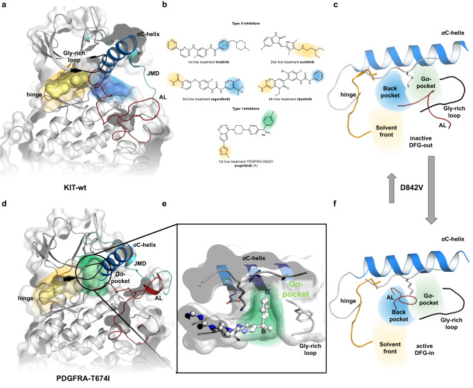
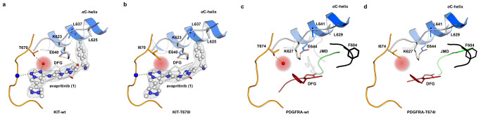
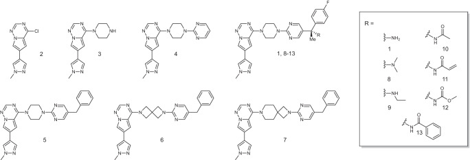
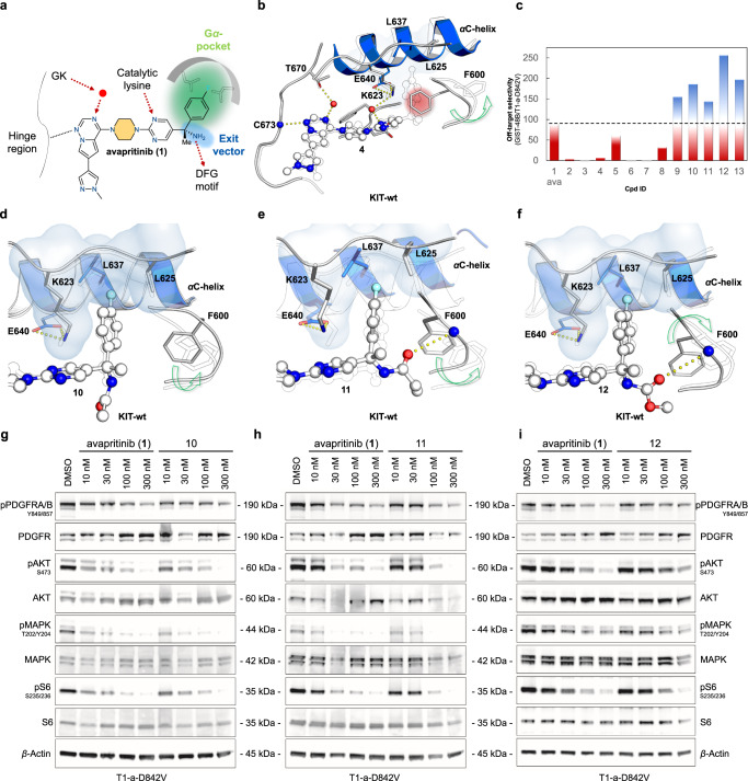
Avapritinib is the only potent and selective inhibitor approved for the treatment of D842V-mutant gastrointestinal stromal tumors (GIST), the most common primary mutation of the platelet-derived growth factor receptor α (PDGFRA). The approval was based on the NAVIGATOR trial, which revealed overall response rates of more than 90%. Despite this transformational activity, patients eventually progress, mostly due to acquired resistance mutations or following discontinuation due to neuro-cognitive side effects. These patients have no therapeutic alternative and face a dismal prognosis. Notable, little is known about this drug's binding mode and its medicinal chemistry development, which is instrumental for the development of the next generation of drugs. Against this background, we solve the crystal structures of avapritinib in complex with wild-type and mutant PDGFRA and stem cell factor receptor (KIT), which provide evidence and understanding of inhibitor binding and lead to the identification of a sub-pocket (Gα-pocket). We utilize this information to design, synthesize and characterize avapritinib derivatives for the determination of key pharmacophoric features to overcome drug resistance and limit potential blood-brain barrier penetration.
© 2024. The Author(s).
Conflict of interest statement
D.R. reports lecture and consulting fees from Pfizer, AZ, Sanofi, BI, and Bayer; is a shareholder of Centessa Pharmaceuticals plc., outside the submitted word. S.B. reports grants and personal fees from Blueprint Medicines, Incyte, and Novartis, PharmaMar, Eli Lilly & Co, Adcendo, Bayer, Blueprint Medicines, Boehringer Ingelheim, Cogent, Daiichi Sankyo, Deciphera, GSK, Exelixis, Novartis, Roche, PharmaMar. J.L. is a shareholder of Centessa Pharmaceuticals plc., outside the submitted work. The remaining authors declare no competing interests.
Figures
Similar articles
-
Avapritinib in advanced PDGFRA D842V-mutant gastrointestinal stromal tumour (NAVIGATOR): a multicentre, open-label, phase 1 trial.Lancet Oncol. 2020 Jul;21(7):935-946. doi: 10.1016/S1470-2045(20)30269-2. Lancet Oncol. 2020. PMID: 32615108 Clinical Trial.
-
Avapritinib in unresectable or metastatic PDGFRA D842V-mutant gastrointestinal stromal tumours: Long-term efficacy and safety data from the NAVIGATOR phase I trial.Eur J Cancer. 2021 Mar;145:132-142. doi: 10.1016/j.ejca.2020.12.008. Epub 2021 Jan 16. Eur J Cancer. 2021. PMID: 33465704 Free PMC article. Clinical Trial.
-
Avapritinib in Patients With Advanced Gastrointestinal Stromal Tumors Following at Least Three Prior Lines of Therapy.Oncologist. 2021 Apr;26(4):e639-e649. doi: 10.1002/onco.13674. Epub 2021 Feb 1. Oncologist. 2021. PMID: 33453089 Free PMC article.
-
Avapritinib in the treatment of PDGFRA exon 18 mutated gastrointestinal stromal tumors.Future Oncol. 2020 Aug;16(22):1639-1646. doi: 10.2217/fon-2020-0348. Epub 2020 Jun 10. Future Oncol. 2020. PMID: 32517495 Review.
-
Avapritinib: First Approval.Drugs. 2020 Mar;80(4):433-439. doi: 10.1007/s40265-020-01275-2. Drugs. 2020. PMID: 32100250 Review.
Cited by
-
Gene Mutations in Gastrointestinal Stromal Tumors: Advances in Treatment and Mechanism Research.Glob Med Genet. 2024 Aug 22;11(4):251-262. doi: 10.1055/s-0044-1789204. eCollection 2024 Dec. Glob Med Genet. 2024. PMID: 39176108 Free PMC article. Review.
-
Mechanistic insights and the clinical prospects of targeted therapies for glioblastoma: a comprehensive review.Exp Hematol Oncol. 2024 Apr 13;13(1):40. doi: 10.1186/s40164-024-00512-8. Exp Hematol Oncol. 2024. PMID: 38615034 Free PMC article. Review.
-
Impact of structural biology and the protein data bank on us fda new drug approvals of low molecular weight antineoplastic agents 2019-2023.Oncogene. 2024 Jul;43(29):2229-2243. doi: 10.1038/s41388-024-03077-2. Epub 2024 Jun 17. Oncogene. 2024. PMID: 38886570 Free PMC article. Review.
-
Clinical and preclinical insights into a novel MDM2::PDGFRA fusion in recurrent glioblastoma.NPJ Precis Oncol. 2025 Aug 16;9(1):289. doi: 10.1038/s41698-025-01076-4. NPJ Precis Oncol. 2025. PMID: 40819143 Free PMC article.
-
Targeted inhibition of PDGFRA with avapritinib, markedly enhances lenvatinib efficacy in hepatocellular carcinoma in vitro and in vivo: clinical implications.J Exp Clin Cancer Res. 2025 May 7;44(1):139. doi: 10.1186/s13046-025-03386-8. J Exp Clin Cancer Res. 2025. PMID: 40336047 Free PMC article.
References
Publication types
MeSH terms
Substances
Grants and funding
LinkOut - more resources
Full Text Sources
Miscellaneous
