. 2024 Jan 2;43(1):9.
doi: 10.1186/s13046-023-02910-y.

A novel PDPN antagonist peptide CY12-RP2 inhibits melanoma growth via Wnt/β-catenin and modulates the immune cells

Affiliations

A novel PDPN antagonist peptide CY12-RP2 inhibits melanoma growth via Wnt/β-catenin and modulates the immune cells

Chunyan Feng et al. J Exp Clin Cancer Res. .

Abstract

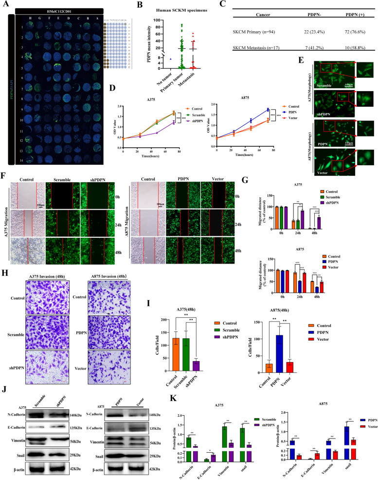
Background: Podoplanin (PDPN) is a highly conserved, mucin-type protein specific to the lymphatic system. Overexpression of PDPN is associated with the progression of various solid tumors, and plays an important roles in the tumor microenvironment by regulating the immune system. However, the role of PDPN-mediated signal activation in the progression of melanoma is still unknown.

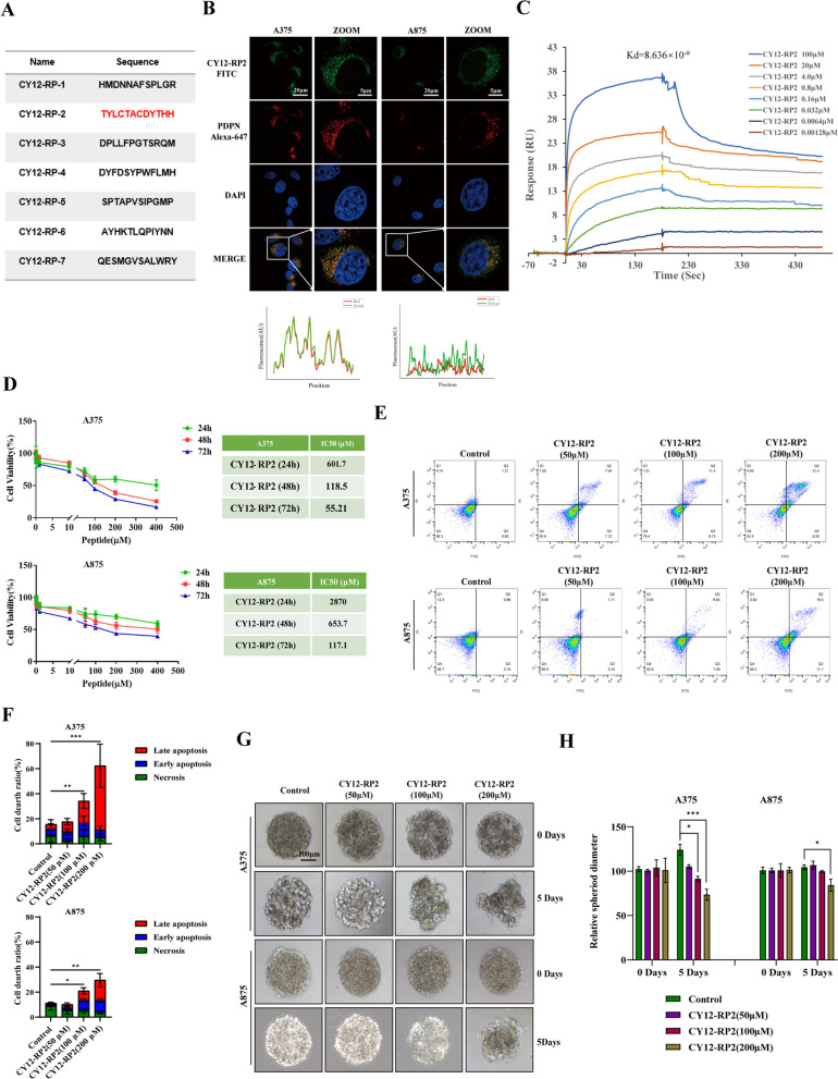
Methods: PDPN expression was first analyzed in 112 human melanoma tissue microarrays and melanoma cell lines. Functional experiments including proliferation, clone formation, migration, and metastasis were utilized to identify the suppressive effects of PDPN. The Ph.D.TM-12 Phage Display Peptide Library was used to obtain a PDPN antagonist peptide, named CY12-RP2. The immunofluorescence, SPR assay, and flow cytometry were used to identify the binding specificity of CY12-RP2 with PDPN in melanoma cells. Functional and mechanistic assays in vivo and in vitro were performed for discriminating the antitumor and immune activation effects of CY12-RP2.

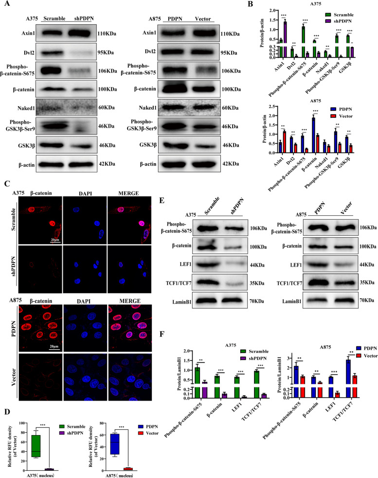
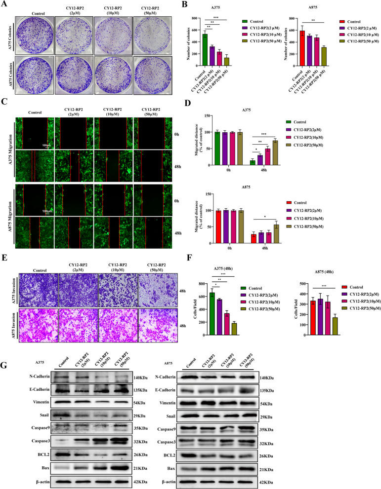
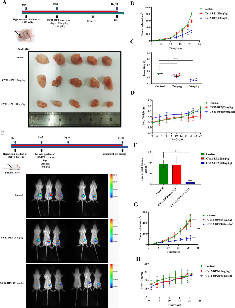
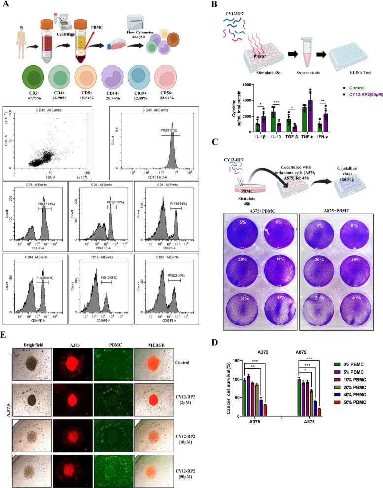
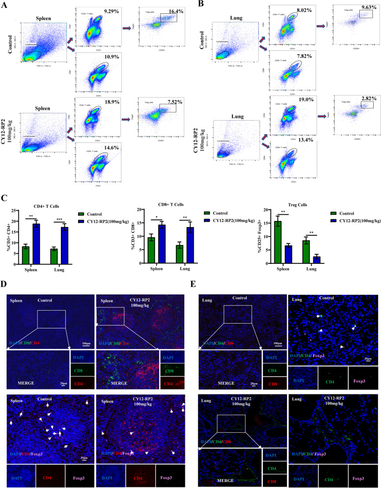
Results: PDPN was overexpressed in melanoma tissue and cells, and inhibited melanoma cells proliferation, migration, and metastasis by blocking the EMT and Wnt/β-catenin pathway. PDPN antagonistic peptide, CY12-RP2, could specifically bind with PDPN, suppressing melanoma various functions inducing apoptosis in both melanoma cells and 3D spheroids. CY12-RP2 also enhanced the anti-tumor capacity of PBMC, and inhibited melanoma cells growth both in xenografts and allogeneic mice model. Moreover, CY12-RP2 could inhibit melanoma lung metastasis, and abrogated the immunosuppressive effects of PDPN by increasing the proportion of CD3 + CD4 + T cells, CD3 + CD8 + T cells, CD49b + Granzyme B + NK cells, and CD11b + CD86 + M1-like macrophages and the levels of IL-1β, TNF-α, and IFN-γ.

Conclusions: This study has demonstrated the important role of PDPN in the progression of melanoma and formation of immunosuppressive environment, and provided a potential approach of treating melanoma using the novel CY12-RP2 peptide. In melanoma, PDPN is overexpressed in the cancer cells, and promotes melanoma cells growth and metastasis through activating the Wnt/β-catenin pathway. Treatment with the PDPN antagonistic peptide CY12-RP2 could not only inhibit the melanoma growth and metastasis both in vitro and in vivo through Wnt/β-catenin pathway blockade, but also abrogate the immunosuppressive effects of PDPN through modulating immune cells.

Keywords: CY12-RP2 peptide; EMT; Immune activation; Melanoma; PDPN; Wnt/β-catenin pathway.


Conflict of interest statement

The authors declare no competing interests.

Figures

Fig. 1
Fig. 1
PDPN is upregulated in melanoma, and is associated with the proliferation and metastasis of melanoma. A Human melanoma tissue microarrays named HMelC112CD01 consisting of nontumor melanoma sample (n = 1), primary melanoma samples (n = 94), and melanoma distant metastasis samples (n = 17). B Human melanoma samples were subjected to immunofluorescence for PDPN with quantitative analyses. C Statistically PDPN expression with different melanoma samples. D PDPN expression correlated with melanoma cell lines (A375, A875) proliferation as measured by CCK8 assay. E Images demonstrated morphological changes after stable knockdown of PDPN in A375 cells and high expression of PDPN in A875 cells. The power field scale bar, 100 μm. F, G Wound healing assays were performed for the migration capability of A375 cells stably knockdown PDPN or A875 cells with PDPN overexpression, and statistical analysis was performed to determine the migrated distance. The power field scale bar, 100 μm. H, I Transwell analysis was performed to quantify the invasive ability of PDPN knockdown or overexpression cells, and statistical analysis was performed to determine the invasion of cells. The power field scale bar, 100 μm. J, K Western blot analysis was performed to identify the effects of PDPN on the EMT markers E-Cadherin, N-Cadherin, vimentin, and snail. β-actin as an internal control was used. The statistical analysis was performed to quantify the relative protein levels. Data are presented as mean ± SD. *p < 0.05, **p < 0.01, ***p < 0.001 versus control
Fig. 2
Fig. 2
Effect of PDPN on the Wnt/β-Catenin signaling pathway in melanoma cells. A Western blot analysis was performed to detect the Wnt/β-catenin signaling-related proteins. β-actin as an internal control was used. B Statistical analysis was performed to quantify the relative protein levels. C Immunofluorescence staining was conducted to quantify PDPN effects on the nuclear β-catenin intensity in melanoma cell lines. The power field scale bar, 20 μm. D Statistical analysis was performed to quantify the immunofluorescence intensity of nuclear β-catenin in A875 cells. E Western blot assay was also performed on nuclear proteins related to the Wnt/β-catenin signaling pathway including β-catenin, phospho-β-catenin, LEF1, and TCF1/TCF7. The LaminB1 was used as an internal control for nuclear proteins. F Statistical analysis was performed to quantify the relative protein levels. Data are presented as mean ± SD. *p < 0.05, **p < 0.01, ***p < 0.001 versus control
Fig. 3
Fig. 3
Biopanning of PDPN antagonist peptides. A summary table of peptide sequences selected from the fourth round phage biopanning of PDPN peptides using a phage display library kit. B Immunofluorescence staining was performed to quantify the colocalization of CY12-RP2 and PDPN protein in melanoma cell lines (A375, A875). The power field scale bar, 20 μm. C CY12-RP2 (100, 20, 4.0, 0.8, 0.16, 0.032, 0.0064, 0.00128 μM) dose-dependent binding to PDPN. D Suppression efficiency of melanoma cells growth by CY12-RP2 was measured by CCK8 assay at various concentrations for 24, 48 or 72 h, respectively. E, F The A375 and A875 cells were treated with various concentrations of CY12-RP2 (0, 50, 100, 200 μM) for 48 h and analyzed using the Annexin V/PI staining flow cytometry (E), and statistical analysis was performed to quantify the apoptosis rates (F). G, H The 3D cellular spheres were treated with the set concentrations of CY12-RP2 for 5 days and cell morphology was assessed (G), relative spheroid diameter was measured (H). Data are presented as mean ± SD. *p < 0.05, **p < 0.01, ***p < 0.001 versus control. The power field scale bar, 100 μm
Fig. 4
Fig. 4
CY12-RP2 suppresses the invasion and migration of melanoma cells via blocking Wnt/β-catenin pathway. A The effect of CY12-RP2 on the colony formation of melanoma cell lines. B The colony formation statistical analysis. C Effects of CY12-RP2 on the migratory capacity of melanoma cell lines were analyzed by wound healing assay. The power field scale bar, 100 μm. D Statistical analysis was performed to determine the migrated distance. E Effects of CY12-RP2 on the invasive ability of melanoma cell lines were analyzed by transwell assays. The power field scale bar, 100 μm. F Statistical analysis was performed to determine the number of invaded cells. G The analysis of CY12-RP2 on EMT and apoptosis signaling pathways by using Western blot assay. Data are presented as mean ± SD. *p < 0.05, **p < 0.01, ***p < 0.001 versus control
Fig. 5
Fig. 5
Effects of CY12-RP2 on melanoma tumorigenesis in vivo. A Melanoma cell line A375 was subcutaneously implanted in BALB/c nude mice, and 5 days later treated with CY12-RP2 (25 mg/kg, 100 mg/kg) every two day for 15 days. Tumor mass was resected after the 15-day treatment period. B Tumor growth curve of subcutaneous melanoma xenograft. C The weight of orthotopic xenografts tumors at the end of the experiment. D Body weight of BALB/c nude mice over the course of the experiment. E Murine melanoma cell line B16F10-luc was subcutaneously implanted in BALB/c nude mice, and 5 days later treated with CY12-RP2 (25 mg/kg, 100 mg/kg) every two day for 15 days. Bioluminescence imaging of tumor growth in BALB/c mice was performed. F Statistical analysis of bioluminescence imaging. G The tumor volume after treating with CY12-RP2. H The body weight of mice after treating with CY12-RP2. Data are presented as mean ± SD. *p < 0.05, **p < 0.01, ***p < 0.001 versus control
Fig. 6
Fig. 6
Influence of CY12-RP2 on the immune-mediated anti-tumor capacity of PBMC in vitro and in vivo. A The procedure of PBMC isolation from donor peripheral blood, and the ratio of different immune cells in PBMC was assayed by flow cytometry. B PBMCs were stimulated with CY12-RP2 for 48 h, and the cytokine levels in the supernatant were assayed by ELISA. C, D PBMCs stimulated by CY12-RP2 were co-cultured with melanoma cells to demonstrate its killing capacity (C), statistical analysis was performed for the killing capacity of PBMC (D). E Brightfield and fluorescent microscopy images of A375 cells (red) co-culture with PBMC (GFP) were stimulated with various concentrations (0, 2, 10, 50 μM) of CY12-RP2. Data are presented as mean ± SD. *p < 0.05, **p < 0.01, ***p < 0.001 versus control
Fig. 7
Fig. 7
Melanoma cells pulmonary metastasis can be alleviated by CY12-RP2. A BALB/c mice were administered intravenously with 5 × 105 melanoma cells B16-F10-Luc and analyzed for lung metastasis for 3 weeks using bioluminescence imaging according to the schematic chart. B Statistical analysis of bioluminescence imaging. C BALB/c mice's body weight was determined at the indicated time. D Organs including heart, liver, spleen, lung, and kidney were imaged using bioluminescence imaging. E Quantification analysis of lung metastases fluorescence in BALB/c mice. F Representative pulmonary nodules images in a B16-F10 lung metastasis model. The power field scale bar, 500 μm, 250 μm and 100 μm. G Cytokine levels including IL-1β, IL-2, IL-10, TGF-β, TNF-α, and IFN-γ in lung metastases model plasma were determined by ELISA. Data are presented as mean ± SD. *p < 0.05, **p < 0.01, ***p < 0.001 versus control
Fig. 8
Fig. 8
CY12-RP2 modifies the proportion of T cells in the spleen and lung of mice. A, B The percentage of CD4 + T cells, CD8 + T cells, and CD4 + CD25 + Foxp3 + Treg cells in the spleen and lung of BALB/c mice were detected by flow cytometry. C Statistical analysis was performed to count the percentage of CD4 + T cells, CD8 + T cells, and CD4 + CD25 + Foxp3 + Treg cells in the spleen and lung of BALB/c mice with lung metastases. D, E Representative triple immunofluorescence of T cells (CD4 + T cells, CD8 + T cells) and CD4 + Foxp3 + Treg cells in the spleen (D) and lung (E) of pulmonary metastasis model. The power field scale bar, 20 μm and 100 μm

Similar articles

Cited by

References

    1. Gupta AK, Bharadwaj M, Mehrotra R. Skin cancer concerns in people of color: risk factors and prevention. Asian Pac J Cancer Prev. 2016;17(12):5257–5264. - PMC - PubMed
    1. van der Weyden L, Brenn T, Patton EE, Wood GA, Adams DJ. Spontaneously occurring melanoma in animals and their relevance to human melanoma. J Pathol. 2020;252(1):4–21. doi: 10.1002/path.5505. - DOI - PMC - PubMed
    1. Domingues B, Lopes JM, Soares P, Populo H. Melanoma treatment in review. Immunotargets Ther. 2018;7:35–49. doi: 10.2147/ITT.S134842. - DOI - PMC - PubMed
    1. Bertrand JU, Steingrimsson E, Jouenne F, Bressac-de Paillerets B, Larue L. Melanoma Risk and Melanocyte Biology. Acta Derm Venereol. 2020;100(11):adv00139. doi: 10.2340/00015555-3494. - DOI - PMC - PubMed
    1. Tyrell R, Antia C, Stanley S, Deutsch GB. Surgical resection of metastatic melanoma in the era of immunotherapy and targeted therapy. Melanoma Manag. 2017;4(1):61–68. doi: 10.2217/mmt-2016-0018. - DOI - PMC - PubMed