. 2024 Jan 2;15(1):10.
doi: 10.1186/s13287-023-03619-7.

Critical contribution of mitochondria in the development of cardiomyopathy linked to desmin mutation

Affiliations

Critical contribution of mitochondria in the development of cardiomyopathy linked to desmin mutation

Yeranuhi Hovhannisyan et al. Stem Cell Res Ther. .

Abstract

Background: Beyond the observed alterations in cellular structure and mitochondria, the mechanisms linking rare genetic mutations to the development of heart failure in patients affected by desmin mutations remain unclear due in part, to the lack of relevant human cardiomyocyte models.

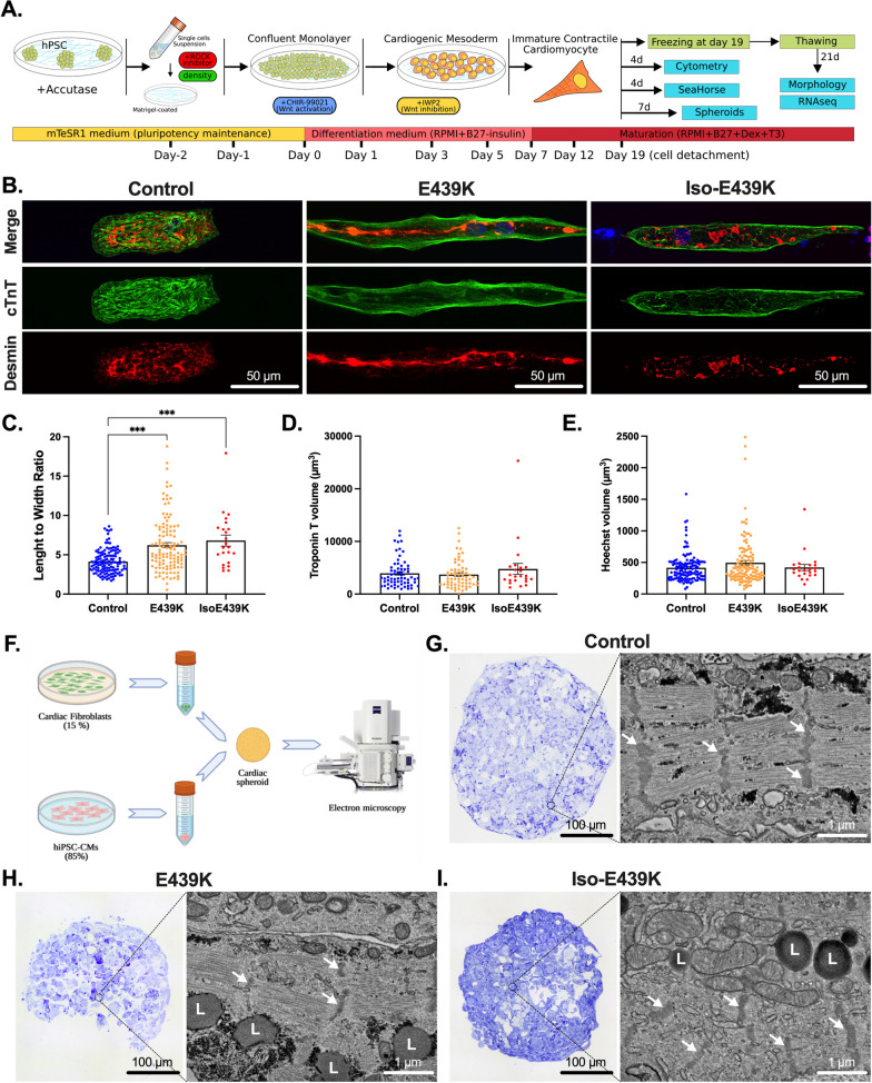
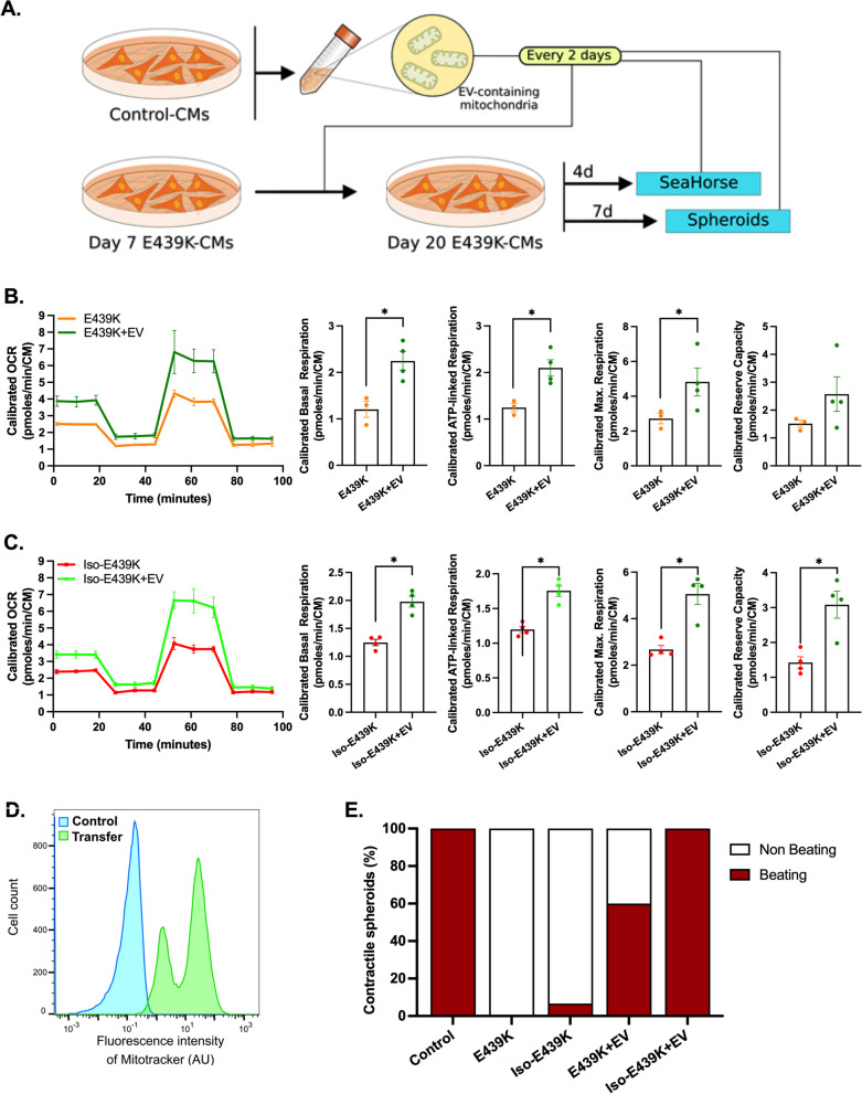
Methods: To shed light on the role of mitochondria in these mechanisms, we investigated cardiomyocytes derived from human induced pluripotent stem cells carrying the heterozygous DESE439K mutation that were either isolated from a patient or generated by gene editing. To increase physiological relevance, cardiomyocytes were either cultured on an anisotropic micropatterned surface to obtain elongated and aligned cardiomyocytes, or as a cardiac spheroid to create a micro-tissue. Moreover, when applicable, results from cardiomyocytes were confirmed with heart biopsies of suddenly died patient of the same family harboring DESE439K mutation, and post-mortem heart samples from five control healthy donors.

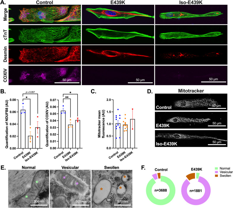
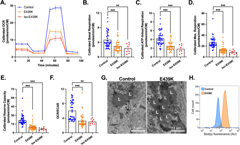
Results: The heterozygous DESE439K mutation leads to dramatic changes in the overall cytoarchitecture of cardiomyocytes, including cell size and morphology. Most importantly, mutant cardiomyocytes display altered mitochondrial architecture, mitochondrial respiratory capacity and metabolic activity reminiscent of defects observed in patient's heart tissue. Finally, to challenge the pathological mechanism, we transferred normal mitochondria inside the mutant cardiomyocytes and demonstrated that this treatment was able to restore mitochondrial and contractile functions of cardiomyocytes.

Conclusions: This work highlights the deleterious effects of DESE439K mutation, demonstrates the crucial role of mitochondrial abnormalities in the pathophysiology of desmin-related cardiomyopathy, and opens up new potential therapeutic perspectives for this disease.

Keywords: Cardiomyocytes; Desmin; Dilated cardiomyopathy; Heart failure; Mitochondria; Myofibrillar myopathy; Stem cells; iPSC.


Conflict of interest statement

The authors declare that they have no competing interests.

Figures

Fig. 1
Fig. 1
Familial myofibrillar myopathy with heterozygous DESE439K variant. A Pedigree of the affected family. Circles and squares represent female and male subjects, respectively. Solid symbols show patients with myopathy and cardiomyopathy. Crossed-out symbols stand for deceased subjects. The + symbol represents patient with presence of the pathogenic heterozygous DES variant. Solid arrow indicates the family member whose cardiac biopsies were used for the histological and biochemical analysis. Empty arrow indicates the family member whose peripheral blood mononuclear cells were used to generate iPSC clones. B Biventricular transversal heart slice of cardiac samples of the Index case CII (solid arrow in A) showing dilation of both ventricles. C Hematoxylin–eosin safran staining of formalin fixed left ventricle section showing extensive fibrosis
Fig. 2
Fig. 2
Morphological characterization of iPSC-derived cardiomyocytes carrying the DESE439K mutation. A Schematic overview of the cardiac differentiation protocol from iPSC. After detachment or freezing at day 19, the cardiomyocytes are cultured for 4, 7 or 21 more days and analyzed. B Cardiac troponin T (Green) and Desmin (Red) immunostaining of cardiomyocytes cultured on gelatin micro-patterns (20 µm-wide lines) for 21 days after thawing. Nuclei were counterstained with Hoechst (blue). Note the marked disorganization of the sarcomeres and the accumulation of desmin in the cytoplasm of mutant cardiomyocytes. C, E Cardiomyocyte length-to-width ratio (C), cardiomyocyte volume (D, troponin T staining) and nuclei volume (E, Hoechst staining) were measured on cardiomyocytes after 21 days of culture on gelatin micro-patterns using ImageJ software. Values are given as means ± SEM. ***, p < 0.001. F Schematic representation of the production of a cardiac spheroid model used for electron microscopy analysis. G-I Bright-Field microscopy images of Toluidine blue-stained semithin sections and representative field emission scanning electron microscopy (FE-SEM) images from the full mapping of ultrathin sections of cardiac spheroids. White arrows indicate Z-lines; L, lipid droplets
Fig. 3
Fig. 3
Structural mitochondrial abnormalities in cardiomyocytes with the DESE439K mutation. A Cardiac troponin T (Green), Desmin (Red) and COXIV (purple) immunostaining of cardiomyocytes cultured on gelatin micro-patterns (20 µm-width lines) for 21 days after thawing (see Fig. 2A). Nuclei were counterstained with Hoechst (blue). Note the absence of COXIV labeling in E439K-CMs and Iso-E439K-CMs. B Quantitative analysis of Western Blot of NDUFB8 and COXIV showing the marked decrease of mitochondrial proteins in E439K-CMs and Iso-E439K-CMs compared to Control-CMs. Results are presented after normalizing to Stain-Free total protein profile. Results are expressed as mean values ± SEM. *, p < 0.05, **, p < 0.01. C Quantification of mitochondria in cardiomyocytes by cytometry using MitoTracker, indicating close levels of mitochondria between mutant and control cardiomyocytes. Values are given as means ± SEM. D Visualization of mitochondria using the mitochondria-specific fluorescent probe MitoTracker in iPSC-derived cardiomyocytes, indicating no significant differences in the cytoplasmic distribution of mitochondria in mutant cardiomyocytes. E Representative transmission electron microscopy (TEM) images of three types of mitochondria, i.e. normal (green asterisks), vesicular (purple asterisks) and swollen (brown asterisks), observed in iPSC-derived cardiomyocytes. F Ring chart showing the proportions of the 3 different types of mitochondria in E439K-CMs (n = 1881: 3.6% normal, 85.8% vesicular and 10.6% swollen mitochondria) and Control-CMs (n = 3688: 91% normal, 5.4% vesicular and 3.6% swollen mitochondria) revealing a switch of mitochondria from the normal to the vesicular type in E439K-CMs
Fig. 4
Fig. 4
Transcriptomic analysis of E439K-CMs compared to Control-CMs reveals a strong signature of mitochondrial impairment. A Principal component analysis (PCA) of gene expression of the different samples from E439K-CMs (n = 6) and Control-CMs (n = 6). B Gene Set Enrichment Analysis (GSEA) of E439K-CMs compared to Control-CMs. (C-G) Non-supervised Weighted gene correlation network analysis (WGCNA) have detected two significant clusters of genes C. The molecular complex determination and enrichment analysis of the cluster 1 D reveals different sets of genes related to mitochondria (EG)
Fig. 5
Fig. 5
Decreased mitochondrial respiration and altered metabolic activity in cardiomyocytes with DESE439K mutation. A Representative mitochondrial oxygen consumption rate (OCR) profiles in Control-CMs, E439K-CMs and Iso-E439K-CMs. OCR profiles are expressed as pmol O2/min normalized to the number of cardiomyocytes and calibrated to the lowest value of the plate after addition of rotenone and antimycin A. B Quantification of basal respiration, C ATP-linked respiration, D maximal respiration and E reserve capacity of cardiomyocytes. F Ratio of OCR to ECAR in iPSC-derived cardiomyocytes. Values are expressed as mean ± SEM. **, p < 0.01, ***, p < 0.001. G Representative transmission electron microscopy images of lipid droplets (L) in iPSC-derived cardiomyocytes. H Flow cytometry analysis of cardiomyocytes after BODIPY staining demonstrating higher lipid levels in E439K-CMs
Fig. 6
Fig. 6
Editing of mtDNA in cardiomyocytes with the DESE439K mutation using the MT-COI gene fragment. A mtDNA editing was analyzed using a denaturation temperature (Td) gradient across the heating block on first round PCR products. 3D-PCR recovered edited MT-COI DNA down to 79.7 °C for E439K-CMs and Iso-E439K-CMs. The vertical white line in the cropped gels indicates the threshold between Control-CMs, E439K-CMs and Iso-E439K-CMs 3D-PCR products in terms of the Td. M: molecular weight markers. Asterisks refer to the samples cloned and sequenced. Full-length gels are presented in Additional file 1: Fig. S6. B Schematic representation of the Td for the last positive 3D-PCR amplifications for 6 E439K-CMs samples (orange circles), 2 Iso-E439K-CMs samples (red circles), 5 Control-CMs samples (blue circles), non-transfected HeLa (dark green circle) and 293 T (light green circle) cells and cells transfected with a plasmid containing MT-COI DNA fragment (purple circle). The arrow indicates the threshold Td (85 °C) at which the samples are hypermutated. C A selection of hypermutated G > A edited E439K-CMs and Iso-E439K-CMs samples (Td = 79.7 °C). The sequences are given with respect to the plus or coding strands. Only differences are shown. All sequences were unique, indicating that they corresponded to distinct molecular events. D Frequency analysis of edited MT-COI fragment gene as a function of the number of edits per sequence at 79,7 °C and 80.7 °C for the E439K-CMs and Iso-E439K-CMs-derived and for the control-CMs derived 3D-PCR products, respectively. The size of the columns indicates the combined numbers of sequences analyzed across the three samples. E Bulk dinucleotide context of MT-COI gene fragment and compared to the expected values. The horizontal line represents the expected frequencies assuming that G→A transitions were independent of the dinucleotide context and correspond to the weighted mean dinucleotide composition of the reference sequence. The blue (Control-CMs), orange (E439K-CMs) and red (Iso-E439K-CMs) bars represent the percentage of G > A transitions occurring within 5′GpN dinucleotides for the hypermutated sequences. Preferential mutations in 5′GpA and 5′GpG contexts correspond to an APOBEC3G signature. F Gene expression quantification of APOBEC3G in Control-CMs evaluated by RT-qPCR after treatment with IFNα (1000 U/mL). G Molecular complex detection of weighted gene correlation network analysis (WGCNA) from RNAseq data of E439K-CMs and Control-CMs have detected sets of genes related to IFN IFNα/β and rRNA processing
Fig. 7
Fig. 7
Long-term effects of the transfer of mitochondria on the function and respiration of cardiomyocytes with the DESE439K mutation. A Experimental set-up. B, C Mitochondrial oxygen consumption rate (OCR) profiles and respiration parameters of the mito stress test of E439K-CMs C or Iso-E439K-CMs D untreated of treated with EVs-containing mitochondria (+ EV). OCR profiles are expressed as pmol O2/min normalized to the number of cardiomyocytes and calibrated to the lowest value of the plate after addition of R/A. D Flow cytometry analysis of Control-CMs untreated (Control) or treated (Transfer) with extracellular vesicles (EV) from Control-CMs previously incubated with MitoTracker. E Percentage of contractile spheroids assessed by video microscopy

Similar articles

Cited by

References

    1. Paulin D, Huet A, Khanamyrian L, Xue Z. Desminopathies in muscle disease. J Pathol. 2004;204:418–427. doi: 10.1002/path.1639. - DOI - PubMed
    1. Capetanaki Y, Papathanasiou S, Diokmetzidou A, Vatsellas G, Tsikitis M. Desmin related disease: a matter of cell survival failure. Curr Opin Cell Biol. 2015;32:113–120. doi: 10.1016/j.ceb.2015.01.004. - DOI - PMC - PubMed
    1. Brodehl A, Gaertner-Rommel A, Milting H. Molecular insights into cardiomyopathies associated with desmin (DES) mutations. Biophys Rev. 2018;10:983–1006. doi: 10.1007/s12551-018-0429-0. - DOI - PMC - PubMed
    1. Agnetti G, Herrmann H, Cohen S. New roles for desmin in the maintenance of muscle homeostasis. FEBS J. 2022;289:2755–2770. doi: 10.1111/febs.15864. - DOI - PubMed
    1. Hnia K, Ramspacher C, Vermot J, Laporte J. Desmin in muscle and associated diseases: beyond the structural function. Cell Tissue Res. 2015;360:591–608. doi: 10.1007/s00441-014-2016-4. - DOI - PubMed