. 2024 Jan 2;24(1):3.
doi: 10.1186/s40644-023-00638-8.

Beyond invasive biopsies: using VASARI MRI features to predict grade and molecular parameters in gliomas

Affiliations

Beyond invasive biopsies: using VASARI MRI features to predict grade and molecular parameters in gliomas

Nurhuda Hendra Setyawan et al. Cancer Imaging. .

Abstract

Background: Gliomas present a significant economic burden and patient management challenge. The 2021 WHO classification incorporates molecular parameters, which guide treatment decisions. However, acquiring these molecular data involves invasive biopsies, prompting a need for non-invasive diagnostic methods. This study aims to assess the potential of Visually AcceSAble Rembrandt Images (VASARI) MRI features to predict glioma characteristics such as grade, IDH mutation, and MGMT methylation status.

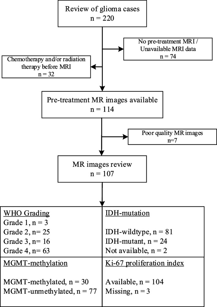
Methods: This study enrolled 107 glioma patients treated between 2017 and 2022, meeting specific criteria including the absence of prior chemotherapy/radiation therapy, and the presence of molecular and MRI data. Images were assessed using the 27 VASARI MRI features by two blinded radiologists. Pathological and molecular assessments were conducted according to WHO 2021 CNS Tumor classification. Cross-validation Least Absolute Shrinkage and Selection Operator (CV-LASSO) logistic regression was applied for statistical analysis to identify significant VASARI features in determining glioma grade, IDH mutation, and MGMT methylation status.

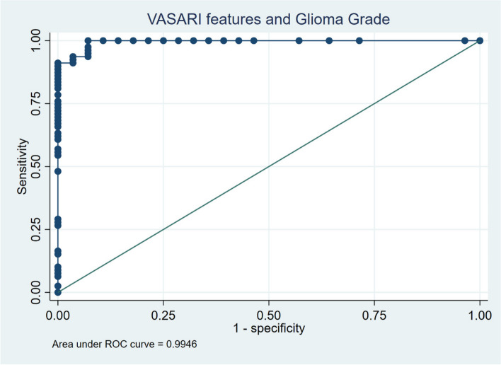
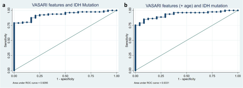
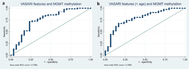
Results: The study demonstrated substantial observer agreement in VASARI feature evaluation (inter- and intra-observer κ = 0.714 - 0.831 and 0.910, respectively). Patient imaging characteristics varied significantly with glioma grade, IDH mutation, and MGMT methylation. A predictive model was established using VASARI features for glioma grade prediction, exhibiting an AUC of 0.995 (95% CI = 0.986 - 0.998), 100% sensitivity, and 92.86% specificity. IDH mutation status was predicted with AUC 0.930 (95% CI = 0.882 - 0.977), and improved slightly to 0.933 with 'age-at-diagnosis' added. A model predicting MGMT methylation had a satisfactory performance (AUC 0.757, 95% CI = 0.645 - 0.868), improving to 0.791 when 'age-at-diagnosis' was added.

Conclusions: The T1/FLAIR ratio, enhancement quality, hemorrhage, and proportion enhancing predict glioma grade with excellent accuracy. The proportion enhancing, thickness of enhancing margin, and T1/FLAIR ratio are significant predictors for IDH mutation status. Lastly, MGMT methylation is related to the longest diameter of the lesion, edema crossing the midline, and the proportion of the non-enhancing lesion. VASARI MRI features offer non-invasive and accurate predictive models for glioma grade, IDH mutation, and MGMT methylation status, enhancing glioma patient management.

Keywords: Glioma; Grade; IDH; MGMT; MRI; VASARI.


Conflict of interest statement

All authors declared no conflict of interest.

Figures

Fig. 1
Fig. 1
Subject selection process
Fig. 2
Fig. 2
Receiver operating characteristic (ROC) curve with an area under the curve (AUC) value of 0.9946 for the model prediction of glioma grade based on VASARI features
Fig. 3
Fig. 3
Receiver operating characteristic (ROC) curve with (a) an area under the curve (AUC) value of 0.930 for the model prediction of IDH mutation status based on VASARI features and (b) AUC value of 0.933 with the addition of age-at-diagnosis variable
Fig. 4
Fig. 4
Receiver operating characteristic (ROC) curve with (a) an area under the curve (AUC) value of 0.757 for the model prediction of MGMT methylation based on VASARI features and (b) AUC value of 0.791 with the addition of age-at-diagnosis variable
Fig. 5
Fig. 5
a Axial T1-weighted, (b) T2-weighted, (c) FLAIR, and (d) post-contrast administration T1-weighted MR images from a 40-year-old male with a WHO CNS Glioma Grade 4. Area of enhancement is visible that is less than 33% of the total area with pathological FLAIR signal intensity.The abnormal areas on T1 and FLAIR being approximately the same (expansive), as well as the enhancement having a vague and thin thickness. Despite the high-grade histopathological appearance, this glioma is IDH-mutant. It is predominantly solid with quite extensive non-enhancing area (68–100% of the total pathological area on FLAIR), accompanied by tumor and edema crossing the midline, and the overall large size of FLAIR abnormalities. This tumor was also proven to have methylation in its MGMT gene
Fig. 6
Fig. 6
a Axial T1-weighted, (b) T2-weighted, (c) FLAIR, and (d) post-contrast administration T1-weighted MR images from a 60-year-old male with a Glioblastoma WHO CNS Grade 4, IDH-wildtype. There is a distinct and relatively thick area of enhancement on the edge of the necrotic area, with a cumulative size of about a third of the total tumor area. The abnormal FLAIR area appears slightly larger than the pathological intensity on the T1-weighted image. This glioma carries the IDH-wildtype marker. The tumor almost has no non-enhancing areas as it primarily consists of necrotic areas with a solid enhancing area at its edge. The longest diameter of the tumor size is relatively not too large, and the tumor's edema area seems limited to one hemisphere. This tumor is also MGMT-unmethylated

Similar articles

Cited by

References

    1. Ostrom QT, Patil N, Cioffi G, Waite K, Kruchko C, Barnholtz-Sloan JS. CBTRUS statistical report: Primary brain and other central nervous system tumors diagnosed in the United States in 2013–2017. Neuro Oncol. 2020;22(Supplement_1):IV1–IV96s. doi: 10.1093/neuonc/noaa200. - DOI - PMC - PubMed
    1. Molinaro AM, Taylor JW, Wiencke JK, Wrensch MR. Genetic and molecular epidemiology of adult diffuse glioma. Nat Rev Neurol. 2019;15(7):405–417. doi: 10.1038/s41582-019-0220-2. - DOI - PMC - PubMed
    1. Haider SA, Asmaro K, Kalkanis SN, et al. The economic impact of glioma survivorship. Neurology. 2020;95(11):e1575–e1581. doi: 10.1212/WNL.0000000000010263. - DOI - PubMed
    1. Kim HJ, Ho SH, Lee S, et al. The Economic Burden of Brain Disability in Korea, 2008-2011. Inquiry. 2020;57:004695802093639. doi: 10.1177/0046958020936396. - DOI - PMC - PubMed
    1. Messali A, Villacorta R, Hay JW. A Review of the Economic Burden of Glioblastoma and the Cost Effectiveness of Pharmacologic Treatments. Pharmacoeconomics. 2014;32(12):1201–1212. doi: 10.1007/s40273-014-0198-y. - DOI - PubMed