. 2024 Jan 2;14(1):222.
doi: 10.1038/s41598-023-50935-2.

Persistent median artery and communicating branch related to the superficial palmar arch

Affiliations

Persistent median artery and communicating branch related to the superficial palmar arch

Marko Simić et al. Sci Rep. .

Abstract

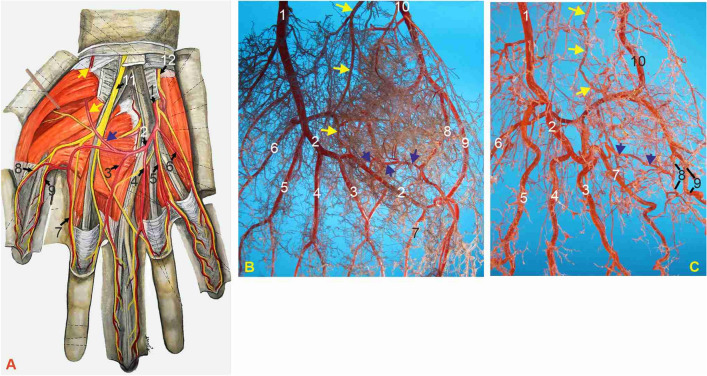
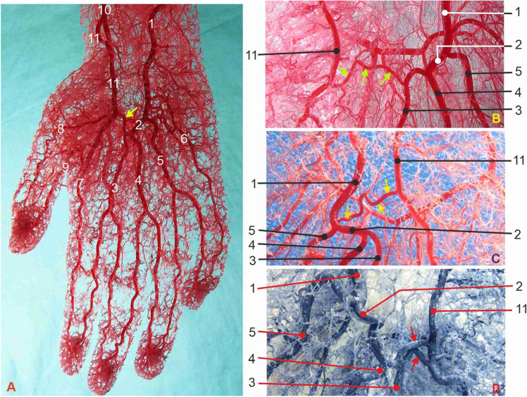
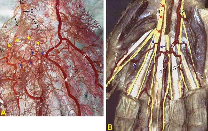
Microvascular surgery, plastic and reconstructive hand surgery, and coronary artery bypass surgery call for a microanatomical study of the branching pattern of the superficial palmar arch (SPA). For the anatomical analysis, we used a group of 20 dissected human hands injected with 4% formaldehyde solution and a 10% mixture of melted gelatin and India ink. The morphometric study was performed on 40 human hands of adult persons injected with methyl-methacrylate fluid into the ulnar and radial arteries simultaneously and afterwards corroded in 40% KOH solution for the preparation of corrosion cast specimens. The mean diameter of the SPA, between the second and third common palmar digital arteries, was 1.86 ± 0.08 mm. We identified the persistent median artery (PMA) in 5% of hands. We distinguished the three main groups of the SPAs according to variations in morphology and branching of the arch: Type 1, the long SPA; Type 2, the middle length SPA; and Type 3, the short SPA found in 27.5% of specimens. The communicating branch (CB), a vessel interconnecting the SPA to the closest branch of the radial artery, is classified into two different morphological groups. The third type of incomplete short arterial arch is the most important of the three groups of SPAs. That short SPA is potentially inadequate for restoring circulation after occlusion or radial artery harvesting for coronary artery bypass.


Conflict of interest statement

The authors declare no competing interests.

Figures

Figure 1
Figure 1
Palmar view of hands with arteries of the superficial neurovascular layer, type 1. (A) Drawing of the typically described final anastomosis of the arching terminal part of the ulnar artery (blue arrow) with the palmar branch of the radial artery (yellow arrows) and the widest field of supply including the thumb (modified from Radojević and Bošković). (B,C) Corrosion casts of left hand palmar arteries: (B) subtype a.; (C) subtype b. 1—ulnar artery; 2—superficial palmar arch (SPA); blue arrows—communicating branch (CB); yellow arrows—superficial palmar branch of the radial artery (SPB); 3—first CPDA; 4—second CPDA; 5—third CPDA; 6—ulnaris digiti minimi palmar artery; 7—radialis indicis artery; 8—radialis pollicis palmar artery; 9—ulnaris pollicis palmar artery; 10—radial artery; 11—median nerve; 12—ulnar nerve.
Figure 2
Figure 2
Corrosion casts of four hands with the medium length SPA, type 2, giving off three CPDAs. (A) and (B) right hand palmar arteries. (C) and (D) left hand palmar arteries. 1—ulnar artery; 2—superficial palmar arch (SPA); yellow arrows—communicating branch (CB); red arrows—doubled CB; 3—first CPDA; 4—second CPDA; 5—third CPDA; 6—ulnaris digiti minimi palmar artery; 7—radialis indicis artery; 8—radialis pollicis palmar artery; 9—ulnaris pollicis palmar artery; 10—radial artery; 11—prominent superficial palmar branch of the radial artery (SPB).
Figure 3
Figure 3
(A) Corrosion casts of type 3, the short SPA giving origin to two CPDAs. 1—ulnar artery; 2—superficial palmar arch (SPA); blue arrows—communicating branch (CB); 3—first CPDA; 4—second CPDA; 5—third CPDA; 6—ulnaris digiti minimi palmar artery; 7—radialis indicis artery; 8—radialis pollicis palmar artery; 9—superficial palmar branch of the radial artery (SPB); yellow arrows—anastomotic connections between the CB and SPB; 10—radial artery; 11—median nerve; 12—ulnar nerve. (B) Superficial dissection of the right hand showing the persistent median artery (red arrows) joining the CB (blue arrows).

References

    1. Waschke J, Bockers T, Paulsen F, editors. Sobotta Anatomy Textbook. Elsevier; 2019. pp. 188–189.
    1. Ross AC. Pectoral girdle and upper limb. In: Standring S, editor. Gray’s Anatomy. The Anatomical Basis of Clinical Practice. 42. Elsevier; 2021. pp. 980–982.
    1. Moore KL, Dalley AF, Agur AMR. Clinically Oriented Anatomy. Lippincott Williams and Wilkins; 2014. pp. 781–782.
    1. Rodriguez-Niedenfuhr M, Burton GJ, Deu J, Sanudo JR. Development of the arterial pattern in the upper limb of staged human embryos: Normal development and anatomic variations. J. Anat. 2001;199:407–417. doi: 10.1046/j.1469-7580.2001.19940407.x. - DOI - PMC - PubMed
    1. Alexander JG, Leal MC, Baptista JS. Persistent median artery inside the carpal tunnel: Description and surgical implications. Autops. Case. Rep. 2020;10:e2020209. doi: 10.4322/acr.2020.209. - DOI - PMC - PubMed