

This is a preprint.
It has not yet been peer reviewed by a journal.
The National Library of Medicine is
running a pilot
to include preprints that result from research funded by NIH in PMC and PubMed.
[Preprint]. 2023 Dec 15:2023.12.15.571797.
doi: 10.1101/2023.12.15.571797.
Pneumococcal Extracellular Vesicles Mediate Horizontal Gene Transfer via the Transformation Machinery
Affiliations
- PMID: 38168155
- PMCID: PMC10760141
- DOI: 10.1101/2023.12.15.571797
Item in Clipboard
Pneumococcal Extracellular Vesicles Mediate Horizontal Gene Transfer via the Transformation Machinery
bioRxiv.
.
Update in
-
Pneumococcal extracellular vesicles mediate horizontal gene transfer via the transformation machinery.mSphere. 2024 Dec 19;9(12):e0072724. doi: 10.1128/msphere.00727-24. Epub 2024 Nov 6. mSphere. 2024. PMID: 39503503 Free PMC article.
Abstract
Bacterial cells secrete extracellular vesicles (EVs), the function of which is a matter of intense investigation. Here, we show that the EVs secreted by the human pathogen Streptococcus pneumoniae (pneumococcus) are associated with bacterial DNA on their surface and can deliver this DNA to the transformation machinery of competent cells. These findings suggest that EVs contribute to gene transfer in Gram-positive bacteria, and in doing so, may promote the spread of drug resistance genes in the population.
Keywords: Extracellular vesicle; Streptococcus pneumoniae; antibiotic resistance; competence; horizontal gene transfer; transformation.
Figures
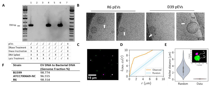
(A) PCR analysis of pEV DNA localization. pEVs underwent a series of treatments to determine the location of DNA (representative gel, n=3). At each treatment step, including the untreated pEVs, a sample was saved for PCR amplification and subsequent gel electrophoresis. The rows below each lane indicate each sample’s treatment. DNase treatment was performed with 1 U Turbo DNase at 37 °C for 30 minutes. DNase inactivation was accomplished with 5 μM EDTA (f.c.) for 10 minutes at 75 °C. Samples that were spiked with DNA had 50 ng of genomic DNA added. Lysis treatment was performed with 1% triton (f.c.) at 65 °C for 10 minutes The negative control in the last lane includes only 1x PBS, the buffer used for PEV elution from SEC. Primers targeted the gene gapdh. First and last lanes include the GeneRuler 1 kb Plus DNA Ladder (Invitrogen). (B) Images selected from cryo-electron micrographs of pEVs from R6 and D39 that display nucleic acid strands surrounding pEVs. White arrows indicate point of association between pEVs and nucleic acid strands. All images are the same scale (scale bar in final image). (C-D) Spatial analysis of DNA and pEV co-localization. (C) Representative image of DNA particles, false colored in green, and pEV particles, false colored in magenta. pEV sample was treated with PicoGreen and DiD to label DNA and the pEV membrane, respectively. Scale bar is indicated. (D) Ripley’s cross-L function for clustering of simulated random (blue) and observed (orange) pEV particles to DNA particles as a function radius, r. Data represent n=4 independent fields of view of DNA and pEV molecules (n=175 pEV particles and n=477 DNA particles). Shading represents the acceptance region for a hypothesis test of complete spatial randomness, with significance level 5%, using the envelopes of the L-functions of 1000 simulations. (E) DNA and pEV co-diffusion. Frechet distances computed for simulated (Random) and experimental (Data) of pEV-DNA particle trajectories. Data represent n=4 pEV-DNA pairs. Random Fréchet distance determined using diffusion coefficients for each pair determined in SFig 2. Inset: representative trajectories (white) from a DNA (green) and pEV (magenta) pair. (F) Sequencing of DNA isolated from pEVs and their parent bacterium from three unencapsulated pneumococcal strains (n=1 per strain).
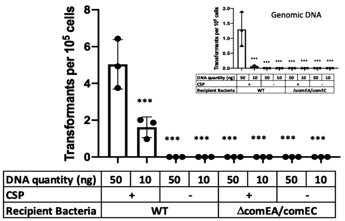
R6 cells (SpecS background) were exposed to pEV DNA (main figure) or genomic DNA (inset) from a R6-SpecR strain. Transformations were performed with and without CSP. Further, we tested recipient cells that do not encode a functional transformasome (ΔcomEAΔcomEC). Given the variability in efficacy between pEV batches, we did not draw conclusions about transformation efficiencies between pEVs or genomic DNA-mediated transformations. We propose that the range of transformation efficiencies reflects pEVs heterogeneity. Bars represent mean α SEM with dots overlayed within a bar representing a data point from each independent experiment (n ≥ 3, *** adjusted p-value < 0.0001 for Dunnett’s multiple comparison test).
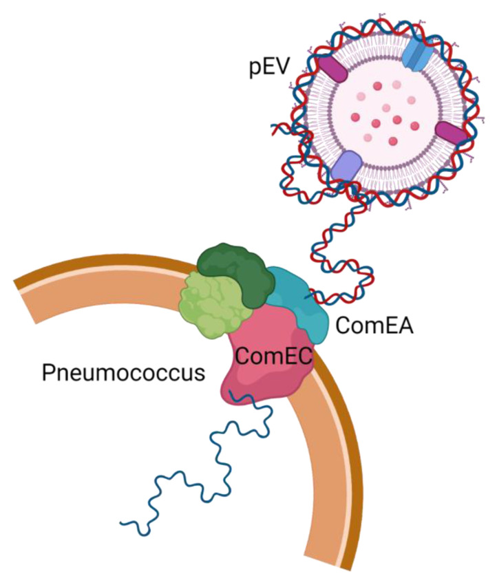
DNA is associated with the extracellular surface of pEVs and can enter pneumococcal cells. Entry requires activation of competence by the competence stimulating peptide as well as a functional transformasome.
References
-
- Donati C, Hiller NL, Tettelin H, Muzzi A, Croucher NJ, Angiuoli SV, Oggioni M, Dunning Hotopp JC, Hu FZ, Riley DR, Covacci A, Mitchell TJ, Bentley SD, Kilian M, Ehrlich GD, Rappuoli R, Moxon ER, Masignani V. 2010. Structure and dynamics of the pan-genome of Streptococcus pneumoniae and closely related species. Genome Biol 11:R107. - PMC - PubMed
-
- Hiller NL, Janto B, Hogg JS, Boissy R, Yu S, Powell E, Keefe R, Ehrlich NE, Shen K, Hayes J, Barbadora K, Klimke W, Dernovoy D, Tatusova T, Parkhill J, Bentley SD, Post JC, Ehrlich GD, Hu FZ. 2007. Comparative genomic analyses of seventeen Streptococcus pneumoniae strains: insights into the pneumococcal supragenome. J Bacteriol 189:8186–8195. - PMC - PubMed
Publication types
Grants and funding
LinkOut - more resources
Full Text Sources