[Preprint]. 2023 Dec 13:rs.3.rs-3639521.
doi: 10.21203/rs.3.rs-3639521/v1.

Artificial intelligence-based morphometric signature to identify ductal carcinoma in situ with low risk of progression to invasive breast cancer

Affiliations

Artificial intelligence-based morphometric signature to identify ductal carcinoma in situ with low risk of progression to invasive breast cancer

Marcelo Sobral-Leite et al. Res Sq. .

Update in

  • A morphometric signature to identify ductal carcinoma in situ with a low risk of progression.
    Sobral-Leite M, Castillo SP, Vonk S, Messal HA, Melillo X, Lam N, de Bruijn B, Hagos YB, van den Bos M, Sanders J, Almekinders M, Visser LL, Groen EJ, Kristel P, Ercan C, Azarang L, van Rheenen J, Hwang ES, Yuan Y; Grand Challenge PRECISION Consortium; Menezes R, Lips EH, Wesseling J. Sobral-Leite M, et al. NPJ Precis Oncol. 2025 Jan 28;9(1):25. doi: 10.1038/s41698-024-00769-6. NPJ Precis Oncol. 2025. PMID: 39875514 Free PMC article.

Abstract

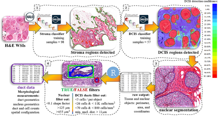
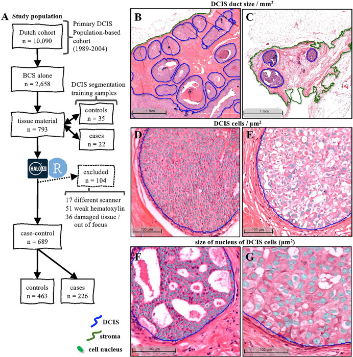
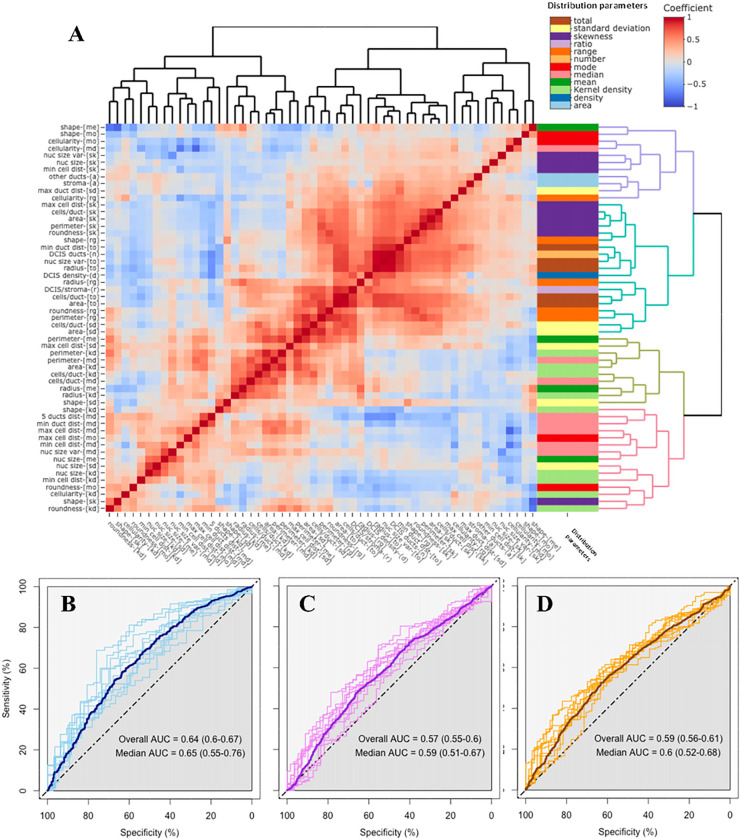
Ductal carcinoma in situ (DCIS) may progress to ipsilateral invasive breast cancer (iIBC), but often never will. Because DCIS is treated as early breast cancer, many women with harmless DCIS face overtreatment. To identify these women that may forego treatment, we hypothesized that DCIS morphometric features relate to the risk of subsequent iIBC. We developed an artificial intelligence-based DCIS morphometric analysis pipeline (AIDmap) to detect DCIS as a pathologist and measure morphological structures in hematoxylin-eosin-stained (H&E) tissue sections. These were from a case-control study of patients diagnosed with primary DCIS, treated by breast-conserving surgery without radiotherapy. We analyzed 689 WSIs of DCIS of which 226 were diagnosed with subsequent iIBC (cases) and 463 were not (controls). The distribution of 15 duct morphological measurements in each H&E was summarized in 55 morphometric variables. A ridge regression classifier with cross validation predicted 5-years-free of iIBC with an area-under the curve of 0.65 (95% CI 0.55-0.76). A morphometric signature based on the 30 variables most associated with outcome, identified lesions containing small-sized ducts, low number of cells and low DCIS/stroma area ratio. This signature was associated with lower iIBC risk in a multivariate regression model including grade, ER, HER2 and COX-2 expression (HR = 0.56; 95% CI 0.28-0.78). AIDmap has potential to identify harmless DCIS that may not need treatment.

Keywords: DCIS; artificial intelligence; biomarkers; digital pathology.


Conflict of interest statement

Conflict of Interests The authors declare that they have no conflicts of interest.

Figures

Figure 1
Figure 1
AIDmap workflow. HALO deep learning neural network was trained to recognize morphological structures in H&E whole-slides images (WSIs). 1: The first classifier was trained to annotate the fibroglandular tissue (stroma), leaving adipocytes outside (green line). 2: DCIS classification was applied within the annotated stroma, by detecting pixels that reached more than 90% of confidence of composing a DCIS duct (red areas in the image heatmap). 3: Next, a nuclear segmentation sensing hematoxylin staining was applied within the DCIS regions to detect all nuclear structures (supplementary appendix section S1.1). After these three steps, HALO provided tables containing the area, perimeter and spatial coordinates of stroma, DCIS and nuclear objects that were imported to R studio. 4: A True/False computational filtering was applied according to the nuclear perimeter, area and circular shape factor in order to eliminate false nuclear objects. And a True/False filtering was applied on DCIS objects, according the density of cells and average minimal nuclear distance (min. nucl. dist.) within the duct, to eliminate false DCIS ducts detected by HALO (supplementary appendix section S1.2). Finally, morphological measurements for each DCIS duct were obtained (supplementary appendix section S1.4).
Figure 2
Figure 2
Sample and classification details. A) Flow chart of the study population of patients diagnosed with primary DCIS in the Netherlands between 1989 and 2004. For training in the HALO AI module, 57 H&E sections from DCIS treated with BCS alone were used for automated stroma and DCIS segmentation. In total, 689 H&E WSI were successfully analyzed and their images revealed the variability on the density of DCIS ducts: large (B) and small (C) duct size (mm2); density of DCIS cells within the ducts: high (D) and low (E) DCIS cells/μm2; and average size of DCIS nucleus of the cells within the DCIS ducts: (F) large and small (G) DCIS nucleus (μm2); among other morphometric variables that varied among the samples.
Figure 3
Figure 3
Analysis of the morphometric variables. A) Heatmap with the Spearman’s rank correlation coefficients between the 55 variables obtained from the AIDmap in each H&E slide. Row side colors represent the parameters used to calculate each morphometric variable. Abbreviations are listed in supplementary appendix (section S1.4). Receiver operating characteristic (ROC) curves and area under the curve (AUC) calculations from the generalized linear models to predict absence of iIBC event during follow up after 5, 10 or 15 years (B-D, respectively).
Figure 4
Figure 4
Morphometric signature of DCIS. A) Volcano plot showing the odds ratios (OR) and the p values (p) of the 55 morphometric variables, obtained from linear regression analysis according the iIBC status during follow-up. B) Heatmap of the hierarchical cluster analysis of the 30 morphometric variables statistically associated in the volcano plot. Row side colors in blue degrees represent the categories of iIBC events during follow-up. The dendrogram colors highlights the 4 groups sharing morphometric similarities.
Figure 5
Figure 5
Characteristics of the 4 morphometric signatures. Differences are illustrated in the violin plots of the distribution of 4 morphometric variables among the morphometric signatures: DCIS/stroma area ratio (A) and the total number of cells inside DCIS ducts (B). The iIBC risk curve for the patients classified with one of the morphometric signatures (C), and the forest plots from the Cox multivariate regression models estimating the risk of iIBC progression during follow-up (D).

References

    1. Ringberg A, Palmer B, Linell F, Rychterova V, Ljungberg O. Bilateral and multifocal breast carcinoma. A clinical and autopsy study with special emphasis on carcinoma in situ. European journal of surgical oncology: the journal of the European Society of Surgical Oncology and the British Association of Surgical Oncology. 1991;17(1):20–9. - PubMed
    1. Maxwell AJ, Clements K, Hilton B, Dodwell DJ, Evans A, Kearins O, et al. Risk factors for the development of invasive cancer in unresected ductal carcinoma in situ. European journal of surgical oncology: the journal of the European Society of Surgical Oncology and the British Association of Surgical Oncology. 2018;44(4):429–35. - PubMed
    1. Ryser MD, Weaver DL, Zhao F, Worni M, Grimm LJ, Gulati R, et al. Cancer Outcomes in DCIS Patients Without Locoregional Treatment. Journal of the National Cancer Institute. 2019;111(9):952–60. - PMC - PubMed
    1. Myers ER, Moorman P, Gierisch JM, Havrilesky LJ, Grimm LJ, Ghate S, et al. Benefits and Harms of Breast Cancer Screening: A Systematic Review. JAMA: the journal of the American Medical Association. 2015;314(15):1615–34. - PubMed
    1. Falk RS, Hofvind S, Skaane P, Haldorsen T. Second events following ductal carcinoma in situ of the breast: a register-based cohort study. Breast cancer research and treatment. 2011;129(3):929–38. - PubMed

Publication types