[Preprint]. 2023 Dec 2:2023.12.01.569650.
doi: 10.1101/2023.12.01.569650.

PIEZO2-dependent rapid pain system in humans and mice

Affiliations

PIEZO2-dependent rapid pain system in humans and mice

Otmane Bouchatta et al. bioRxiv. .

Abstract

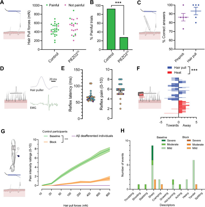
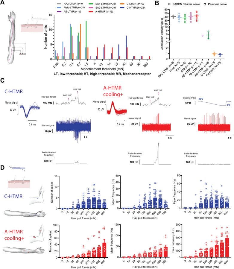
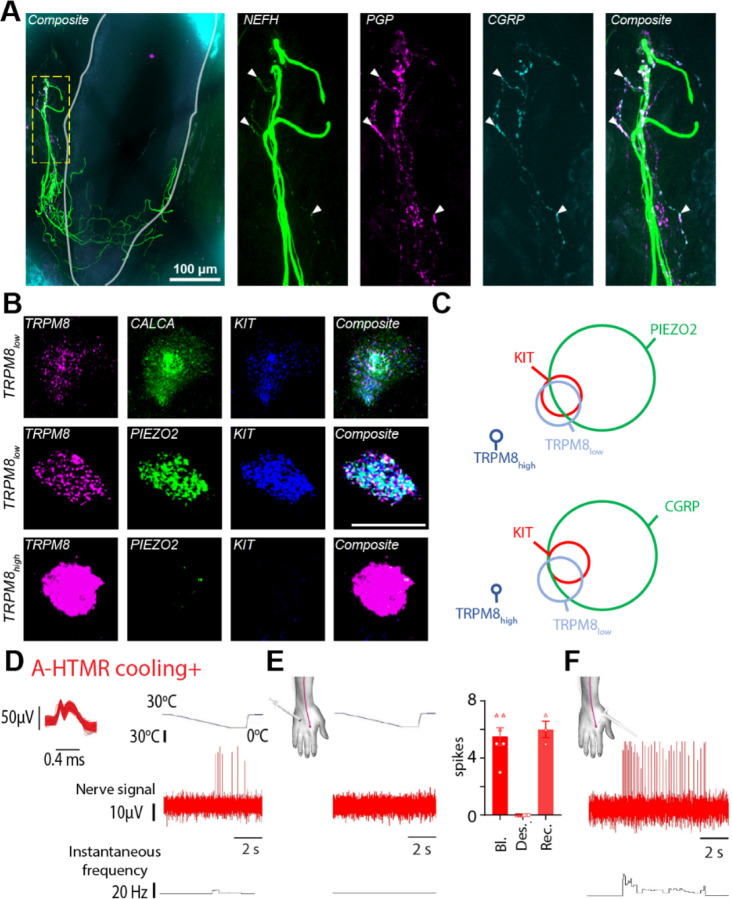
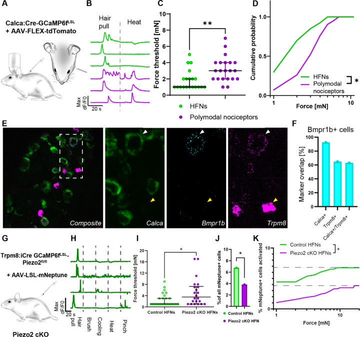
The PIEZO2 ion channel is critical for transducing light touch into neural signals but is not considered necessary for transducing acute pain in humans. Here, we discovered an exception - a form of mechanical pain evoked by hair pulling. Based on observations in a rare group of individuals with PIEZO2 deficiency syndrome, we demonstrated that hair-pull pain is dependent on PIEZO2 transduction. Studies in control participants showed that hair-pull pain triggered a distinct nocifensive response, including a nociceptive reflex. Observations in rare Aβ deafferented individuals and nerve conduction block studies in control participants revealed that hair-pull pain perception is dependent on Aβ input. Single-unit axonal recordings revealed that a class of cooling-responsive myelinated nociceptors in human skin is selectively tuned to painful hair-pull stimuli. Further, we pharmacologically mapped these nociceptors to a specific transcriptomic class. Finally, using functional imaging in mice, we demonstrated that in a homologous nociceptor, Piezo2 is necessary for high-sensitivity, robust activation by hair-pull stimuli. Together, we have demonstrated that hair-pulling evokes a distinct type of pain with conserved behavioral, neural, and molecular features across humans and mice.


Conflict of interest statement

Conflict of interest. None

Figures

Figure 1:
Figure 1:. Hair-pulling pain is a distinct PIEZO2-dependent pain submodality mediated by rapidly conducting fibers.
A–B. Individuals with PIEZO2 deficiency syndrome (PIEZO2DS) exhibited a profound deficit in hair-pull pain perception (control participants: n=7, PIEZO2DS individuals: n=6). A. At comparable forces, most hair-pull trials were reported as painful by control participants and nonpainful by PIEZO2DS individuals. Dots represent individual trials (control participants: n=28 trials, PIEZO2DS individuals: n=21 trials). B. The histograms represent the percentage of trials reported as painful (control participants: 92.8% vs. PIEZO2DS individuals: 28.57%, p<0.0001; Fisher’s exact test). C. Hair-pulling evoked a readily discriminable percept. Control participants consistently distinguished between hair-pull pain and pinprick pain at equivalent pain intensity ratings (correct responses: hair pull 93.2 ± 6.0%, pinprick 86.2 ± 7.1%; n=15 trials per stimulus per participant; n=5 participants). Data are presented as individual- and pooled-mean (± SEM) responses in percentages. D–E. Hairpull pain elicited a nociceptive reflex. D. An example EMG recording of a nociceptive reflex response evoked by multi-hair pulling from a posterior arm muscle. E. Latency distribution of reflex responses recorded in anterior and posterior muscle compartments of the upper arm and corresponding pain ratings. Since no differences were found in reflex latencies and pain ratings (p>0.05; t-test) between the two compartments, the data were pooled. All reflex responses (n=23) were evoked at stimulus intensities rated as painful. Data are shown as individual and mean (±SEM) responses from 3 control participants (color-coded). F. Hair pulling evoked a distinct urge-to-move psychophysical response. Control participants chose to preferentially move towards the pain source in response to multi-hair pulling contrary to heating where they chose to move away (n=11 participants, p<0.001; Mann Whitney test, with hair pull urge vs. baseline: p=0.0585 and heat urge vs. baseline: p<0.001; Paired t-test). Data are presented as mean ± SEM. G–H. Hair-pull pain was scalable and mediated by Aβ fibers. G. Pain intensity ratings in response to single-hair pulling forces were assessed in control participants (n=20) at ‘Baseline’ and during preferential Aβ-fiber conduction ‘Block’, as well as in individuals with selective Aβ deafferentation (n=2). Pain intensity ratings were significantly affected by hair-pulling forces (F(7,438) = 13.50, p<0.0001) and whether large myelinated fibers were conducting or not (F(2,438) = 266.5, p<0.0001). During the conduction block, the hair-pull pain ratings decreased significantly across a range of pull forces (Baseline vs. Block: [50–800 mN]: p<0.0001). In addition, the Aβ-deafferented individuals were unable to perceive hair-pull pain. Statistical differences were assessed using a two-way ANOVA followed by Tukey’s multiple comparisons test. Data are presented as mean ± SEM. H. Quality of hair-pull pain assessed using the short-form McGill questionnaire in Baseline and Block conditions. The data show the number of occurrences (events) for each pain descriptor.
Figure 2:
Figure 2:. Specialized A-fiber nociceptors encode hair-pull pain.
A. Mechanical threshold distribution of HTMRs and LTMRs in the recorded sample. For RA1 afferents, the preferred stimulus is hair movement, so monofilament thresholds were not measured. B. Conduction velocities of HTMRs and LTMRs in response to surface electrical stimulation and/or mechanical stimulation using an electronic monofilament with force-feedback. Conduction velocities of Aβ-HTMRs (42.8 ± 8.9 m/s, n=11) were statistically indistinguishable from Aβ-LTMRs ([SA1]: 37.2 ± 7.8 m/s, q=2.74, p=0.5318, n=9; [SA2]: 40.4 ± 8.3 m/s, q=1.21, p=0.9890, n=10; [Field]: 39.2 ± 6.9 m/s, q=1.726, p=0.922, n=8). Statistical differences were assessed using a one-way ANOVA with Tukey’s multiple comparisons test. Data are shown as individual and mean (±SEM) responses. PABCN, posterior antebrachial cutaneous nerve. C. Representative traces displaying hair-pull responses of a C-HTMR and a cooling-responsive A-HTMR. Single hairs were pulled from the receptive field of the recorded afferent until the hair was extracted. D. Receptive field locations of C-HTMRs (n=11) and cooling-responsive A-HTMRs (n=10) with hair present in the receptive field for hair-pull testing (left). The data show the individual trials and mean (±SEM) responses of C-HTMRs and cooling+ A-HTMRs to hair pulling at different forces. For C-HTMRs, hair pulling forces had a significant effect on the number of spikes (F(8,190) = 29.36, p<0.0001), as well as on mean (F(8,190) = 20.21, p<0.0001) and peak frequencies (F(8,190) = 20.81, p<0.0001). The response of C-HTMRs to increasing pull forces plateaued or dropped with further increases in pull forces. Accordingly, the peak frequency at the highest pulling force of 800 mN was not significantly different from other forces tested (p>0.05). For cooling+ A-HTMRs, hair pulling forces also had a significant effect on the number of spikes (F(8,135) = 17.39, p<0.0001), as well as on mean (F(8,135) = 31.34, p<0.0001) and peak frequencies (F(8,135) = 28.19, p<0.0001). However, in contrast to C-HTMRs, the activity in A-HTMRs was tuned to increasing hair-pull forces with the most robust response observed at the highest pulling force. Accordingly, the peak frequency at the highest pulling force of 800 mN was significantly higher than all other forces tested (at least p<0.001). Statistical differences were assessed by a one-way ANOVA with Tukey’s multiple comparisons test. The mechanical threshold, conduction velocity (where tested), and cooling response of five A-HTMRs from this sample were published in a preprint.
Figure 3:
Figure 3:. Hair follicle-associated myelinated nociceptors belonged to a distinct transcriptomic class.
A. Human skin immunofluorescence showed colocalization of Nefh, PGP and CGRP markers in nerve endings associated with hair follicles, confirming the existence of myelinated hair-follicle nociceptors in humans. Solid grey line marks the hair follicle. B. Fluorescent in situ hybridization (FISH) in human DRG neurons showed co-expression of TRPM8low with genetic markers delineating the hair-follicle nociceptor population. Scale bar = 50 μm. C. Quantification of FISH data (n=180 cells from 2 donors for KIT-PIEZO2-TRPM8 and 204 cells from 2 donors for KIT-CGRP-TRPM8). KITPIEZO2-TRPM8: KIT 8.3%, PIEZO2 40.6%, TRPM8low 12.2%, TRPM8high 3.3% of all neurons. KIT-CALCA-TRPM8: KIT 6.4%, CALCA 45.6%, TRPM8low 10.8%, TRPM8high 2.0% of all neurons. D–F. Cooling responsiveness of cooling+ A-fiber nociceptors is dependent on TRPM8. Recording traces showing the response of a cooling+ A-fiber nociceptor to a drop in temperature before (D) and after menthol injections (E), inducing TRPM8 desensitization. Individual and mean (±SEM) responses of cooling+ A-fiber nociceptors to a drop in temperature under ‘Baseline (Bl)’ and ‘TRPM8 desensitization (Des.)’ conditions (2 units, tested in triplicate). In one case, the ‘Recovery (Rec.)’ phase was also recorded. F. Recording trace showing the response of a cooling+ A-fiber nociceptor to high-threshold mechanical stimulation (600 mN von Frey) under ‘TRPM8 desensitization’ condition.
Figure 4:
Figure 4:. Piezo2 set the sensitivity of hair follicle nociceptors to hair pulling.
A. Diagram of the trigeminal in vivo calcium imaging experiment with single hair-pull stimuli and genetic model for labelling Calca nociceptors. CalcaCre mice were crossed with Ai95 Cre dependent GCamp6f strain. To postnatally limit the labelling to persistent Calca nociceptors offspring was injected at P1–P3 with AAV/PHP.S-CAG-Fl-tdTomato virus. B. Representative calcium transients recorded in Calca+ heat-insensitive hair follicle nociceptors (green) and heatresponsive polymodal nociceptors (magenta) neurons evoked by single hair-pull and heat stimuli. C. Forces activating HFNs (green) were markedly smaller (median threshold: 1 mN, n=20) than for polymodal nociceptors (magenta, median threshold: 3 mN, p=0.0037, n=21 for polymodal nociceptors, unpaired Student’s t-test). Data is shown as individual replicates with median and interquartile range overlay. D. Cumulative distribution of activation thresholds for HFNs (green) and polymodal nociceptors (magenta) neurons evoked by single hair-pull shows a markedly higher sensitivity to hair-pull in HFNs. Statistical differences were assessed by two-way ANOVA (F (1, 10)=6.521, p=0.0287). E. Mouse HFNs express low levels of Trpm8. Sample images of fluorescence in situ hybridization co-localization of HFN’s markers Bmrp1b, Calca and Trpm8. F. Bmpr1b+ neurons express Calca and majority of them expresses low levels of Trpm8 transcripts. G. Genetic model of Piezo2 loss of function in HFNs. GCamp6f expressing conditional Piezo2 KO was generated by crossing the Trpm8iCre mouse strain with double transgenic Ai95-Piezo2fl/fl line to obtain homozygous Piezo2 cKO. To limit the labelling to persistent Calca nociceptors offspring was injected at P1–P3 with AAV/PHP.S-CAG-LSL-mNeptune. H. Representative calcium transients of functionally isolated HFNs recorded in control mice. Demonstrating high selectivity of HFNs. I. Hair follicle nociceptors (green) sensitivity to hair pull force (median threshold: 1 mN, n=29) was markedly diminished in the absence of Piezo2 (magenta, median threshold: 3.5 mN, n=21, p=0.0158, upaired Student’s t-test). Data is shown as individual replicates with median and interquartile range overlay. J. Percentage of cells responding to hair-pull in Piezo2cKO was diminished by almost half (3.8% in Piezo2 cKO vs 6.8% in control, p=0.0418, Fisher’s exact test). K. Additive effect of Piezo2 on hair follicle nociceptor responses during hair-pull. Cumulative distribution of hair follicle nociceptors activation scaled to total recruitment during hair-pull trial, (two-way ANOVA (F(1, 19)=3556, p<0.0001, n=29 neurons from 7 mice for control and n=22 neurons from 9 mice for Piezo2KO).

References

    1. Horch K.W., Tuckett R.P., and Burgess P.R. (1977). A key to the classification of cutaneous mechanoreceptors. J Invest Dermatol 69, 75–82. 10.1111/1523-1747.ep12497887. - DOI - PubMed
    1. Wall P.D., McMahon S.B., and Koltzenburg M. (2006). Wall and Melzack’s textbook of pain, 5th Edition (Elsevier/Churchill Livingstone; ).
    1. Nagi S.S., Marshall A.G., Makdani A., Jarocka E., Liljencrantz J., Ridderstrom M., Shaikh S., O’Neill F., Saade D., Donkervoort S., et al. (2019). An ultrafast system for signaling mechanical pain in human skin. Sci Adv 5, eaaw1297. 10.1126/sciadv.aaw1297. - DOI - PMC - PubMed
    1. Yu H., Usoskin D., Nagi S.S., Hu Y., Kupari J., Bouchatta O., Cranfill S.L., Gautam M., Su Y., Lu Y., et al. (2023). Single-Soma Deep RNA Sequencing of Human Dorsal Root Ganglion Neurons Reveals Novel Molecular and Cellular Mechanisms Underlying Somatosensation. bioRxiv, 2023.2003.2017.533207. 10.1101/2023.03.17.533207. - DOI
    1. Tavares-Ferreira D., Shiers S., Ray P.R., Wangzhou A., Jeevakumar V., Sankaranarayanan I., Cervantes A.M., Reese J.C., Chamessian A., Copits B.A., et al. (2022). Spatial transcriptomics of dorsal root ganglia identifies molecular signatures of human nociceptors. Sci Transl Med 14, eabj8186. 10.1126/scitranslmed.abj8186. - DOI - PMC - PubMed

Publication types