[Preprint]. 2023 Dec 13:2023.12.12.571321.
doi: 10.1101/2023.12.12.571321.

Simple and Highly Specific Targeting of Resident Microglia with Adeno-Associated Virus

Affiliations

Simple and Highly Specific Targeting of Resident Microglia with Adeno-Associated Virus

Carolina Serrano et al. bioRxiv. .

Update in

Abstract

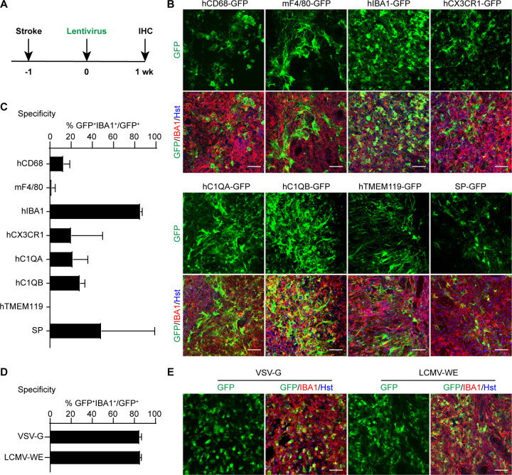
Microglia, as the immune cells of the central nervous system (CNS), play dynamic roles in both health and diseased conditions. The ability to genetically target microglia using viruses is crucial for understanding their functions and advancing microglia-based treatments. We here show that resident microglia can be simply and specifically targeted using adeno-associated virus (AAV) vectors containing a 466-bp DNA fragment from the human IBA1 (hIBA1) promoter. This targeting approach is applicable to both resting and reactive microglia. When combining the short hIBA1 promoter with the target sequence of miR124, up to 95% of transduced cells are identified as microglia. Such a simple and highly specific microglia-targeting strategy may be further optimized for research and therapeutics.

Keywords: AAV; adeno-associated virus; human IBA1; lentivirus; microglia; virus transduction.


Conflict of interest statement

Conflict of interests A patent application was filed on using viruses to target microglia/macrophages.

Figures

Figure 1.
Figure 1.. In vivo screens for microglia-targeting lentivirus.
(A) Schematic diagram of the experimental procedure. One week post MCAO-induced stroke, lentiviruses with various promoter-driven GFP were injected into the striata and examined after another week (wk). (B) Representative confocal images showing marker expression for the indicated lentiviruses. Scale bars, 50 μm. (C) Quantifications showing high microglia-specificity of GFP expression in mice injected with the lenti-hIBA1-GFP (mean ± SEM; n=3 mice per group). (D) Quantifications showing comparable specificity of lentivirus packaged with either VSV-G or LCMV-WE envelope (mean ± SEM; n=3 mice per group). (E) Representative confocal images showing marker expression for the indicated lentiviruses. Scale bars, 50 μm.
Figure 2.
Figure 2.. In vivo screens for microglia-targeting AAVs.
(A) Schematic diagram of the experimental procedure. One week post L-NIO-induced stroke, scAAVs with various serotypes were injected into the striata and examined after another week (wk). (B) Representative confocal images showing marker expression for the indicated scAAVs. Scale bars, 50 μm. (C) Quantifications showing microglia-specificity of GFP expression for the indicated scAAVs (mean ± SEM; n=3 mice per group). (D) Quantifications showing microglia transduction efficiency for the indicated scAAVs (mean ± SEM; n=3 mice per group). (E) Schematic diagram of the experimental procedure. One week post L-NIO-induced stroke, scAAV5 or scAAV8 was injected into the striata and examined after another 4 week (wk). (F) Representative confocal images showing marker expression for the indicated scAAVs. Scale bars, 50 μm. (G) Quantifications showing the specificity and transduction efficiency for microglia (mean ± SEM; n=3 mice per group).
Figure 3.
Figure 3.. In vivo screens for the minimal microglia-targeting promoter.
(A) Schematic diagram of the experimental procedure. ssAAV5 viruses with different promoters were injected into sham mice or mice with L-NIO-induced stroke. Brains were analyzed one week later. (B) Diagram of the examined ssAAVs with different lengths of hIBA1 promoter. (C) Representative confocal images showing marker expression for the indicated ssAAV5s. Scale bars, 50 μm. (D) Quantifications showing microglia-specificity of GFP expression for the indicated ssAAV5s (mean ± SEM; n=3 mice per group). (E) Quantifications showing microglia transduction efficiency for the indicated ssAAV5s (mean ± SEM; n=3 mice per group).
Figure 4.
Figure 4.. Increased long-term specificity and efficiency for the minimal hIBA1a promoter when packaged in scAAV.
(A) Schematic diagram of the experimental procedure. scAAV5 virus with the hIBA1a promoter was injected into sham mice or mice with L-NIO-induced stroke. Brains were analyzed 1 week or 4 weeks later. (B) Representative confocal images showing marker expression for the indicated conditions. Scale bars, 50 μm. (C) Quantifications showing microglia-specificity of GFP expression for the indicated conditions (mean ± SEM; n=3 mice per group). (E) Quantifications showing microglia transduction efficiency for the indicated conditions (mean ± SEM; n=3 mice per group).
Figure 5.
Figure 5.. miR124T confers high specificity of ssAAV under the hIBA1a promoter.
(A) Schematic diagram of the experimental procedure. The miR124T sequence was inserted into the 3’ end of the GFP gene. ssAAV5 virus was then injected into the striatum of adult wildtype mouse and analyzed 4 weeks later. (B) Representative confocal images showing marker expression in the injection area and an area away from the injection site. Neurons are marked by the NeuN staining. Scale bars, 50 μm. (C) Quantifications showing high microglia-specificity of GFP expression (mean ± SEM; n=3 mice per group). (D) Quantifications showing high microglia transduction efficiency (mean ± SEM; n=3 mice per group).

References

    1. Colonna M., Butovsky O., Microglia Function in the Central Nervous System During Health and Neurodegeneration. Annu Rev Immunol 35, 441–468 (2017). - PMC - PubMed
    1. Kanmogne M., Klein R. S., Neuroprotective versus Neuroinflammatory Roles of Complement: From Development to Disease. Trends Neurosci 44, 97–109 (2021). - PMC - PubMed
    1. Navarro V. et al., Microglia in Alzheimer’s Disease: Activated, Dysfunctional or Degenerative. Front Aging Neurosci 10, 140 (2018). - PMC - PubMed
    1. Wu Y., Dissing-Olesen L., MacVicar B. A., Stevens B., Microglia: Dynamic Mediators of Synapse Development and Plasticity. Trends Immunol 36, 605–613 (2015). - PMC - PubMed
    1. Leng F., Edison P., Neuroinflammation and microglial activation in Alzheimer disease: where do we go from here? Nat Rev Neurol 17, 157–172 (2021). - PubMed

Publication types