[Preprint]. 2023 Dec 15:2023.12.15.571740.
doi: 10.1101/2023.12.15.571740.

Improved bladder cancer antitumor efficacy with a recombinant BCG that releases a STING agonist

Affiliations

Improved bladder cancer antitumor efficacy with a recombinant BCG that releases a STING agonist

Peter K Um et al. bioRxiv. .

Abstract

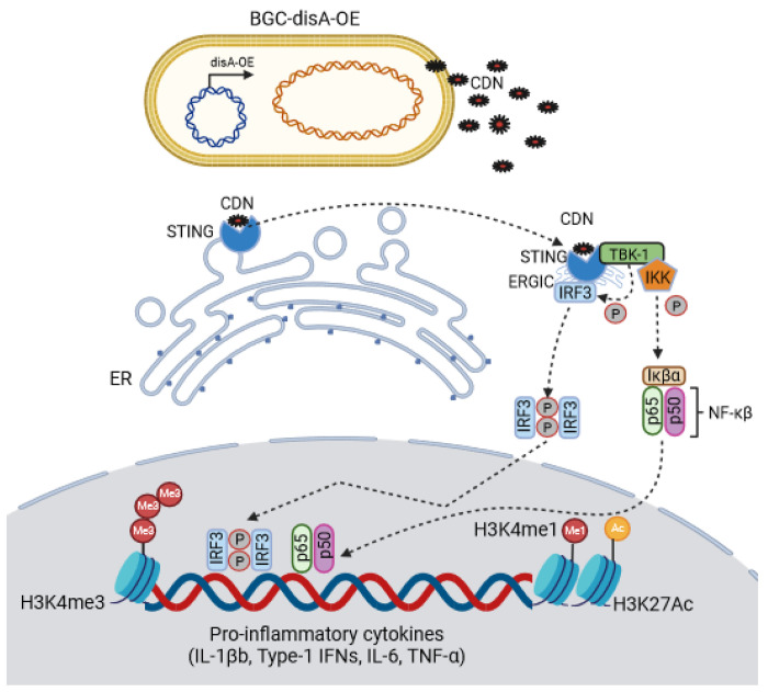
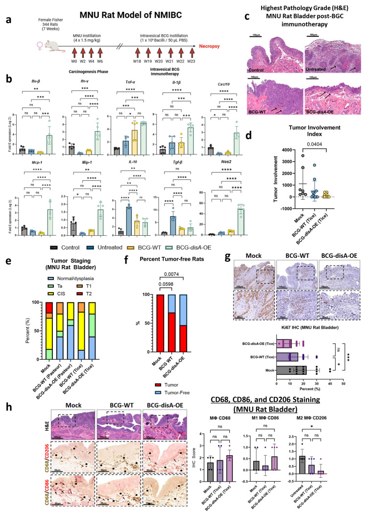
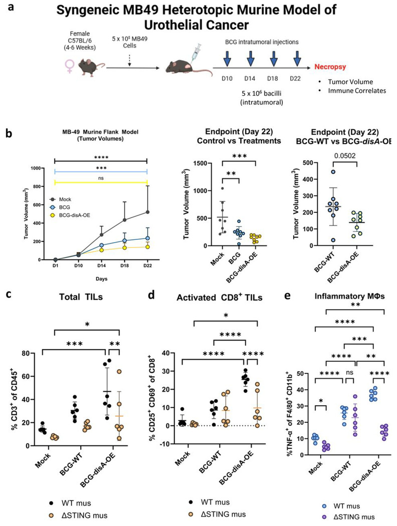
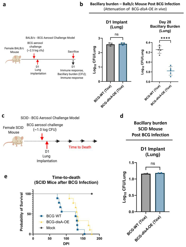
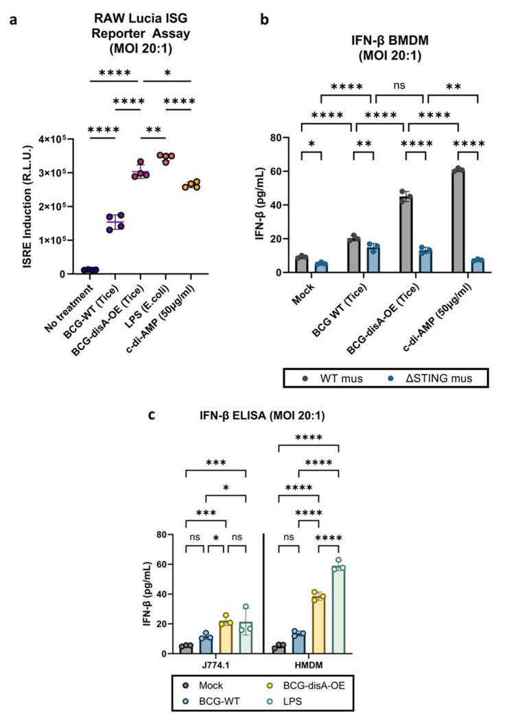
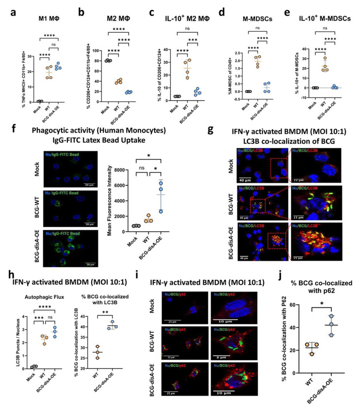
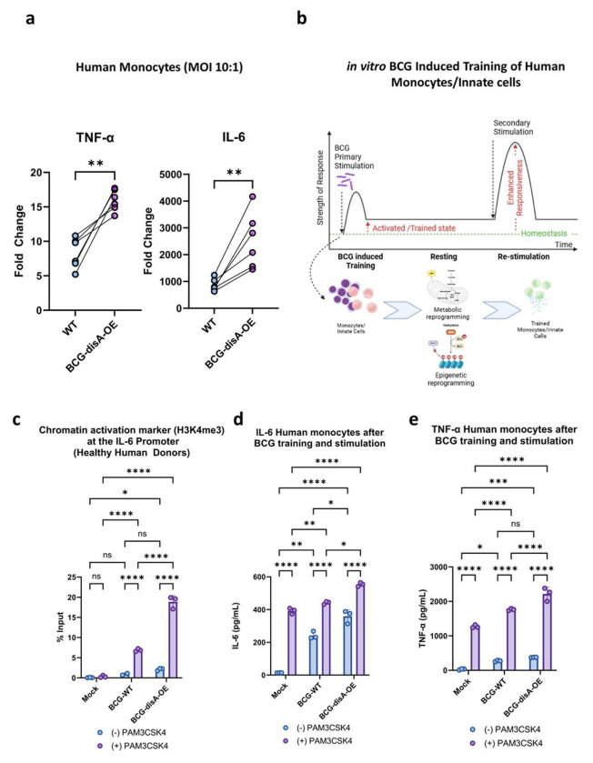
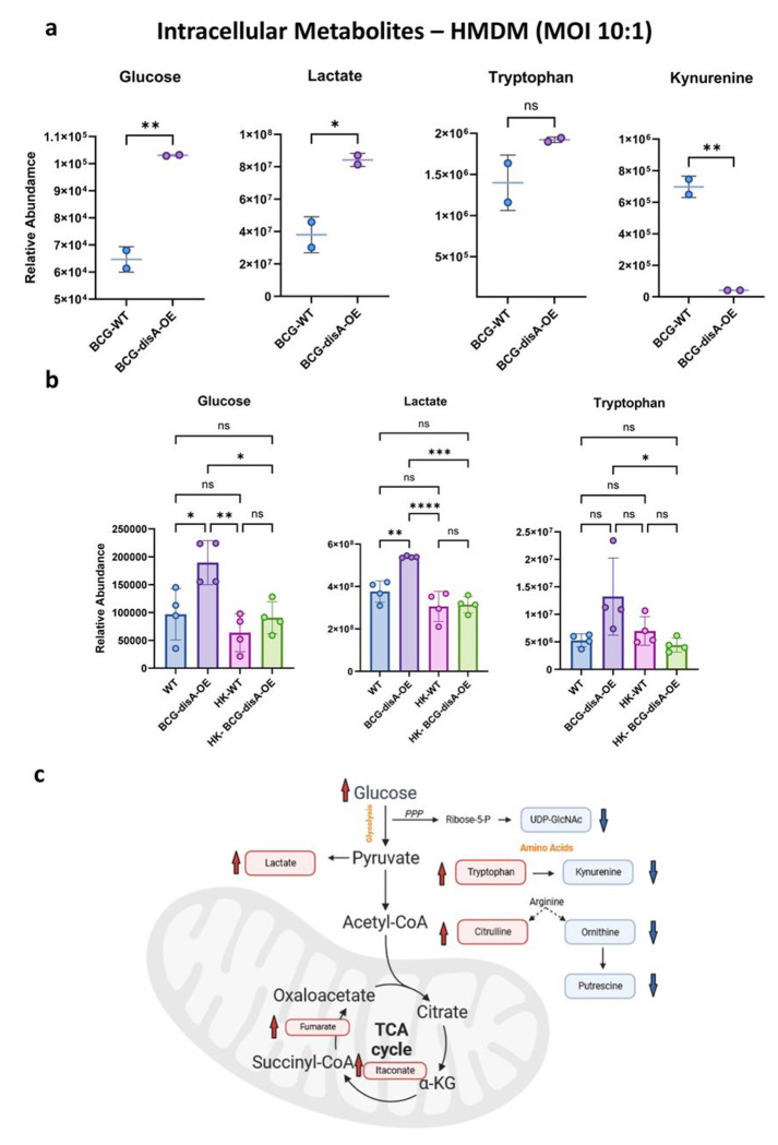
Despite the introduction of several new agents for the treatment of bladder cancer (BC), intravesical BCG remains a first line agent for the management of non-muscle invasive bladder cancer. In this study we evaluated the antitumor efficacy in animal models of BC of a recombinant BCG known as BCG-disA-OE that releases the small molecule STING agonist c-di-AMP. We found that compared to wild-type BCG (BCG-WT), in both the orthotopic, carcinogen-induced rat MNU model and the heterotopic syngeneic mouse MB-49 model BCG-disA-OE afforded improved antitumor efficacy. A mouse safety evaluation further revealed that BCG-disA-OE proliferated to lesser degree than BCG-WT in BALB/c mice and displayed reduced lethality in SCID mice. To probe the mechanisms that may underlie these effects, we found that BCG-disA-OE was more potent than BCG-WT in eliciting IFN-β release by exposed macrophages, in reprogramming myeloid cell subsets towards an M1-like proinflammatory phenotypes, inducing epigenetic activation marks in proinflammatory cytokine promoters, and in shifting monocyte metabolomic profiles towards glycolysis. Many of the parameters elevated in cells exposed to BCG-disA-OE are associated with BCG-mediated trained innate immunity suggesting that STING agonist overexpression may enhance trained immunity. These results indicate that modifying BCG to release high levels of proinflammatory PAMP molecules such as the STING agonist c-di-AMP can enhance antitumor efficacy in bladder cancer.


Conflict of interest statement

COMPETING INTERESTS M.P., W.R.B., and T.J.B. are co-inventors on patent applications involving BCG-disA-OE. W.R.B. and T.J.B. are co-founders of OncoSTING, LLC, which holds rights to commercialize BCC-disA-OE. The remaining authors declare no competing interests.

Figures

Figure 1.
Figure 1.
A schematic diagram of BCG-disA-OE’s intra-cellular delivery of cyclic di-nucleotides (CDNs) and subsequent binding to the STING homodimer; STING-CDN trafficking to the Golgi via ER-Golgi intermediate compartment (ERGIC) and then TBK-1 recruitment and subsequent phosphorylation of IRF3 and NFκβ for downstream transcriptional activation of pro-inflammatory cytokines.
Figure 2.
Figure 2.. BCG-disA-OE elicits improved antitumor efficacy over BCG-WT in an orthotopic carcinogen-induced MNU rat model of urothelial cancer.
a. Schematic diagram of the MNU rat model of NMIBC. b. mRNA levels for proinflammatory cytokines (IFN-β, IFN-γ, TNF-α, IL-1β), regulatory chemokines (CXCL10, Mcp-1, MIP-1α), immunosuppressive M2-like macrophage cytokines (IL-10, TGF-β), and the M1-like tumoricidal effector (Nos2) in whole bladders at necropsy (wk 23) measured by RT-qPCR relative to GAPDH (n= 5 animals / group). c. Representative H & E staining showing highest pathology grade for each group (control, untreated MNU bladder). d. Tumor involvement values at necropsy e. Tumor stage at necropsy. f. Percent of rats which were cancer-free at necropsy; BCG-WT (Pasteur and Tice), and BCG-disA-OE (Pasteur and Tice). g. Representative immunohistochemistry and bar graph of rat bladder tissue at necropsy stained for Ki67. h. Representative immunohistochemical co-staining and graph for CD68 (brown), CD86 (M1-like macrophages; red) and CD206 (M2-like macrophages; red) in rat bladder tissues at necropsy. Tumor staging and involvement index was performed by a pathologist trained urothelial cancers who was blinded to sample identities. The MNU model was conducted twice with BCG strains from the Tice background and the Pasteur background. Data shown represent pooled results from the two studies (n = 11-16 animals per group). Data are represented as mean values ± S.D. Statistical analyses were done using one-way ANOVA with Tukey’s test for multiple comparisons in panels b, g, & h; one-way ANOVA with Dunnett’s Test for multiple comparisons in panel d; two-sided Fisher’s Exact test in panel f (* p < 0.05, ** p < 0.01, *** p < 0.001, ****p < 0.0001).
Figure 3.
Figure 3.. BCG-disA-OE elicits improved antitumor efficacy over BCG-WT in the syngeneic MB49 heterotopic mouse model of urothelial cancer.
a. Schematic diagram of the MB49 syngeneic mouse model of urothelial cancer. b. MB49 tumor volumes and at time of necropsy on day 22 (8 animals/group). c. Tumor infiltrating lymphocytes (TILs, percent CD3+ of CD45) at necropsy, d. Activated CD8+ TILs (percent CD25+ CD69+ of CD8+), and e. inflammatory macrophages (percent TNFα+ of F4/80+ CD11b+). The flow cytometry experiments were performed with treatment on days 10, 14, 17, and 21, with necropsy on day 22 as shown in Fig. S3a (6 animals/group). Data are represented as mean values ± S.D. Statistical analyses done using one-way ANOVA with Dunnett’s multiple comparisons test in panel b (control vs treatments); two-tailed student’s T-test in panel b (BCG-WT vs BCG-disA-OE); two-way ANOVA with Tukey’s multiple comparisons test in panels b, c, d, & e (* p < 0.05, ** p < 0.01, *** p < 0.001, ****p < 0.0001).
Figure 4.
Figure 4.. BCG-disA-OE is less pathogenic than BCG-WT in two mouse models.
a. Schematic diagram of the immunocompetent BALB/c mouse challenge model. b. BALB/c lung colony forming unit (CFU) counts at day 1 (n= 3 animals/group) and day 28 (n= 5 animals/group). c. Schematic diagram of the immunocompromised SCID mouse challenge model. d. SCID mouse lung colony forming unit (CFU) counts at day 1 (n= 2 animals/group). e. Percent survival of SCID mice following low dose challenge (n=10 animals/group). The experiment was performed with BCG strains in the Tice background. Similar results were obtained using the Pasteur background as shown in Fig. S4. Data are represented as mean values ± S.D. Statistical analyses done using 2-tailed Student’s t-test in panels b and d, Kaplan-Meier survival curve in panel e (**** p < 0.0001).
Figure 5.
Figure 5.. BCG-disA-OE elicits greater interferon-β (IFN-β) responses than BCG-WT in primary murine and human macrophages in vitro.
a. IRF3 induction measured in RAW-Lucia ISG reporter murine (Balb/c) macrophages. b. IFN-β levels in murine BMDM from wild-type and STING−/− mice (C57BL/6 background). C. IFN-β levels in J774.1 macrophages and human monocyte-derived macrophages (HMDM) following exposure to BCG strains. Cytokine levels were measured by ELISA after 24 hours exposure at and MOI of 20:1. Data are presented as mean values ± SD (n=3 biological replicates). Statistical analyses done using one-way ANOVA w/Tukey’s multiple comparisons test in panel a ; two-way ANOVA with Tukey’s test for multiple comparisons in panels b and d (* p < 0.05, ** p < 0.01, *** p < 0.001, **** p < 0.0001).
Figure 6.
Figure 6.
BCG-disA-OE elicits greater macrophage re-programming, phagocytic activity, and autophagy than BCG-WT in human and murine macrophages. Percentages of cells arising from primary murine macrophages exposed to BCG (Tice) strains at an MOI of 20:1 at 24 hours post-exposure: a. M1-like macrophages (TNFα-expressing of MHCII+ CD11b+ F4/80+ cells), b. M2-like macrophages (CD206−, CD124-expressing of CD11b+ F4/80+ cells), c. IL-10 expressing M2-like macrophages (IL-10 expressing of M2-like macrophages), d. monocytic myeloid-derived macrophages (M-MDSCs, Ly6Chi, Ly6G- of CD11b+ F4/80+ cells), e. IL-10 expressing M-MDSCs (IL-10-expressing of M-MDSCs) (FigS9b). Flow cytometry studies shown are for BCG strains in the Pasteur background. Data are presented as mean values ± SD (n = 3 biological replicates). Gating schemes and data acquisition examples are shown in Fig. S6–S9. f. Phagocytic activity in human primary macrophages in representative confocal photomicrographs showing intracellular uptake of FITC-labeled IgG-opsonized latex beads (green) with nuclei stained blue. g. Autophagy induction measured by LC3B puncta co-localization with BCG strains, and h. quantification of BCG-LC3B co-localization in primary murine macrophages shown by representative confocal photomicrographs. i. Autophagy induction measured by p62 puncta co-localization with BCG strains and p62, and j. quantification of BCG-p62 co-localization. FITC-labeled BCG strains are stained green, LC3B or p62 autophagic puncta (red), nuclei blue, and co-localization (yellow). Cells were fixed using 4% paraformaldehyde 6 h after infection (MOI 10:1), and images obtained with an LSM700 confocal microscope and Fiji software processing. Quantification was measured by mean fluorescence intensity. Co-localization studies shown are for BCG strains in the Tice background. Data shown for the confocal microscopy studies are mean values ± SD (n= 3 biological replicates). Statistical analyses done using one-way ANOVA w/Tukey’s multiple comparisons test in panels a-f, & h; 2-tailed Student’s t-test in panels h and j (* p < 0.05, ** p < 0.01, *** p < 0.001, **** p < 0.0001).
Figure 7.
Figure 7.. Compared with BCG-WT, BCG-disA-OE is a more potent inducer of epigenetic changes characteristic of trained immunity in primary human monocytes.
a. Fold change in mRNA levels of TNF-a and IL-6 in primary human monocytes (n=6 healthy donors) relative to the RNU6A transcript after 24 hr exposures at a MOI of 10:1. b. Schematic diagram of in vitro monocyte training. c. Relative levels of the H3K4me3 chromatin activation mark in the IL-6 promoter region in the primary human monocytes of one healthy donor determined by ChIP-PCR assay on day 6 following initial stimulation on day 0 with no treatment (NT) or one of the BCG strains and a second stimulation on day 6 with NT or the TLR1/2 agonist PAM3CSK4. d. Secreted levels of the cytokine IL-6 and e. TNF-a from primary human monocytes (3 healthy donors) following BCG training and re-stimulation by the same protocol. Monocytes were initially challenged on day 0 with a 24 hr exposure to the BCG strains at a MOI of 10:1 followed by washing. After 5 days of rest, they were treated for 24 hr with either no treatment (RPMI) or the TLR1/2 agonist Pam3CSK4. Data are represented as mean values ± SD (n= 3 biological replicates). Statistical analyses done using a paired 2-tailed Student’s t-test on panel A; two-way ANOVA w/Tukey’s multiple comparisons test on panels c, d, & e (* p < 0.05, ** p < 0.01, *** p < 0.001, **** p < 0.0001).
Figure 8.
Figure 8.. Compared with BCG-WT, BCG-disA-OE is a more potent inducer of metabolomic changes characteristic of trained immunity in primary human monocytes.
a-b. Metabolite levels determined by LCMS in human or murine MDM determined 24 hr after exposure to BCG (Tice) strains or heat-killed (HK) controls. Schematic diagram (c) showing key metabolites significantly upregulated (red arrow upward) or downregulated (blue arrow downward) in BCG-disA-OE infected macrophages relative to BCG-WT infected macrophages. Data are represented as mean values ± SD (n= 4 biological replicates) Statistical analyses done using two-tailed Student’s t-test on panel A; one-way ANOVA w/Tukey’s test for multiple comparisons in panel b (* p < 0.05, ** p < 0.01, *** p < 0.001, **** p < 0.0001).

References

    1. Saginala K., et al. Epidemiology of Bladder Cancer. Med Sci (Basel) 8(2020). - PMC - PubMed
    1. Grabe-Heyne K., et al. Intermediate and high-risk non-muscle-invasive bladder cancer: an overview of epidemiology, burden, and unmet needs. Front Oncol 13, 1170124 (2023). - PMC - PubMed
    1. Morales A., Eidinger D. & Bruce A.W. Intracavitary Bacillus Calmette-Guerin in the treatment of superficial bladder tumors. J Urol 116, 180–183 (1976). - PubMed
    1. Fankhauser C.D., Teoh J.Y. & Mostafid H. Treatment options and results of adjuvant treatment in nonmuscle-invasive bladder cancer (NMIBC) during the Bacillus Calmette-Guérin shortage. Curr Opin Urol 30, 365–369 (2020). - PubMed
    1. Roumiguié M., et al. International Bladder Cancer Group Consensus Statement on Clinical Trial Design for Patients with Bacillus Calmette-Guérin-exposed High-risk Non-muscle-invasive Bladder Cancer. Eur Urol 82, 34–46 (2022). - PubMed

Publication types