[Preprint]. 2023 Dec 12:2023.12.11.571076.
doi: 10.1101/2023.12.11.571076.

Biased cell adhesion organizes a circuit for visual motion integration

Affiliations

Biased cell adhesion organizes a circuit for visual motion integration

Yannick Carrier et al. bioRxiv. .

Update in

Abstract

Layer specific computations in the brain rely on neuronal processes establishing synaptic connections with specific partners in distinct laminae. In the Drosophila lobula plate neuropile, the axons of the four subtypes of T4 and T5 visual motion direction-selective neurons segregate into four layers, based on their directional preference, and form synapses with distinct subsets of postsynaptic neurons. Four bi-stratified inhibitory lobula plate intrinsic cells exhibit a consistent synaptic pattern, receiving excitatory T4/T5 inputs in one layer, and conveying inhibitory signals to an adjacent layer. This layered arrangement establishes motion opponency. Here, we identify layer-specific expression of different receptor-ligand pairs belonging to the Beat and Side families of Cell Adhesion Molecules (CAMs) between T4/T5 neurons and their postsynaptic partners. Genetic analysis reveals that Beat/Side mediated interactions are required to restrict T4/T5 axonal innervation to a single layer. We propose that Beat/Side contribute to synaptic specificity by biasing adhesion between synaptic partners before synaptogenesis.


Figures
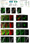
Figure 1.
Figure 1.. Lobula plate Motion Opponency Circuit
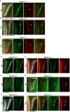
(A) Morphology of the four T4/T5 subtypes. All T4/T5 neurons with the same directional tuning preference project their axons retinotopically to the same layer of the lobula plate neuropile, producing four layers tuned for motion in each of the cardinal directions: layer 1, front-to-back; layer 2, back-to-front, layer 3, upward; layer 4, downward. (B) The connectome of T4/T5c and d subtypes with more than 10 synapses with a single T4 or T5 neurons, after (Shinomiya et al., 2022). (C) Motion Opponency Circuit in the lobula plate layer 4. Direction-selective wide-field motion responses in postsynaptic partners VS-Lobula Plate Tangential Cells (LPTCs) match the direction selectivity of their upstream T4/T5d neurons. LPi4-3 neurons receive input from T4/T5d neurons, and convey inhibitory signals to LPTC cells in the neighboring motion-opponent layer 3. LPi3-4 neurons contribute to a complementary circuit in the other direction (also, Figure S2). (D) T4/T5 neurons expressing membrane-GFP (green) (E) LPi3-4 neurons. (F) LPi4-3 neurons. (G) HS (layer 1) and VS (layer 4) LPTC neurons. In (D-G). Anti-Brp immunostaining (gray) labels the neuropiles. In (E) and (F) Syt::GFP labels axonal terminals (arrowhead) and the somatodendritic DenMARK::RFP labels the neurons, with stronger dendritic expression (arrow). (H-L) Sequential segregation of T4/T5 axons in four lobula plate layers during development. (I-K) MCFO-sparsely labeled T4/T5s during development. (M-O) In green, endogenous Syd1 expression in T4/T5 neurons during development through combination of a T4/T5 driver (R42F06-Gal4)>uas-flp and Frt-Stop-Frt-Syd1::GFP. In grey anti-Brp immunostaining. Scheme in (C) after (Mauss et al., 2015). In (I) and (K) grey dashed lines delineate the lobula plate, white dashed lines delineate a/b and c/d layers. Scale bars are 20μm, except in (I) and (K) where they are 5μm.
Figure 2.
Figure 2.. Layer specific expression of matching expression of Beat and Side proteins between synaptic partners
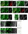
(A) Log-fold change for Differentially expressed genes (DEGs) between T4c and T4d (x axis), and T5c and T5d (y axis). See Methods for details and thresholds. (B) Average expression level of DEGs (from (A)). (C) IgSF CAMs in T4/T5 and LPi neurons. Average expression levels are shown for five time points after pupal formation and in the Adult. (D) Interactome of Beat/Side families after (Li et al., 2017) (E) Line plots with average expression levels of Side-IV, Side-II in T4/T5s and Beat-IIa/b and Beat-VI in LPi neurons.
Figure 3.
Figure 3.. Beat/Side localization in the developing lobula plate
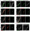
(A-C) Localization of endogenously tagged Side-II::GFP (green) and Side-IV (Red) proteins during development. (D-I) Localization of endogenously GFP tagged Beat-IIa (D,E), Beat-IIb (F,G) and Beat-IV (H,I) during development. Open arrowhead indicates layer 1, closed arrowhead layer 2, closed arrowhead layer 3 and open arrow layer 4). DNcad (grey) labels the neuropiles. Scale bars are 20μm.
Figure 4.
Figure 4.. Side/Beat receptor-ligand pair interactions between synaptic partners are required for T4/T5 axons to differentiate between layers c and d.
(A-D) Morphology of T4/T5s in controls, mutants and upon T4/T5>RNAi specific downregulation. T4/T5 neurons express membrane tethered CD4-tdGFP (green). Side-IV protein (red) (arrow in A and D), Brp (gray) labels the neuropiles. (A) Control. (B) side-IInull homozygous mutants. (C) beat-VInull mutants. (D, E) Side-II downregulation in T4/T5 neurons with side-II RNAi (D) and sgRNA against side-II (E). In (B-E) axons of T4c/T5c and T4d/T5d form a single merged c/d layer. Upon side-II downregulation in T4/T5, Side-IV localization (arrow) remained restricted to half of the merged 3/4 layer (compare A with D). In (D) open arrow points to the half of the merged 3/4 layer where Side-IV is not localized. (F) beat-VIRI in T4/T5 neurons. (G) Pan-neuronal beat-VI downregulation with Elav-Gal>beat-VIRI. (H-I) MARCM clones of T4/T5d neurons. (H) Wild type T4/T5d subtype neuron innervates layer 4 (closed arrow) (N=6). (I) side-IInull (N=12) d subtype MARCM clones always innervate both layers 3 (arrowheads) and 4 (arrows). Closed arrow/arrowhead identify projections of a single d subtype neuron innervating both layer 3 (closed arrowhead) and layer 4 (closed arrow). All controls for figures 4 and 5 are compiled in Figure S8. Scale bars are 20μm, and 5μm in (H-I).
Figure 5.
Figure 5.. Beats and Sides are required for T4/T5 axons to differentiate between layers c and d
Morphology of T4/T5s in controls, mutants and upon T4/T5>RNAi specific downregulation. T4/T5 neurons express membrane tethered CD4-tdGFP (green). Side-IV protein (red), Brp (gray) labels the neuropiles. (A) Wild type control. (B-C) T4/T5-specific side-IV downregulation with RNAi (B), and sgRNA (C). Side-IV is absent from the lobula plate upon T4/T5>side-IVRI (arrow, compare with Figure 4 and Figure S8) (D-G) Morphology of T4/T5s in Side-IVnull (D), Beat-IVnull (E), Side-VIInull (F) and Side-IVnull, Side-VIInull double mutants (G). T4/T5 layer segregation was impaired in the double mutant but not in the single mutants. All controls for figures 4 and 5 are compiled in Fig S8. Scale bars are 20μm
Figure 6.
Figure 6.. Beat/Side overexpression is sufficient to change layer specific innervation
LPi4-3 visualized with R38G02-LexA or -Gal4 in the wild type and upon manipulation of Beat/Side interactions. In all panels, arrow points to dendritic terminals and arrowhead to axonal terminals. Brp (grey) labels the neuropiles. (A-B) LPi4-3 neurons expressing membrane tethered CD8tdGFP (green) in the wild type (A), and upon side-IIRI mediated downregulation in T4/T5 neurons (B). (C-D) Wild type LPi4-3 neurons expressing Syt::GFP in the axonal terminals (arrowhead) in layer 3, and Denmark::RFP that labelled the neurons, with stronger expression in the dendrites (arrow) in layer 4 (C), and upon beat-VIRI downregulation (D). (E-F) Wild type LPi4-3 expressing the nicotinic acetylcholine receptor subunit Dα7, which localized to LPi4-3-cholinergic synapses (Ammer et al., 2023) (E), and upon beat-IIa overexpression (F). Overexpression of Beat-IIa in LPi4-3 induced the innervation of only layer 3, where the neurons developed Dα7+ dendritic processes (arrow). (G) Overexpression of Side-VII in LPi4-3 neurons expressing Syt::GFP and Denmark::RFP. Side-VII overexpression promoted the development of dendritic processes in layer 2 (arrow), where the partner of Side-VI, Beat-IV, is strongly expressed in T4/T5 axons. LPi4-3 neurons maintained their dendritic and axonal innervations in layers 4 and 3, respectively. (H) t-GRASP activity-independent GRASP between T4/T5 neurons (R42F06-Lexa) and LPi4-3 (R38G02-Gal4) neurons in the wild type (left), and upon beat-IIa overexpression in LPi4-3 neurons (right). Scale bars are 20μm.
Figure 7.
Figure 7.. Beat/Side interactions mediate cell adhesion before synaptogenesis
LPi4-3-Split-Gal4 driving membrane tethered CD4-tdGFP (green). DNcad (grey) labels de neuropiles. LPi4-3 lobula plate innervation in the wildtype. Arrow point to the right edge of the lobula plate where layer 4 develops. (A-B) wild type. (C-D) LPi4-3 overexpressing beat-IIa. Beat-IIa overexpression promotes LPi4-3 exclusive layer 3 innervation as early as P25, ~40h before synaptogenesis. Scale bars are 20μm.

References

    1. Ammer G., Serbe-Kamp E., Mauss A.S., Richter F.G., Fendl S., and Borst A. (2023). Multilevel visual motion opponency in Drosophila. Nature neuroscience 26, 1894–1905. - PMC - PubMed
    1. Apitz H., and Salecker I. (2015). A region-specific neurogenesis mode requires migratory progenitors in the Drosophila visual system. Nature neuroscience 18, 46–55. - PMC - PubMed
    1. Baier H. (2013). Synaptic laminae in the visual system: molecular mechanisms forming layers of perception. Annual review of cell and developmental biology 29, 385–416. - PubMed
    1. Bekkers J.M., and Stevens C.F. (1991). Excitatory and inhibitory autaptic currents in isolated hippocampal neurons maintained in cell culture. Proceedings of the National Academy of Sciences of the United States of America 88, 7834–7838. - PMC - PubMed
    1. Butler A., Hoffman P., Smibert P., Papalexi E., and Satija R. (2018). Integrating single-cell transcriptomic data across different conditions, technologies, and species. Nat Biotechnol 36, 411–420. - PMC - PubMed

Publication types