[Preprint]. 2023 Dec 19:2023.12.12.571338.
doi: 10.1101/2023.12.12.571338.

Securin Regulates the Spatiotemporal Dynamics of Separase

Affiliations

Securin Regulates the Spatiotemporal Dynamics of Separase

Christopher G Sorensen Turpin et al. bioRxiv. .

Update in

  • Securin regulates the spatiotemporal dynamics of separase.
    Sorensen Turpin CG, Sloan D, LaForest M, Klebanow L, Mitchell D, Severson AF, Bembenek JN. Sorensen Turpin CG, et al. J Cell Biol. 2025 Feb 3;224(2):e202312099. doi: 10.1083/jcb.202312099. Epub 2024 Nov 18. J Cell Biol. 2025. PMID: 39556062 Free PMC article.

Abstract

Separase is a key regulator of the metaphase to anaphase transition with multiple functions. Separase cleaves cohesin to allow chromosome segregation and localizes to vesicles to promote exocytosis in mid-anaphase. The anaphase promoting complex/cyclosome (APC/C) activates separase by ubiquitinating its inhibitory chaperone, securin, triggering its degradation. How this pathway controls the exocytic function of separase has not been investigated. During meiosis I, securin is degraded over several minutes, while separase rapidly relocalizes from kinetochore structures at the spindle and cortex to sites of action on chromosomes and vesicles at anaphase onset. The loss of cohesin coincides with the relocalization of separase to the chromosome midbivalent at anaphase onset. APC/C depletion prevents separase relocalization, while securin depletion causes precocious separase relocalization. Expression of non-degradable securin inhibits chromosome segregation, exocytosis, and separase localization to vesicles but not to the anaphase spindle. We conclude that APC/C mediated securin degradation controls separase localization. This spatiotemporal regulation will impact the effective local concentration of separase for more precise targeting of substrates in anaphase.


Figures

Figure 1.
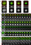
Figure 1.. Separase and Securin dynamics in meiosis I.
(A-D) Representative images SEP-1::GFP (green) with H2B::mCherry (red) during meiosis I. (A) During prophase I, SEP-1::GFP is cytoplasmic and excluded from the nucleus. (B) At NEBD, SEP-1::GFP accumulates in the nucleus, on chromosomes (caret) and to cytoplasmic kinetochore linear elements throughout the cortex (white arrows), where it remains throughout prometaphase (C). (D) By mid-anaphase, separase localizes between separating chromosomes (caret) and to vesicles (arrowheads). (E-L) Embryos expressing H2B::mCherry (red) with (E-H) IFY-1WT::GFP (green) or (I-L) IFY-1DM::GFP (green) in meiosis I. (E) IFY-1WT::GFP and (I) IFY-1DM::GFP are present in both the cytoplasm and the nucleus in prophase oocytes. IFY-1WT::GFP (F) and IFY-1DM::GFP (J) display identical localization patterns as separase at NEBD and through prometaphase I (G, K). In anaphase I, IFY-1WT::GFP (H) is mostly degraded and is not observed on vesicles. In contrast, IFY-1DM::GFP (L) is not degraded in anaphase and accumulates on chromosomes (caret) and the anaphase I spindle (white arrowhead) but does not enrich on cortical granules. (M) Quantification of IFY-1WT::GFP spindle-associated and cytoplasmic signal showing rapid degradation (t = 0 is chromosome separation at anaphase onset). (N) Quantification of endogenously tagged SEP-1::mCherry and IFY-1WT::GFP localized to linear elements in the cortex, (t = 0 is separase localization to vesicles). Securin levels equilibrate with cytoplasmic signal before separase leaves linear elements and appears on vesicles. Scale bar: 10μm.
Figure 2.
Figure 2.. Spatiotemporal dynamics of Separase, Securin and Cohesin at Anaphase Onset.
(A-B) Representative localization pattern of SEP-1::mScarlet (green) co-expressed with cohesin subunit COH-3::GFP (red) at the spindle in anaphase I. (A) In prometaphase I, spindle SEP-1::mScarlet localizes to kinetochore cups around the bivalents and is spatially isolated from COH-3::GFP at the midbivalent. (B) Just before anaphase onset, SEP-1::mScarlet accumulates on spindle poles, still not enriched with COH-3::GFP. (C) Seconds before chromosome segregation, SEP-1::mScarlet invades the midbivalent, colocalizing briefly with COH-3::GFP (caret). (D) COH-3::GFP is quickly lost from chromosomes right after SEP-1::mScarlet invades the midbivalent region and chromosomes immediately move poleward at anaphase onset (caret). (E) Montage showing rapid SEP-1::mScarlet localization changes on the spindle and abrupt loss of COH-3::GFP at anaphase onset (time is shown in seconds relative to t=0, which indicates the first timepoint with significant midbivalent accumulation of SEP-1::mScarlet). (F-H) Montages showing the dynamics of SEP-1::GFP, IFY-1WT::GFP and GFP::IFY-1DM (green) co-expressed with chromosome marker H2B::mCherry (red) during the metaphase-to-anaphase I transition (t=0 marks when chromosome move apart in F, G, or when midbivalent signal appears in H). (F) SEP-1::GFP appears prominently at the midbivalent when chromosomes move apart at anaphase onset. (G) IFY-1WT::GFP signal is significantly reduced by anaphase onset. (H) GFP::IFY-1DM remains high on the spindle throughout anaphase I and localizes similar to separase. (I) In the cortex, SEP-1::GFP relocalizes from linear elements (white arrow) to cortical granules labeled with mScarlet::RAB-11.1 (arrowhead) by 30 seconds after anaphase onset. Scale bars: 2μm.
Figure 3.
Figure 3.. The APC/C and securin control SEP-1 localization in meiosis I.
Images of the germline in worms expressing SEP-1:::GFP (green) with chromosome marker H2B::mCherry and cortical granule cargo protein mCherry::CPG-2 (red). Insets show high magnification of the spindle in A’-D’ or vesicles in A”-D”. Numbers correspond to relative position of embryos in the uterus, which also corresponds to their increasing age as each ovulation occurs every 25–30 minutes. (A) In control animals fed OP50, prometaphase I embryos (embryo 0) have mCherry::CPG-2 in vesicles, while older embryos (+1 to +3) have mCherry::CPG-2 incorporated into the eggshell (denoted by yellow asterisk in the +1 embryo). Prometaphase I embryos in the spermatheca have SEP-1::GFP localized to kinetochores at the spindle (A’) and linear elements in the cortex (A”). (B) Intermediate ify-1 RNAi animals have prometaphase I embryos in the spermatheca (0) with SEP-1::GFP localized abnormally to chromosomes in the midbivalent region (B’) and localization to vesicles (yellow arrowhead in C, cortical region in B”). Multiple older embryos (+1-+2) also have SEP-1::GFP localized to cortical granules (yellow arrowheads). Early embryos lack eggshell signal but eventually some mCherry::CPG-2 signal appears outside older embryos (+3, asterisk). (C) Severe ify-1 RNAi causes SEP-1::GFP to remain on vesicles in older embryos (+1, +2, yellow arrowheads). mCherry::CPG-2 remains trapped in cortical granules and no eggshell is detected in any of the embryos at any age. SEP-1::GFP displays mislocalized chromosome (C’, caret denotes signal at midbivalent) and vesicle (C”) localization in prometaphase I embryos. (D) After apc/c RNAi, separase localization is normal in prometaphase I embryos in the spermatheca (embryo 0), appearing on the kinetochore cups at the spindle (B’) and cortical linear elements (B”). Arrested embryos (+1-+4) in the uterus all show SEP-1:::GFP on kinetochore structures (white arrows). mCherry::CPG-2 remains in cortical granules (white arrowheads) and does not incorporate into the eggshell. Scale bars: 10μm for A-D; 5μm for insets.
Figure 4.
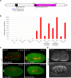
Figure 4.. Generation and Characterization of GFP::IFY-1DM in C. elegans.
(A) Schematic of IFY-1 indicating the APC/C recognition motif (destruction box), within the unstructured region at the N- terminus. Separase binding domain indicates the region that was resolved in the structure of the separase/securin complex. Conserved residues in the D-box were mutated to alanine to prevent APC/C recognition. (B) Embryonic lethality of multiple independently generated transgenic IFY-1WT::GFP and IFY-1DM::GFP lines at 20°C and 25°C in homozygous or heterozygous animals. (C) In IFY-1WT::GFP, two-celled embryos always have two polar bodies. GFP::IFY-1DM expression causes a spectrum of polar body extrusion defects with most embryos lacking any polar bodies, indicating defects in meiotic divisions. (D) IFY-1WT::GFP embryos that complete meiosis are not permeable, while IFY-1DM::GFP embryos all shrink in hyperosmotic solution, indicating permeability barrier defects. N = number of embryos scored. Scale bar: 10μm.
Figure 5.
Figure 5.. GFP::IFY-1DM inhibits chromosome segregation during anaphase I.
(A) IFY-1WT::GFP or (B) IFY-1DM::GFP (green) expressed together with H2B::mCherry (red) during meiosis I. Chromosomes move apart quickly in IFY-1WT::GFP, but are severely delayed in IFY-1DM::GFP embryos. Time (seconds) is relative to midbivalent localization. (C) Average distance between segregating chromosomes during anaphase I showing a significant delay in IFY-1DM::GFP embryos. The IFY-1DM::GFP curve starts after an extended delay of chromosome movement after midbivalent localization. N = 9 for IFY-1WT::GFP, N > 18 for IFY-1DM::GFP, asterisks denote a statistically significant difference. P-value = <0.05, error bars are standard deviations of the mean. (D) Frequency and representative images of ultrafine, chromosome bridging (caret) and severely impaired chromosome segregation defects observed in embryos expressing IFY-1DM::GFP (green, H2B::mCherry is red). N = number of embryos scored. Scale bar: 5μm.
Figure 6.
Figure 6.. GFP::IFY-1DM blocks cortical granule exocytosis in anaphase I.
(A-C) IFY-1WT::GFP and (D-F) IFY-1DM::GFP (green) were co-expressed with the cortical granule cargo protein, mCherry::CPG-2 (red, white arrows). Time (seconds) is relative to midbivalent localization. In wildtype, the majority of cortical granules are exocytosed by 120 seconds after anaphase onset (B) and mCherry::CPG-2 is extracellular (yellow asterisk), and by the end of anaphase I (C) mCherry::CPG-2 is completely extracellular. In contrast, (E-F) in IFY-1DM::GFP embryos, cortical granules are not exocytosed even after several minutes (yellow asterisks indicate mCherry::CPG-2 labeled vesicles). (G) Quantification of cortical granules in a single spindle plane over time after anaphase onset. N = 6 for IFY-1WT::GFP, N = 7 for IFY-1DM::GFP. Asterisks denote a statistically significant difference, P-value < .0001. Error bars represent standard error of the mean. Scalebar: 10μm.
Figure 7.
Figure 7.. Separase localization to vesicles is reduced by GFP::IFY-1DM.
(A) Representative spindle localization pattern of SEP-1::mScarlet (green) co-expressed with GFP::IFY-1WT (red). Securin is rapidly degraded while separase moves from kinetochore cups to poles to the midbivalent (caret) at anaphase onset. (B) SEP-1::mScarlet colocalizes with stable GFP::IFY-1DM, in a similar pattern to wildtype at the midbivalent (arrowhead) and on the spindle in anaphase. (C) Max projections of cortical images of SEP-1::mScarlet (green) with the vesicle marker CAV-1::GFP (red) and either IFY-1WT::GFP or IFY-1DM::GFP (red). In prometaphase I SEP-1 localizes to linear elements (white arrow) but not vesicles (white arrowheads) in both conditions. At anaphase I onset, SEP-1::mScarlet begins to enrich on vesicles (yellow arrowhead) in GFP::IFY-1WT but not GFP::IFY-1DM embryos. By mid-anaphase I, SEP-1::mScarlet is fully enriched on vesicles in GFP::IFY-1WT embryos. In GFP::IFY-1DM embryos, SEP-1::mScarlet does not enrich on vesicles at anaphase I onset and only shows trace vesicle localization during anaphase I. (D) Magnified images of SEP-1::mScarlet vesicle localization from the 5μm2 regions indicated in (C) at mid-anaphase I in wildtype and mutant. SEP-1::mScarlet signal is reduced on vesicles when co-expressed with IFY-1DM::GFP, with only partial vesicle localization in rare cases (yellow arrowhead). (E) Quantification of vesicle-associated SEP-1::mScarlet signal in cortical planes at mid-anaphase I in GFP::IFY-1WT (N = 11) and GFP::IFY-1DM (N = 12) embryos. Asterisk denotes a statistically significant difference, P-value < .0001. Error bars represent standard error of the mean. Scale bar: 5μm.

References

    1. Agarwal R., & Cohen-Fix O. (2002). Phosphorylation of the mitotic regulator Pds1/securin by Cdc28 is required for efficient nuclear localization of Esp1/separase. Genes & Development, 16(11), 1371–1382. 10.1101/gad.971402 - DOI - PMC - PubMed
    1. Agircan F. G., & Schiebel E. (2014). Sensors at Centrosomes Reveal Determinants of Local Separase Activity. PLOS Genetics, 10(10), e1004672. 10.1371/journal.pgen.1004672 - DOI - PMC - PubMed
    1. Alfieri C., Zhang S., & Barford D. (2017). Visualizing the complex functions and mechanisms of the anaphase promoting complex/cyclosome (APC/C). Open Biology, 7(11), 170204. 10.1098/rsob.170204 - DOI - PMC - PubMed
    1. Bacac M., Fusco C., Planche A., Santodomingo J., Demaurex N., Leemann-Zakaryan R., Provero P., & Stamenkovic I. (2011). Securin and Separase Modulate Membrane Traffic by Affecting Endosomal Acidification. Traffic, 12(5), 615–626. 10.1111/j.1600-0854.2011.01169.x - DOI - PubMed
    1. Bai X., & Bembenek J. N. (2017). Protease dead separase inhibits chromosome segregation and RAB-11 vesicle trafficking. Cell Cycle, 16(20), 1902–1917. 10.1080/15384101.2017.1363936 - DOI - PMC - PubMed

Publication types