

This is a preprint.
Excessive Lipid Production Shapes Glioma Tumor Microenvironment
- PMID: 38168422
- PMCID: PMC10760230
- DOI: 10.21203/rs.3.rs-3694185/v1
Excessive Lipid Production Shapes Glioma Tumor Microenvironment
Update in
-
Excessive lipid production shapes glioma tumor microenvironment.Ultrastruct Pathol. 2024 Sep 2;48(5):367-377. doi: 10.1080/01913123.2024.2392728. Epub 2024 Aug 19. Ultrastruct Pathol. 2024. PMID: 39157967
Abstract
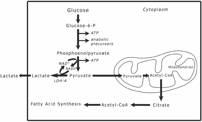
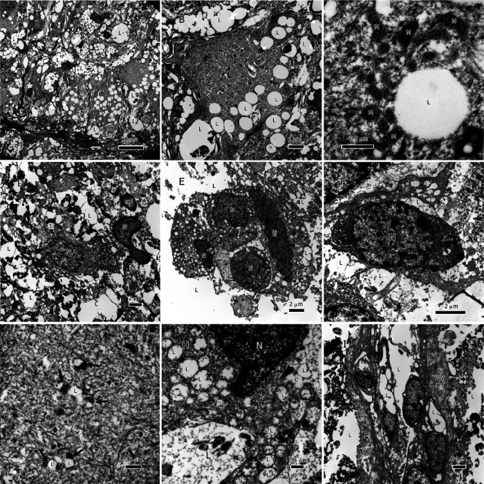
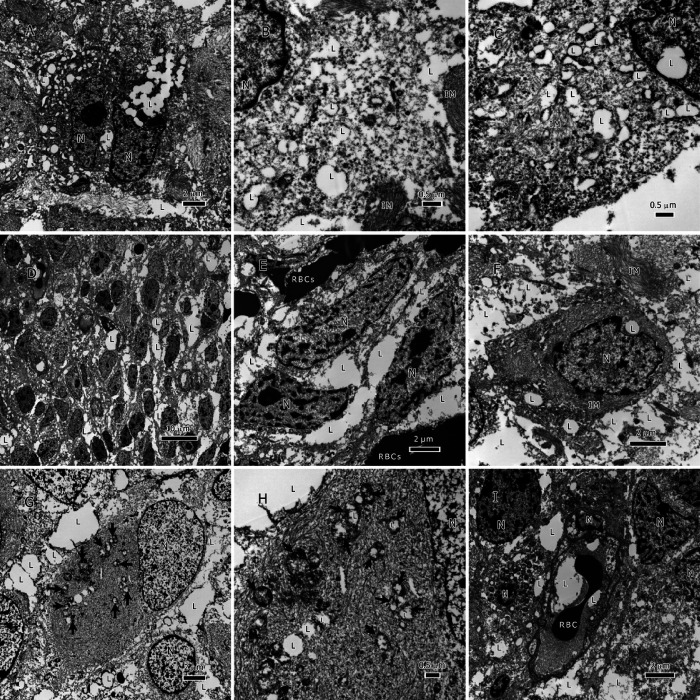
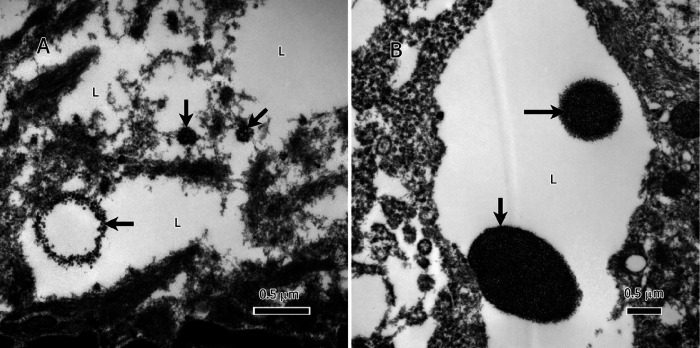
Disrupted lipid metabolism is a characteristic of gliomas. This study utilizes an ultrastructural approach to characterize the prevalence and distribution of lipids within gliomas. This study made use of tissue from IDH1 wild type (IDH1-wt) glioblastoma (n = 18) and IDH1 mutant (IDH1-mt) astrocytoma (n = 12) tumors. We uncover a prevalent and intriguing surplus of lipids. The bulk of the lipids manifested as sizable cytoplasmic inclusions and extracellular deposits in the tumor microenvironment (TME); in some tumors the lipids were stored in the classical membraneless spheroidal lipid droplets (LDs). Frequently, lipids accumulated inside mitochondria, suggesting possible dysfunction of the beta-oxidation pathway. Additionally, the tumor vasculature have lipid deposits in their lumen and vessel walls; this lipid could have shifted in from the tumor microenvironment or have been produced by the vessel-invading tumor cells. Lipid excess in gliomas stems from disrupted beta-oxidation and dysfunctional oxidative phosphorylation pathways. The implications of this lipid-driven environment include structural support for the tumor cells and protection against immune responses, non-lipophilic drugs, and free radicals.
Keywords: astrocytoma; glioblastoma; glycolysis; immune evasion; lipid accumulation; mitochondrial dysfunction; oxidative phosphorylation; therapeutic strategies; tumor microenvironment; ultrastructural analysis.
Conflict of interest statement
Conflicts of Interest: The authors declare no conflict of interest.
Figures
Similar articles
-
Excessive lipid production shapes glioma tumor microenvironment.Ultrastruct Pathol. 2024 Sep 2;48(5):367-377. doi: 10.1080/01913123.2024.2392728. Epub 2024 Aug 19. Ultrastruct Pathol. 2024. PMID: 39157967
-
Comparative survey of mitochondrial ultrastructure in IDH1-mutant astrocytoma and IDH1-wildtype glioblastoma (GBM).Ultrastruct Pathol. 2023 Feb 25:1-6. doi: 10.1080/01913123.2023.2175942. Online ahead of print. Ultrastruct Pathol. 2023. PMID: 36841772 Free PMC article.
-
Enrichment of branched chain amino acid transaminase 1 correlates with multiple biological processes and contributes to poor survival of IDH1 wild-type gliomas.Aging (Albany NY). 2021 Jan 20;13(3):3645-3660. doi: 10.18632/aging.202328. Epub 2021 Jan 20. Aging (Albany NY). 2021. PMID: 33493139 Free PMC article.
-
Tumor Microenvironment in Gliomas: A Treatment Hurdle or an Opportunity to Grab?Cancers (Basel). 2023 Feb 7;15(4):1042. doi: 10.3390/cancers15041042. Cancers (Basel). 2023. PMID: 36831383 Free PMC article. Review.
-
Rediscovering Potential Molecular Targets for Glioma Therapy Through the Analysis of the Cell of Origin, Microenvironment and Metabolism.Curr Cancer Drug Targets. 2021;21(7):558-574. doi: 10.2174/1568009621666210504091722. Curr Cancer Drug Targets. 2021. PMID: 33949933 Review.
References
Publication types
Grants and funding
LinkOut - more resources
Full Text Sources
Miscellaneous
