Review
. 2024 Mar;230(3):279-294.e2.
doi: 10.1016/j.ajog.2023.11.1253. Epub 2024 Jan 2.

Pelvic floor injury during vaginal birth is life-altering and preventable: what can we do about it?

Affiliations
Review

Pelvic floor injury during vaginal birth is life-altering and preventable: what can we do about it?

John O L DeLancey et al. Am J Obstet Gynecol. 2024 Mar.

Abstract

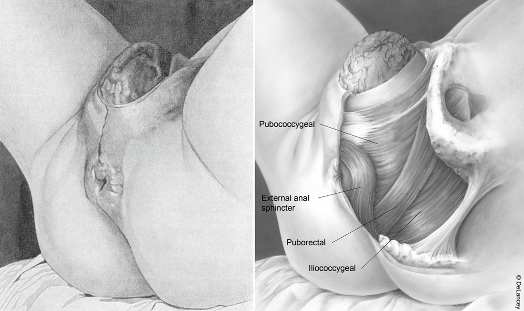
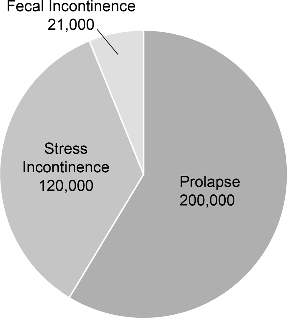
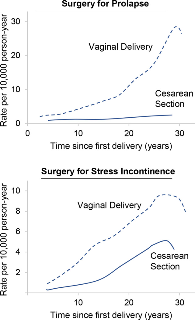
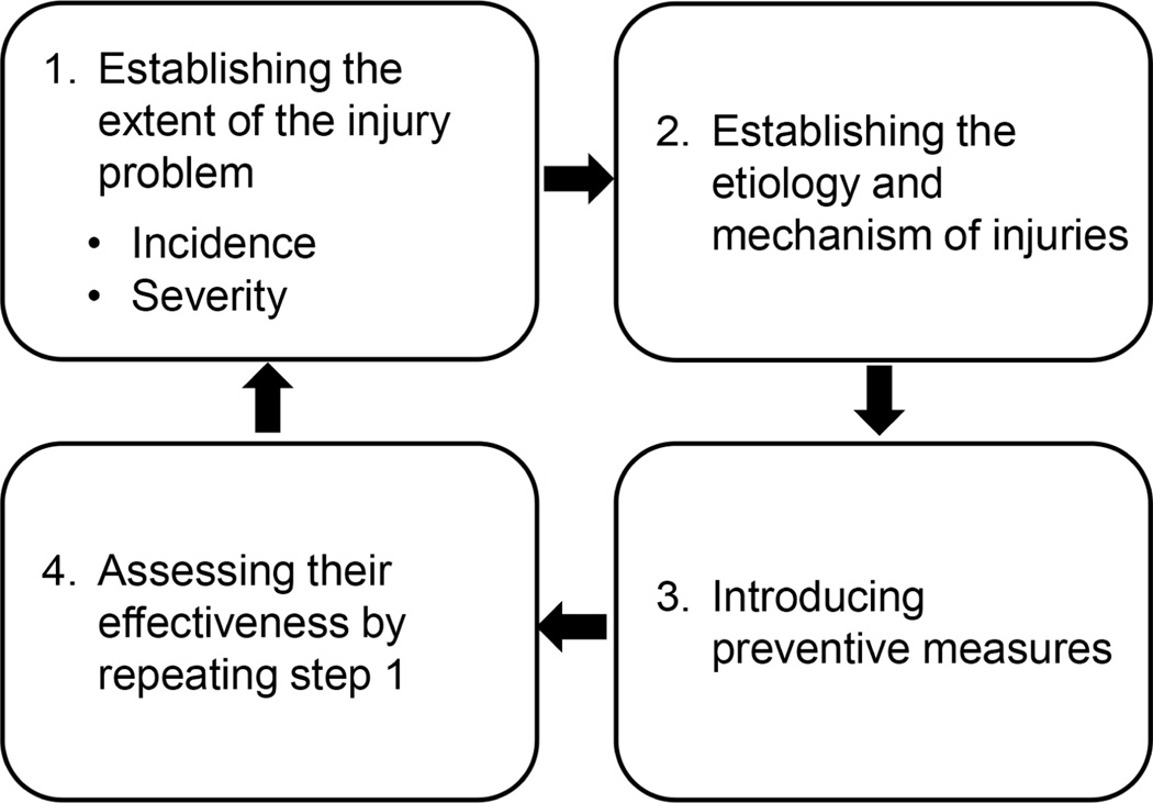
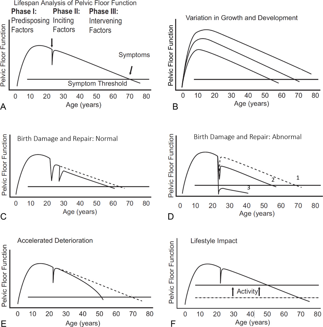
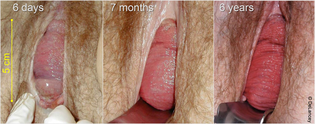
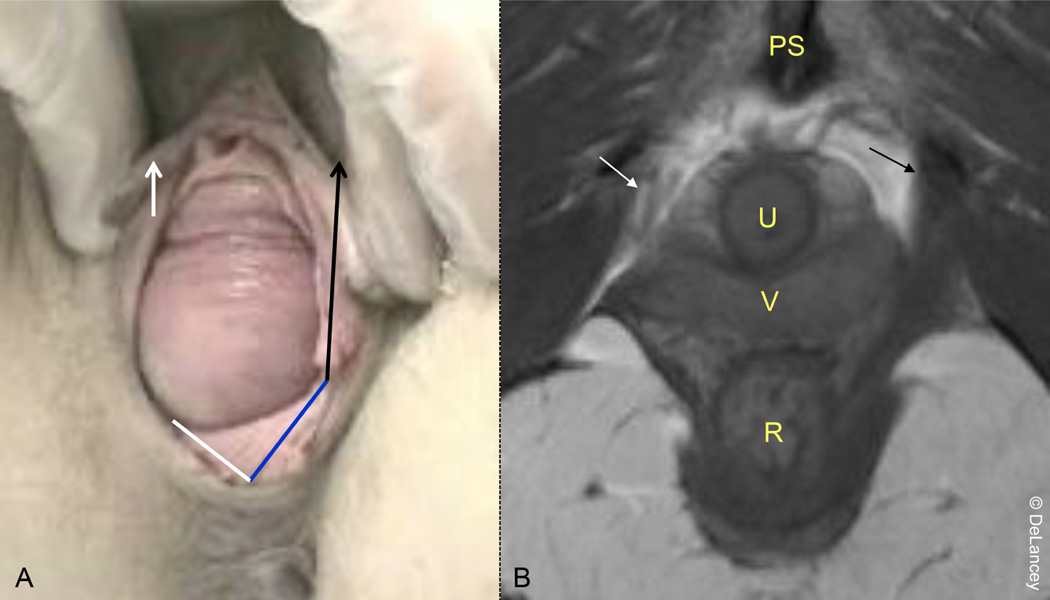
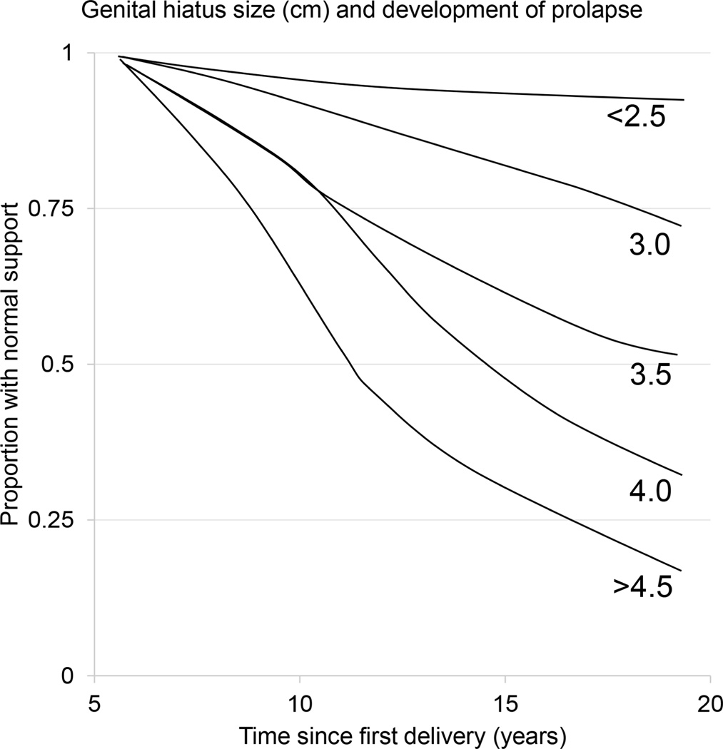
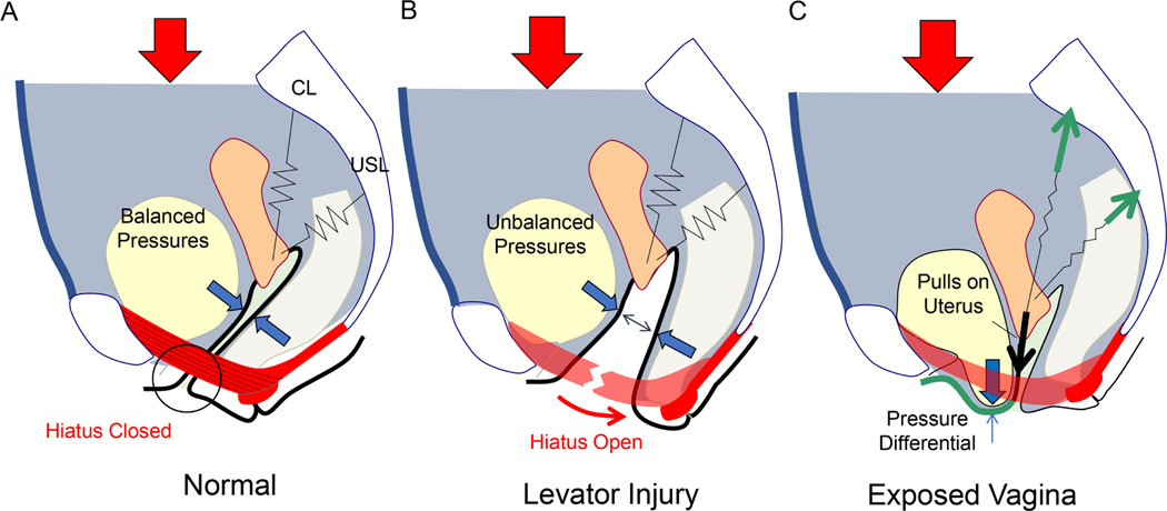
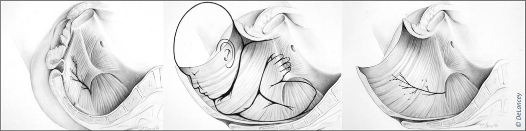
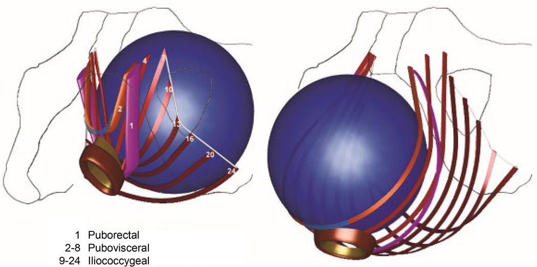
Pelvic floor disorders after childbirth have distressing lifelong consequences for women, requiring more than 300,000 women to have surgery annually. This represents approximately 10% of the 3 million women who give birth vaginally each year. Vaginal birth is the largest modifiable risk factor for prolapse, the pelvic floor disorder most strongly associated with birth, and is an important contributor to stress incontinence. These disorders require 10 times as many operations as anal sphincter injuries. Imaging shows that injuries of the levator ani muscle, perineal body, and membrane occur in up to 19% of primiparous women. During birth, the levator muscle and birth canal tissues must stretch to more than 3 times their original length; it is this overstretching that is responsible for the muscle tear visible on imaging rather than compression or neuropathy. The injury is present in 55% of women with prolapse later in life, with an odds ratio of 7.3, compared with women with normal support. In addition, levator damage can affect other aspects of hiatal closure, such as the perineal body and membrane. These injuries are associated with an enlarged urogenital hiatus, now known as antedate prolapse, and with prolapse surgery failure. Risk factors for levator injury are multifactorial and include forceps delivery, occiput posterior birth, older maternal age, long second stage of labor, and birthweight of >4000 g. Delivery with a vacuum device is associated with reduced levator damage. Other steps that might logically reduce injuries include manual rotation from occiput posterior to occiput anterior, slow gradual delivery, perineal massage or compresses, and early induction of labor, but these require study to document protection. In addition, teaching women to avoid pushing against a contracted levator muscle would likely decrease injury risk by decreasing tension on the vulnerable muscle origin. Providing care for women who have experienced difficult deliveries can be enhanced with early recognition, physical therapy, and attention to recovery. It is only right that women be made aware of these risks during pregnancy. Educating women on the long-term pelvic floor sequelae of childbirth should be performed antenatally so that they can be empowered to make informed decisions about management decisions during labor.

Keywords: enlarged hiatus; forceps delivery; levator ani avulsion; occiput posterior; pelvic floor disorders; pelvic floor injury; pelvic organ prolapse; postpartum care; prenatal education; prevention; rehabilitation; stress urinary incontinence; vaginal birth.


Conflict of interest statement

Conflict of interest: The University of Michigan received partial salary support for JOLD, MM, and JAAM from Materna Medical though a research contract regarding biomechanical analyses. The remaining authors report no conflicts of interest.

Figures

Figure 1.
Figure 1.. Hidden pelvic floor muscles late in the second stage
Note: perineal membrane not shown ©DeLancey
Figure 2.
Figure 2.. Annual number of women undergoing pelvic floor surgeries in the USA,–
Figure 3.
Figure 3.. Incidence of operations for prolapse (top) and stress urinary incontinence (bottom) in relation to mode of delivery and time since first birth
From Leijonhufvud et al.
Figure 4.
Figure 4.. Van Mechelen Model for preventing injury,
Figure 5.
Figure 5.. Graphical display of the concept of pelvic floor function
A. Phases of a woman’s life span B. Different degrees of functional reserve C, D. Variations in birth damage and repair E. Accelerated deterioration F. Lifestyle impact From DeLancey et al. ©DeLancey
Figure 6.
Figure 6.. Enlarged urogenital hiatus (5 cm) six days after birth showing anterior vaginal wall prolapse with no recovery at 7 months or 6 years
©DeLancey
Figure 7.
Figure 7.. Anterior wall prolapse in a woman with a unilateral levator muscle tear
A. Intact muscle seen with black arrow in the MRI scan B. Missing muscle (expected location indicated with white arrow) that results in the asymmetry of the perineal body (Panel A) that is attached on one side (solid black arrow) and not on the other (separated white arrow). ©DeLancey
None
Figure 8. The proportion of individuals maintaining normal support when followed longitudinally
Each plot relates to the size of the urogenital hiatus during straining on examination from 2.5cm to 4.5 cm. Decline in normal support represents increase in prolapse at or below the hymen. From Handa et al.
Figure 9.
Figure 9.. Diagrammatic representation of interactions between levator ani muscle (red), anterior vaginal wall prolapse, and cardinal/uterosacral ligament suspension
Red arrows represent the force created by gravity and abdominal pressure. With normal levator function (A), the hiatus is closed, and the vaginal walls are in apposition; the anterior and posterior pressures are equal and cancel (blue arrows). Levator damage (B) results in hiatal opening, and the vagina becomes exposed to a pressure differential between abdominal and atmospheric pressures. This pressure differential (C) makes the vaginal wall protrude and creates a traction force on the cardinal ligament and uterosacral ligament. ©DeLancey
Figure 10.
Figure 10.. Pelvic floor seen from below after removal of vulvar structures
EAS, external anal sphincter; ICM, iliococcygeal muscle; PM, perineal membrane; PRM, puborectal muscle; PVM, pubovisceral muscle. Urogenital hiatus, red outline; levator hiatus; green outline.
Figure 11.
Figure 11.. Birth-associated changes in the perineal membrane showing separation of the two sides and the resulting “swinging door” rotation and descent
Normal view, light blue; postpartum view, dark blue. From Pipitone et al.
Figure 12.
Figure 12.. Mid-urethral axial MR images in the region were the pubococcygeal muscle is normally seen lateral to the vagina
A. Proton density scan where the solid arrow heads mark the pubococcygeal muscle and the open arrow heads show the obturator internus. Signal intensity is lower (lighter) in the pubococcygeal (arrowhead) than the adjacent internal obturator (open arrowhead). B. Fluid-sensitive scan; this difference is more apparent and asterisk marks pubic bone edema and fracture. C. Normal pubococcygeal muscle (black arrow) is seen between the vagina and internal obturator (black arrow), while it is absent on the left. This pattern persists in the late scan (C). ©DeLancey
Figure 13.
Figure 13.. Illustration of both the circumferential and downward stretch of the levator ani muscle and its innervations during the second stage of labor
Similar changes could occur to the innervation of the external anal sphincter. ©DeLancey
Figure 14.
Figure 14.. The effect of birth on length of the levator ani muscle fibers
Representative muscle bands for different components shown before and after dilation during the second stage. Note that the pubovisceral muscle fibers (slings 2–8) are the shortest before birth and undergo the most elongation (and therefore are at highest risk for stretch injury). Modified from Lien et al.
Figure 15.
Figure 15.. Relationship between age and maximal urethral closure pressure in nulliparas with advancing age

Similar articles

Cited by

References

    1. Nygaard I, Barber MD. Prevalence of Symptomatic Pelvic Floor Disorders in US Women. JAMA. 2008;300(11):1311–1316. doi:10.1001/jama.300.11.1311.Prevalence - DOI - PMC - PubMed
    1. Dieter AA, Wilkins MF, Wu JM. Epidemiological trends and future care needs for pelvic floor disorders. Curr Opin Obstet Gynecol. 2015;27(5):380–384. doi:10.1097/GCO.0000000000000200 - DOI - PMC - PubMed
    1. Patel UJ, Godecker AL, Giles DL, Brown HW. Updated Prevalence of Urinary Incontinence in Women: 2015–2018 National Population-Based Survey Data. Female Pelvic Med Reconstr Surg. 2022;28(4):181–187. doi:10.1097/SPV.0000000000001127 - DOI - PubMed
    1. Boyles SH, Weber AM, Meyn L. Procedures for pelvic organ prolapse in the United States, 1979–1997. Am J Obstet Gynecol. 2003;188(1):108–115. doi:10.1067/mob.2003.101 - DOI - PubMed
    1. Okeahialam NA, Sultan AH, Thakar R. The prevention of perineal trauma during vaginal birth. Am J Obstet Gynecol. Published online August 11, 2023. doi:10.1016/j.ajog.2022.06.021 - DOI - PubMed

Publication types