Review
. 2024 Jan 3;9(1):6.
doi: 10.1038/s41392-023-01679-y.

Cellular zinc metabolism and zinc signaling: from biological functions to diseases and therapeutic targets

Affiliations
Review

Cellular zinc metabolism and zinc signaling: from biological functions to diseases and therapeutic targets

Bonan Chen et al. Signal Transduct Target Ther. .

Abstract

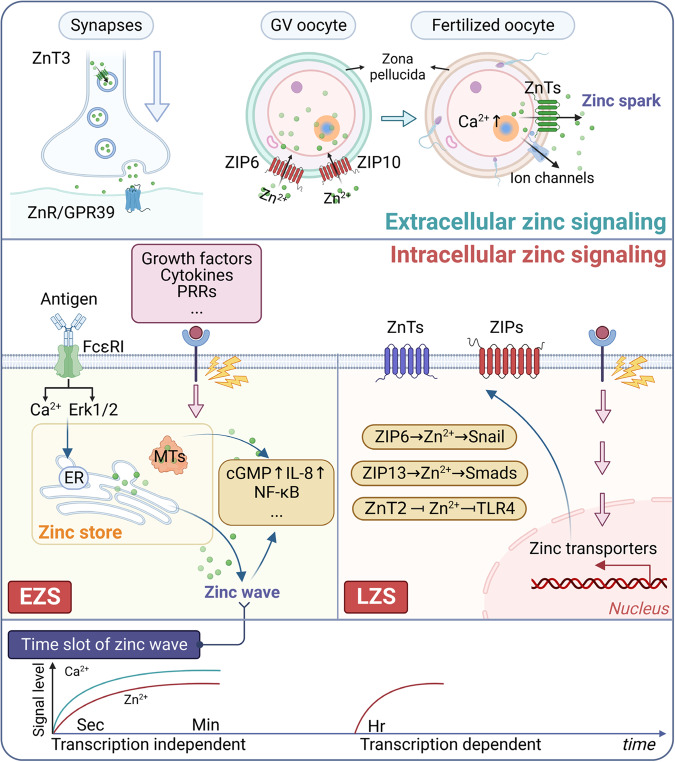
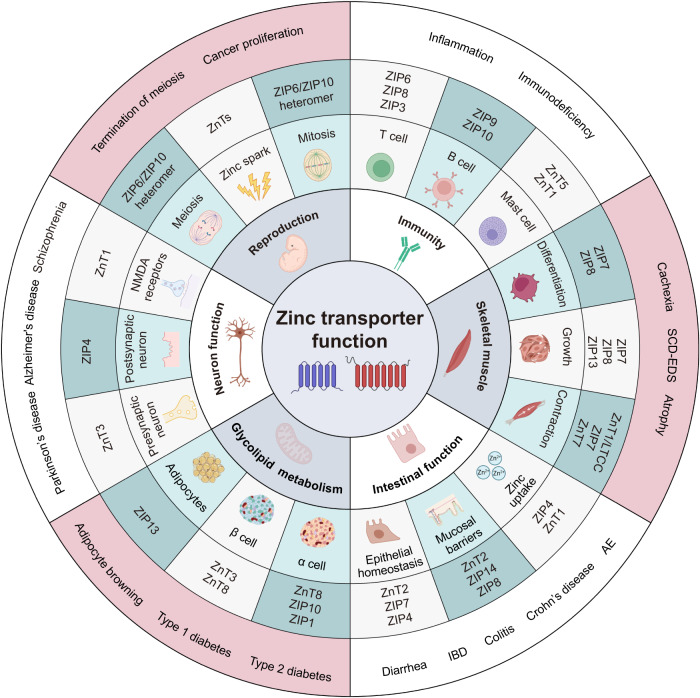
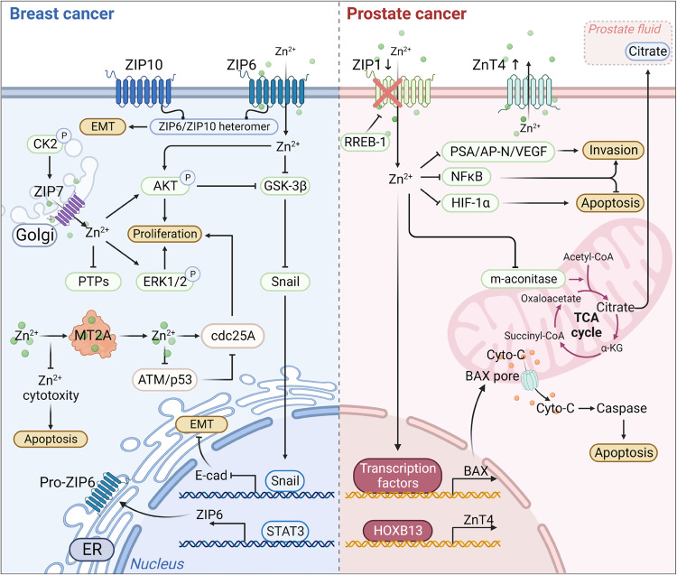
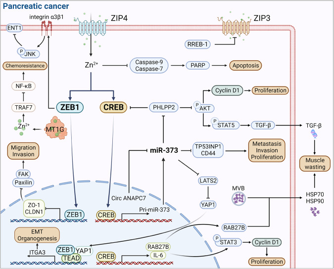
Zinc metabolism at the cellular level is critical for many biological processes in the body. A key observation is the disruption of cellular homeostasis, often coinciding with disease progression. As an essential factor in maintaining cellular equilibrium, cellular zinc has been increasingly spotlighted in the context of disease development. Extensive research suggests zinc's involvement in promoting malignancy and invasion in cancer cells, despite its low tissue concentration. This has led to a growing body of literature investigating zinc's cellular metabolism, particularly the functions of zinc transporters and storage mechanisms during cancer progression. Zinc transportation is under the control of two major transporter families: SLC30 (ZnT) for the excretion of zinc and SLC39 (ZIP) for the zinc intake. Additionally, the storage of this essential element is predominantly mediated by metallothioneins (MTs). This review consolidates knowledge on the critical functions of cellular zinc signaling and underscores potential molecular pathways linking zinc metabolism to disease progression, with a special focus on cancer. We also compile a summary of clinical trials involving zinc ions. Given the main localization of zinc transporters at the cell membrane, the potential for targeted therapies, including small molecules and monoclonal antibodies, offers promising avenues for future exploration.


Conflict of interest statement

The authors declare no competing interests.

Figures

Fig. 1
Fig. 1
Zinc signaling in the intracellular and extracellular regions. Zinc extracellular signaling is mainly involved in the physiological functions of neurosynapses and germ cells. In contrast, intracellular zinc signaling is primarily divided into two parts, EZS and LZS, which exert biological functions by activating downstream pathways, such as inflammatory signaling. Interestingly, the endoplasmic reticulum releases zinc to generate a specific zinc wave, observed within several minutes after FcεRI stimulation in mast cells. EZS early zinc signaling, LZS late zinc signaling. Green dots represent zinc
Fig. 2
Fig. 2
The protein structure and gene family evolution of zinc transporters. a Cartoon of predicted structures of ZnT and ZIP transporter proteins. The picture on the left shows an atomic model of ZnT, which is the helical reconstruction of YiiP based on X-ray structure (PDB ID code: 7y5g). In detail, the schematic topology of ZnT transporters is proposed based on the three-dimensional structure of Escherichia coli homolog YiiP. ZnTs most likely have six TM domains divided into two bundles. Specifically, one of the ZnT’s bundles contains four TM domains (MI, MII, MIV, and MV), and the other one comprises two TM domains (MIII and MVI). Each of the former bundle’s domains can independently bind zinc, tetrahedrally coordinated by two D (aspartate) and two H (histidine) in the mammalian homologs. Similarly, the figure on the right presents putative TM domains of the ZIP family (PDB ID code: 7z6m). Moreover, the topology structure of ZIP is displayed, composed of 8 TM domains with a large N-terminal domain and a small C-terminal. The spatial distribution shows that it consists of three parts, the left and right parts each contain three TM domains (red), and in the middle are two TM domains (blue). Zinc could bind to the active site of TM domain IV and V, containing conserved HND (histidine, asparagine, aspartate) and HEH (two histidines and one glutamic acid) motifs, respectively. b Gene family evolutionary tree and isoform of the ZnTs and ZIPs. The lengths of the different isoforms are labeled in the front of the isoforms, and the color lines indicate the functional domain locations of each isoform. ZnTs belong to the Zn-cation diffusion facilitator (CDF) family, responsible for transporting zinc from intracellular to extracellular. ZIPs are divided into four subfamilies, namely ZIP subfamily I (ZIP9), GufA subfamily (ZIP11), ZIP subfamily II (ZIP1-3), and LIV-1 subfamily (ZIP4-8, ZIP10, ZIP12-14)
Fig. 3
Fig. 3
The protein structure and gene family evolution of MTs. a Diagram of the predicted structure of the MT2 protein, which is modeled from the reconstructed X-ray structure (PDB ID code: 4mt2). The crystallographic structure of rat liver metallothionein has been accurately determined at a resolution of 2.0 Å, achieving a low R-value of 0.176 for all observed data. b Schematic representation of zinc binding in MTs. MTs contain abundant thiol groups capable of binding with heavy metals. Due to the high thiol content, MTs can bind up to 7 zinc atoms, with 3 zinc atoms located in the β domain and 4 zinc atoms in the α domain. c Gene family evolutionary tree and isoforms of MTs are depicted in Figure X. The isoform lengths are labeled in front of each isoform, and color lines indicate the position of the functional domain of each isoform. MTs are categorized into four subfamilies: MT1 (including MT1A, MT1B, MT1E, MT1F, MT1G, MT1H, MT1M, and MT1X), MT2 (including MT2A), MT3, and MT4. While MT1 and MT2 are universally expressed, MT3 is primarily expressed in the central nervous system, and MT4 is predominantly expressed in the skin and other stratified epithelium tissues
Fig. 4
Fig. 4
The main physiological functions of zinc transporters. The zinc transporter functions are basically classified into six parts: immunity, reproduction, muscle, intestinal function, glycolipid metabolism, and neuron function. Further, the function represented by each sector is mainly divided into three parts. Each part corresponds to specific zinc transporters. The outermost circle represents diseases and cancers caused by malfunctioning zinc transporters. SCD-EDS spondylocheirodysplastic ehlers-danlos syndrome, AE acrodermatitis enteropathica, IBD inflammatory bowel disease
Fig. 5
Fig. 5
The molecular mechanism of zinc transporters and MTs in BC and prostate cancers. The left figure represents the mechanism of ZIP-mediated proliferation and EMT procession in BC. ZIP7 locates on the endoplasmic reticulum and is highly expressed in tamoxifen-resistant BC cells. After CK2 phosphorylation, ZIP7 was stimulated to transport zinc from intracellular stores, for example, the Golgi apparatus. Subsequently, the increasing zinc concentration can promote proliferation by activating the downstream PTPs, AKT, and ERK1/2 signaling. ZIP6 and ZIP10 locate on the cytomembrane. In addition, ZIP6 is induced by STAT3 and then translocated to the plasma membrane, promoting the accumulation of cellular zinc. The zinc influx caused by ZIP6 and ZIP6/ZIP10 heteromer triggers the AKT pathway and inhibits GSK-3β, finally boosting the EMT process by reducing the nuclear translocation of Snail. MT2A play a dual role in zinc homeostasis and BC cell proliferation. They can chelate zinc ions to reduce zinc cytotoxicity-induced apoptosis, while also releasing zinc ions to promote cancer cell proliferation through cdc25A activation. The figure on the right elucidates the mechanism of zinc transporter involved in prostate cancer. RREB-1 downregulates ZIP1 expression, leading to zinc homeostasis imbalance in prostate cells. ZIP1 downregulation reduced zinc influx, thus degrading the Bax pore expression level, which is the channel for cyto-C releasing into the cytoplasm. Consequently, the apoptosis induced by cyto-C is inhibited. Moreover, decreasing zinc concentration attenuates the inhibition of m-aconitase, which drives citrate oxidization in the TCA cycle. Meanwhile, the inhibitory effect of zinc on the NF-κB signaling pathway was diminished, as well as the inhibitory effect on the expression of HIF-1α, PSA, AP-N, and VEGF, which contributes to the invasion and proliferation. Besides, HOXB13 upregulates the expression of ZnT4 in prostate cancer through transcriptional regulation. EMT epithelial-mesenchymal transition, CK2 casein kinase 2, PTPs protein tyrosine phosphatases, RREB-1 Ras-responsive element binding protein 1, m- aconitase mitochondrial aconitase, cyro-C cytochrome C, PSA prostate-specific antigen, AP-N activity of urokinase-type plasminogen activator and aminopeptidase N, VEGF vascular endothelial growth factor, TCA tricarboxylic acid
Fig. 6
Fig. 6
The molecular mechanism of zinc transporters and MTs in PC. ZIP4 promotes PC carcinogenesis mainly through two transcription factors, CREB and ZEB1. ZEB1 promotes the procession of EMT by suppressing the expression of ZO-1 and CLDN1 and inducing the transcription of ITGA3. Moreover, the ZEB1 induces integrin α3β1 to phosphorylate JNK and ultimately blocks ENT1, a gemcitabine transporter, which results in chemoresistance. Besides, cellular zinc released by MT1G inhibits NF-κB, suppressing PC chemoresistance. CREB transcripts miR-373 to increase metastasis, invasion, and proliferation by activating the Hippo pathway yet inhibiting the expression of TP53INP1 and CD44. Besides, PHLPP2, inhibited by miR-373, forms a malignant cycle through the suppression of CREB. However, the small molecule, circ ANAPC7, can block miR-373. As a target for PHLPP2 dephosphorylation, AKT increases the proliferation by upregulating cyclin D1 and promotes muscle wasting by phosphorylating STAT5. Another CREB-mediated downstream promoting muscle wasting is RAB27B. Mechanically, RAB27B promotes the release of HSP70 and HSP90 from MVB. Additionally, the CREB-mediated IL-6/STAT3/cyclin D1 pathway leads to proliferation in PC. ZIP4 could restrain apoptosis by inhibiting the activity of caspase9 and caspase7. The expression of ZIP3 is reduced by RREB-1. ZO-1, zonula occludens-1; ITGA3, integrin subunit alpha 3; JNK, c-Jun N-terminal kinase; MVB, multivesicular body; EMT, epithelial-mesenchymal transition
Fig. 7
Fig. 7
The molecular mechanism of zinc transporters and metallothioneins in CRC, GC, and ESCC. In CRC, the binding of ZnT9 and β-catenin triggers the transcription of CCND1, MYC, and MMP7, resulting in proliferation and migration. KCTD9 can replace the binding of ZnT9 and β-catenin. Moreover, ZIP14 contains two alternative splicings, ZIP14-4A and ZIP14-4B. ZIP14-4B is upregulated by SRPK1 and SRSF1, two downstream targets of Wnt signaling, leading to increased Cd2+ uptake. Concerning the GC microenvironment, ZIP7 is the upstream target of the AKT/mTOR signaling pathway. In GC, autophagic degradation of MT1E, MT1M, and MT1X initiated by USP2-E2F4 interaction leads to increased intracellular zinc storage vesicles, promoting GC cell growth. In contrast, MT2A inhibits NF-κB by releasing cellular zinc and thus ultimately suppresses GC cell proliferation. As for ESCC, ZIP6 activates PI3K/AKT and MAPK/ERK signaling pathways, which leads to the overexpression of downstream oncogenes such as MMP1, MMP3, MYC, and SLUG. Meanwhile, the cellular zinc released by MT2A promotes the oncogenic function of IGFBP2. NF-κBIA, NF-κB inhibitor alpha; IGFBP2, insulin-like growth factor binding protein 2
Fig. 8
Fig. 8
Genetic and mRNA alterations of zinc transporter and MT family genes in pan-cancer patients. The upper figure illustrates the gene alterations of zinc transporters. Out of the queried pan-cancer samples, 1526 (59%) showed copy number aberrations, mutations, and mRNA expression changes. The lower figure displays the gene alterations of MTs, where 324 (13%) of the queried pan-cancer samples demonstrated copy number aberrations, mutations, and mRNA expression changes. This diagram includes a total of 30 cancer types, marked with different colors. The data source is from the pan-cancer analysis of whole genomes dataset in cbioportal (https://www.cbioportal.org/study/summary?id=pancan_pcawg_2020)

Similar articles

Cited by

References

    1. Huang L, Tepaamorndech S. The SLC30 family of zinc transporters - a review of current understanding of their biological and pathophysiological roles. Mol. Asp. Med. 2013;34:548–560. doi: 10.1016/j.mam.2012.05.008. - DOI - PubMed
    1. Kambe T, Tsuji T, Hashimoto A, Itsumura N. The Physiological, Biochemical, and Molecular Roles of Zinc Transporters in Zinc Homeostasis and Metabolism. Physiol. Rev. 2015;95:749–784. doi: 10.1152/physrev.00035.2014. - DOI - PubMed
    1. Kimura T, Kambe T. The Functions of Metallothionein and ZIP and ZnT Transporters: An Overview and Perspective. Int J. Mol. Sci. 2016;17:336. doi: 10.3390/ijms17030336. - DOI - PMC - PubMed
    1. Hu H, et al. New anti-cancer explorations based on metal ions. J. Nanobiotechnol. 2022;20:457. doi: 10.1186/s12951-022-01661-w. - DOI - PMC - PubMed
    1. Stockwell BR, Jiang X, Gu W. Emerging mechanisms and disease relevance of ferroptosis. Trends Cell Biol. 2020;30:478–490. doi: 10.1016/j.tcb.2020.02.009. - DOI - PMC - PubMed