Level-set based adaptive-active contour segmentation technique with long short-term memory for diabetic retinopathy classification
- PMID: 38169636
- PMCID: PMC10758353
- DOI: 10.3389/fbioe.2023.1286966
Level-set based adaptive-active contour segmentation technique with long short-term memory for diabetic retinopathy classification
Abstract
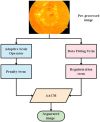
Diabetic Retinopathy (DR) is a major type of eye defect that is caused by abnormalities in the blood vessels within the retinal tissue. Early detection by automatic approach using modern methodologies helps prevent consequences like vision loss. So, this research has developed an effective segmentation approach known as Level-set Based Adaptive-active Contour Segmentation (LBACS) to segment the images by improving the boundary conditions and detecting the edges using Level Set Method with Improved Boundary Indicator Function (LSMIBIF) and Adaptive-Active Counter Model (AACM). For evaluating the DR system, the information is collected from the publically available datasets named as Indian Diabetic Retinopathy Image Dataset (IDRiD) and Diabetic Retinopathy Database 1 (DIARETDB 1). Then the collected images are pre-processed using a Gaussian filter, edge detection sharpening, Contrast enhancement, and Luminosity enhancement to eliminate the noises/interferences, and data imbalance that exists in the available dataset. After that, the noise-free data are processed for segmentation by using the Level set-based active contour segmentation technique. Then, the segmented images are given to the feature extraction stage where Gray Level Co-occurrence Matrix (GLCM), Local ternary, and binary patterns are employed to extract the features from the segmented image. Finally, extracted features are given as input to the classification stage where Long Short-Term Memory (LSTM) is utilized to categorize various classes of DR. The result analysis evidently shows that the proposed LBACS-LSTM achieved better results in overall metrics. The accuracy of the proposed LBACS-LSTM for IDRiD and DIARETDB 1 datasets is 99.43% and 97.39%, respectively which is comparably higher than the existing approaches such as Three-dimensional semantic model, Delimiting Segmentation Approach Using Knowledge Learning (DSA-KL), K-Nearest Neighbor (KNN), Computer aided method and Chronological Tunicate Swarm Algorithm with Stacked Auto Encoder (CTSA-SAE).
Keywords: adaptive-active counter model; diabetic retinopathy; gray level co-occurrence matrix; level set method with improved boundary indicator function; long short term memory.
Copyright © 2023 Bhansali, Patra, Abouhawwash, Askar, Awasthy and Rao.
Conflict of interest statement
The authors declare that the research was conducted in the absence of any commercial or financial relationships that could be construed as a potential conflict of interest. The author(s) declared that they were an editorial board member of Frontiers, at the time of submission. This had no impact on the peer review process and the final decision.
Figures


Similar articles
-
Deep long and short term memory based Red Fox optimization algorithm for diabetic retinopathy detection and classification.Int J Numer Method Biomed Eng. 2022 Mar;38(3):e3560. doi: 10.1002/cnm.3560. Epub 2021 Dec 15. Int J Numer Method Biomed Eng. 2022. PMID: 34865312
-
A novel deep learning model for diabetic retinopathy detection in retinal fundus images using pre-trained CNN and HWBLSTM.J Biomol Struct Dyn. 2024 Feb 19:1-19. doi: 10.1080/07391102.2024.2314269. Online ahead of print. J Biomol Struct Dyn. 2024. PMID: 38373067
-
Segmentation of retinal blood vessels by a novel hybrid technique- Principal Component Analysis (PCA) and Contrast Limited Adaptive Histogram Equalization (CLAHE).Microvasc Res. 2023 Jul;148:104477. doi: 10.1016/j.mvr.2023.104477. Epub 2023 Feb 4. Microvasc Res. 2023. PMID: 36746364
-
Analysis on diagnosing diabetic retinopathy by segmenting blood vessels, optic disc and retinal abnormalities.J Med Eng Technol. 2020 Aug;44(6):299-316. doi: 10.1080/03091902.2020.1791986. Epub 2020 Jul 30. J Med Eng Technol. 2020. PMID: 32729345 Review.
-
A review on computer-aided recent developments for automatic detection of diabetic retinopathy.J Med Eng Technol. 2019 Feb;43(2):87-99. doi: 10.1080/03091902.2019.1576790. Epub 2019 Jun 14. J Med Eng Technol. 2019. PMID: 31198073 Review.
References
-
- Alajlan A. M., Razaque. A. (2023). ESOA-HGRU: egret swarm optimization algorithm-based hybrid gated recurrent unit for classification of diabetic retinopathy. Artif. Intell. Rev., 1–30. 10.1007/s10462-023-10532-1 - DOI
-
- Ali G., Dastgir A., Iqbal M. W., Anwar M., Faheem M. (2023). A hybrid convolutional neural network model for automatic diabetic retinopathy classification from fundus images. IEEE J. Transl. Eng. Health Med. 11, 341–350. 10.1109/JTEHM.2023.3282104 - DOI
-
- Atli I., Gedik O. S. (2021). Sine-Net: a fully convolutional deep learning architecture for retinal blood vessel segmentation. Eng. Sci. Technol. Int. J. 24 (2), 271–283. 10.1016/j.jestch.2020.07.008 - DOI
-
- Atwany M. Z., Sahyoun A. H., Yaqub M. (2022). Deep learning techniques for diabetic retinopathy classification: a survey. IEEE Access 10, 28642–28655. 10.1109/ACCESS.2022.3157632 - DOI
LinkOut - more resources
Full Text Sources
Miscellaneous

