. 2023 Dec 13;7(2):e202302422.
doi: 10.26508/lsa.202302422. Print 2024 Feb.

The structural basis for deubiquitination by the fingerless USP-type effector TssM

Affiliations

The structural basis for deubiquitination by the fingerless USP-type effector TssM

Thomas Hermanns et al. Life Sci Alliance. .

Abstract

Intracellular bacteria are threatened by ubiquitin-mediated autophagy, whenever the bacterial surface or enclosing membrane structures become targets of host ubiquitin ligases. As a countermeasure, many intracellular pathogens encode deubiquitinase (DUB) effectors to keep their surfaces free of ubiquitin. Most bacterial DUBs belong to the OTU or CE-clan families. The betaproteobacteria Burkholderia pseudomallei and Burkholderia mallei, causative agents of melioidosis and glanders, respectively, encode the TssM effector, the only known bacterial DUB belonging to the USP class. TssM is much shorter than typical eukaryotic USP enzymes and lacks the canonical ubiquitin-recognition region. By solving the crystal structures of isolated TssM and its complex with ubiquitin, we found that TssM lacks the entire "Fingers" subdomain of the USP fold. Instead, the TssM family has evolved the functionally analog "Littlefinger" loop, which is located towards the end of the USP domain and recognizes different ubiquitin interfaces than those used by USPs. The structures revealed the presence of an N-terminal immunoglobulin-fold domain, which is able to form a strand-exchange dimer and might mediate TssM localization to the bacterial surface.


Conflict of interest statement

The authors declare that they have no conflict of interest.

Figures

Figure 1.
Figure 1.. TssM is a ubiquitin specific DUB.
(A) Structure-guided alignment of TssM and representative members of the USP-family. Residues printed on black or grey background are invariant or conservatively replaced in at least 50% of the sequences. The active site residues are highlighted in red. Colored letters indicate the localization of the three USP subdomains Thumb (T), Fingers (F), and Palm (P) and the respective secondary structure elements of USP7. The internal TssM deletion is marked with red arrows. The sequence of the CYLD BBOX was removed from the alignment to get a better overview and the site is marked with a red BBOX. USP family members exhibit a highly conserved glutamic residue which is important for binding of ubiquitin’s Arg72. This residue is marked by a green arrow. Blocking loop 1 of USP14 and Littlefinger loop of TssM are highlighted by purple and pink letters respectively. (B) Reaction of TssM with Ub and UbL activity-based probes. Asterisks mark the shifted bands after reaction. (C, D) Activity of 5 (C) or 50 nM (D) TssM against ubiquitin– and NEDD8–AMC (C) or against NEDD8–AMC (D). The RFU values are the means of triplicates. (C) The initial velocities (i. velocity) are the mean velocities derived from measurements shown in (C). (E) Linkage specificity analysis of TssM. A panel of homotypic di-ubiquitin chains was incubated with TssM for the indicated time points. Source data are available for this figure.
Figure S1.
Figure S1.. TssM preferentially reacts with Ub-PA.
(A) Domain scheme of TssM. (B) Reaction of TssM with Ub- and Nedd8-PA. The reactions were stopped by the addition of Laemmli buffer after the indicated time points. Source data are available for this figure.
Figure 2.
Figure 2.. TssM is a fingerless USP.
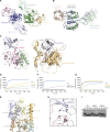
(A) Crystal structure of TssM193–490 shown in cartoon representation. The structure is divided in an Ig-like domain (light blue) and a DUB domain (green). The first β-strand undergoes a strand swap within the dimer. The respective β-strand of the other dimer is shown in cartoon representation and colored pink. The active site residues are shown as sticks. (B) The DUB domain of TssM adopts a USP-like fold. The USP-typical Thumb and Palm subdomains are conserved and colored green and light pink, respectively. The Fingers subdomain (yellow) is missing and replaced by a short α-helix (α-6). The active site residues are shown as sticks. (C) Magnification of the active site formed by Cys-308, His-442, and Asp-457. Cys-308 belongs to the Thumb subdomain and is colored green; His-442 and Asp-457 are located within the Palm Subdomain and colored light pink. (D) Activity of WT TssM compared with the active site mutants. 1.5 μM TssM were incubated with K48-linked di-ubiquitin and the reaction was stopped by the addition of Laemmli buffer after the indicated timepoints. (E) Activity of 5 nM TssM193–490 and the Ig-lacking truncation TssM292–490 against ubiquitin–AMC. The RFU values are the means of triplicates. (F) Activity of 0.5 μM TssM193–490 and TssM292–490 against K63-linked di-ubiquitin. The reaction was stopped by the addition of Laemmli buffer after the indicated time points, resolved by SDS–PAGE and Coomassie-stained. Source data are available for this figure.
Figure S2.
Figure S2.. TssM superpositions.
(A) The ASU of the apo TssM193–490 structure contains a dimer, exhibiting a β-strand swap between the two Ig-like domains. The dimer is shown in cartoon representation, the Ig-like domains are colored light blue (chain A)/dark blue (chain B) and the DUB domains of chain A and B are colored orange and purple, respectively. (B) Chain A (green) and chain B (blue) are shown in cartoon representation and are superimposed with an RMSD of 0.63 Å over 3,221 atoms. (C, D, E, F) Different structural comparisons of TssM (orange) and USP7 (light blue), Fingers region, (green), shown in cartoon representation. (C, D, E, F) Superpositions were based on the entire catalytic domain (C, D), the Thumb subdomain (E) and the Palm subdomain (F). Missing secondary structure elements in TssM are indicated by their numbering based on the USP7 structure. (G) Superposition of TssM (orange) with USP12 (light blue) and USP46 (green) with an RMSD of 4.5 (USP12, 354 atoms) and 4.4 Å (USP46, 414 atoms).
Figure 3.
Figure 3.. Novel Littlefinger loop functionally replaces the Fingers subdomain.
(A) Crystal structure of the TssM193–490/Ub-PA complex shown in cartoon representation. The catalytic domain is divided in the Thumb and Palm subdomains and colored green and light pink, respectively. A flexible loop located between α-7 and β-6 is located close to ubiquitin (light blue) and named Littlefinger (pink). (A, B) Structural comparison of apo (colored light orange) and Ub-PA-bound (subdomains colored as in panel (A)) TssM. The catalytic domains are shown in cartoon representation and were superimposed with a RMSD of 0.35 Å over 1,115 atoms. (C) Recognition of ubiquitin’s Ile-44 patch by residues of the Littlefinger loop and the Palm subdomain. Ubiquitin (light blue) and TssM are shown in cartoon representation. The Littlefinger is colored pink and the Palm subdomain is colored light pink. Key residues are shown as sticks and hydrophobic interactions are indicated by dotted lines. (D) Littlefinger (colored pink) residues stabilize ubiquitin C-terminus (light blue). Key residues are shown as sticks and hydrogen bonds are indicated by dotted lines. (E) Interaction between Gly75 of ubiquitin and the “aromatic motif” represented by Tyr443, shown as light pink stick, is conserved in TssM. Hydrophobic interactions are indicated by dotted lines. (F) Recognition of ubiquitins Arg72/Arg74 by TssM. The conserved residue Glu378 forms a salt bridge with Arg74, in contrast to its role in classic USPs, where it contacts Arg72. TssM stabilizes Arg72 instead via Glu485 and Gln375. Key residues are shown as sticks and colored according to the subdomains: Littlefinger/pink, Thumb/green. (G) Mutational analysis of residues contacting the Ile-44 patch. 1.5 μM WT TssM292–490 and the respective point mutants were incubated with K48-linked di-ubiquitin. The reaction was stopped by the addition of Laemmli buffer after the indicated timepoints, resolved by SDS–PAGE and Coomassie-stained. (H) Comparison of WT TssM and the aromatic motif mutant Y443A. 1.5 μM TssM292–490 were incubated with K48-linked di-ubiquitin and the reaction was stopped by the addition of Laemmli buffer after the indicated timepoints. (I) Mutational analysis of residues stabilizing ubiquitin’s C-terminal Arg72/Arg74 residues. 1.5 μM WT TssM292–490 and the respective point mutants were incubated with K48-linked di-ubiquitin. The reaction was stopped by the addition of Laemmli buffer after the indicated timepoints, resolved by SDS–PAGE and Coomassie-stained. Source data are available for this figure.
Figure S3.
Figure S3.. TssM/Ub-PA complex structure.
(A) The ASU of the TssM193–490/Ub-PA complex structure contains a dimer shown in cartoon representation. The Ig-like domains are colored light green (chain A)/light blue (chain C), the DUB domains are colored dark green (chain A)/dark blue (chain C), and the covalent bound ubiquitins are colored orange (chain B)/light pink (chain D). (B) The catalytic domains of chain A (green) and chain C (blue) are shown in cartoon representation and are superimposed with an RMSD of 0.24 Å over 1,144 atoms. (C) Side-by-side comparison of Ub-bound TssM and USP14 (PDB:2AYO). TssM and USP14 were superimposed based on the catalytic domains. The left panel shows TssM and the right panel USP14. The Littlefinger and blocking loop 1 are highlighted by pink and purple coloring. (D) Activity of 5 nM WT TssM and Ile44-patch contacting mutants against ubiquitin–AMC. The RFU values are the means of triplicates. (E) Comparison of Ile-44 patch recognition by TssM and ATX3L. The ubiquitin-bound structures of TssM (teal) and ATX3L (PDB: 3O65, orange) were superimposed and are shown in cartoon representation. The respective ubiquitins are colored light blue (TssM) and yellow (ATX3L). Key residues forming the Ile44 patch are highlighted as sticks. (F, G) Activity of 5 nM WT TssM against ubiquitin–AMC compared with alanine subtitutions of the aromatic motif (F) or residues stabilizing ubiquitins C-terminus (G). The RFU values are the means of triplicates. (H) Hydrogen bonding between ubiquitin (light blue) and residues of the Palm subdomain (light pink) are indicated by dotted lines. Residues forming the hydrogen bonds are highlighted as sticks. (I) Mutational analysis of residues forming hydrogen bonds with ubiquitin Gln49. 1.5 μM WT TssM292–490 and the respective point mutants were incubated with K48-linked di-ubiquitin. The reaction was stopped by the addition of Laemmli buffer after the indicated timepoints, resolved by SDS–PAGE and Coomassie-stained. Source data are available for this figure.
Figure 4.
Figure 4.. Conserved Ig-like domain.
(A) Arrangement of the TssM molecules in the ASU of the apo (left) and the ubiquitin complex crystals (right). The catalytic domains are colored in grey and the Ig-like domains are colored pink (chain A) and orange (chain B or C). All ubiquitins have been omitted for the sake of the comparison of the TssM molecules. Both ASUs were aligned on their chain A-TssM (bottom). The other TssM (top) is rotated by ∼70°. Hence, both Ig-like domains are separated from each other in the complex crystal, although they are in close proximity swapping their first β-strand in the apo one. (B) Size exclusion chromatography to determine the dimer formation in solution. TssM193–490 (teal), myoglobin (18 kD, red), ovalbumin (44 kD, orange), and bovine serum albumin (66 kD, green) were individually subjected to size exclusion chromatography and the resulting UV curves were merged. (C) Structural superposition of the Ig-like domains derived from the TssM apo and complex structure. Each Ig-like domain was superimposed on the chain A Ig-like domain from the apo structure. RMSD: 1.42 (apo A/apo B; 1,330 atoms), 1.33 (apo A/complex A; 563 atoms), and 1.08 Å (Comp C/apo A 522 atoms). Secondary structure elements differing in the structures are numbered. (D) Structural superposition of TssM193–490 from the apo and complex structure, based on the respective catalytic domains. (E) Close-up view on the superposition shown in (E) highlighting the inter-domain salt bridges. The left panel shows TssM from the apo structure and the right panel shows the complex structure. Residues forming salt bridges are shown as sticks and the resulting salt bridges are indicated by yellow dotted lines. (F) Structure-guided alignment of TssM-derived Ig-like domains from different species, together with a structurally characterized transpeptidase (PDB:3VYP). Residues printed on black or grey background are invariant or conservatively replaced in at least 50% of the sequences. Secondary structure elements are indicated on top (TssM) or bottom (3VYP) of the alignment by colored letters (E = β-strand, H = α-helix).
Figure S4.
Figure S4.. TssM family members.
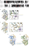
(A) Multiple-sequence alignment of TssM and homologues from different species. Residues printed on black or grey background are invariant or conservatively replaced in at least 50% of the sequences. The active site residues are highlighted in red. Purple L letters indicate the localization of the Littlefinger region. (B) Structural superposition of the TssM/Ub-PA complex structure (light orange) and the AF2-models for TssM from B. thailandensis (green) and B. mayonis (light blue), shown as cartoon representation. The left panel shows an overview of the complete catalytic domains superimposed with RMSD of 0.82 (over 563 atoms) and 0.47 Å (over 890 atoms) respectively. The right panel shows a close-up view of the Littlefinger region with key residues shown as sticks. Asterisks mark the Ile-patch residues of ubiquitin. (C) Structural superposition of the TssM/Ub-PA complex structure (light orange) and the AF2 models of TssM from B. humptydooensis (teal). The left panel shows an overview of the complete catalytic domains superimposed with RMSD of 0.39 Å (over 913 atoms). The right panel shows a close-up view of the Littlefinger region with key residues shown as sticks, indicating that F475 and Y418 are functionally replaced by L304 and F284 which are located at different positions in the B. humptydooensis catalytic domain. (D, E) Structural superposition of the TssM/Ub-PA complex structure (light orange) and the AF2 models for TssM from C. sinusclupearum (blue) and A0A1J5QGM2 (derived from an unidentified metagenomics sequence; green), shown as cartoon representation. The catalytic domains were superimposed with RMSD of 0.82 (over 563 atoms) and 1.64 Å (over 718 atoms), respectively. The Littlefinger (purple) is not conserved in these models, leading to a different orientation of the bound ubiquitin molecules.

References

    1. Adams PD, Afonine PV, Bunkóczi G, Chen VB, Davis IW, Echols N, Headd JJ, Hung LW, Kapral GJ, Grosse-Kunstleve RW, et al. (2010) PHENIX: A comprehensive python-based system for macromolecular structure solution. Acta Crystallogr D Biol Crystallogr 66: 213–221. 10.1107/S0907444909052925 - DOI - PMC - PubMed
    1. Allwood EM, Devenish RJ, Prescott M, Adler B, Boyce JD (2011) Strategies for intracellular survival of Burkholderia pseudomallei. Front Microbiol 2: 170. 10.3389/fmicb.2011.00170 - DOI - PMC - PubMed
    1. Berrow NS, Alderton D, Sainsbury S, Nettleship J, Assenberg R, Rahman N, Stuart DI, Owens RJ (2007) A versatile ligation-independent cloning method suitable for high-throughput expression screening applications. Nucleic Acids Res 35: e45. 10.1093/nar/gkm047 - DOI - PMC - PubMed
    1. Bodelón G, Palomino C, Fernández LÁ (2013) Immunoglobulin domains in Escherichia coli and other enterobacteria: From pathogenesis to applications in antibody technologies. FEMS Microbiol Rev 37: 204–250. 10.1111/j.1574-6976.2012.00347.x - DOI - PubMed
    1. Böth D, Steiner EM, Stadler D, Lindqvist Y, Schnell R, Schneider G (2013) Structure of LdtMt2, an L,D-transpeptidase from Mycobacterium tuberculosis. Acta Crystallogr D Biol Crystallogr 69: 432–441. 10.1107/S0907444912049268 - DOI - PMC - PubMed

Publication types