

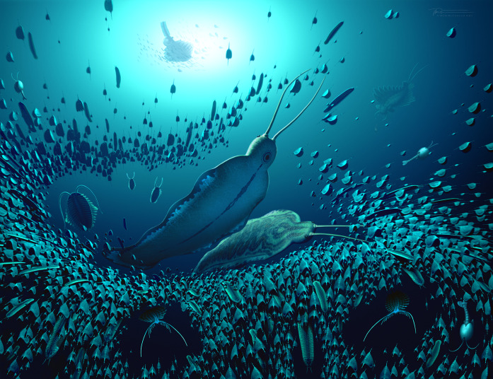
A giant stem-group chaetognath
- PMID: 38170772
- PMCID: PMC10796117
- DOI: 10.1126/sciadv.adi6678
A giant stem-group chaetognath
Abstract
Chaetognaths, with their characteristic grasping spines, are the oldest known pelagic predators, found in the lowest Cambrian (Terreneuvian). Here, we describe a large stem chaetognath, Timorebestia koprii gen. et sp. nov., from the lower Cambrian Sirius Passet Lagerstätte, which exhibits lateral and caudal fins, a distinct head region with long antennae and a jaw apparatus similar to Amiskwia sagittiformis. Amiskwia has previously been interpreted as a total-group chaetognathiferan, as either a stem-chaetognath or gnathostomulid. We show that T. koprii shares a ventral ganglion with chaetognaths to the exclusion of other animal groups, firmly placing these fossils on the chaetognath stem. The large size (up to 30 cm) and gut contents in T. koprii suggest that early chaetognaths occupied a higher trophic position in pelagic food chains than today.
Figures


References
-
- Bush A. M., Bambach R. K., Paleoecologic megatrends in marine metazoa. Annu. Rev. Earth Planet. Sci. 39, 241–269 (2011).
-
- Butterfield N., Oxygen, animals and aquatic bioturbation: An updated account. Geobiology 16, 3–16 (2018). - PubMed
-
- G. J. Vermeij, Evolution and Escalation: An Ecological History of Life (Princeton Univ. Press, 1993).
Publication types
MeSH terms
LinkOut - more resources
Full Text Sources
