. 2024 Jan 17;146(2):1374-1387.
doi: 10.1021/jacs.3c10132. Epub 2024 Jan 3.

Lipid Peroxidation Drives Liquid-Liquid Phase Separation and Disrupts Raft Protein Partitioning in Biological Membranes

Affiliations

Lipid Peroxidation Drives Liquid-Liquid Phase Separation and Disrupts Raft Protein Partitioning in Biological Membranes

Muthuraj Balakrishnan et al. J Am Chem Soc. .

Abstract

The peroxidation of membrane lipids by free radicals contributes to aging, numerous diseases, and ferroptosis, an iron-dependent form of cell death. Peroxidation changes the structure and physicochemical properties of lipids, leading to bilayer thinning, altered fluidity, and increased permeability of membranes in model systems. Whether and how lipid peroxidation impacts the lateral organization of proteins and lipids in biological membranes, however, remains poorly understood. Here, we employ cell-derived giant plasma membrane vesicles (GPMVs) as a model to investigate the impact of lipid peroxidation on ordered membrane domains, often termed membrane rafts. We show that lipid peroxidation induced by the Fenton reaction dramatically enhances the phase separation propensity of GPMVs into coexisting liquid-ordered (Lo) and liquid-disordered (Ld) domains and increases the relative abundance of the disordered phase. Peroxidation also leads to preferential accumulation of peroxidized lipids and 4-hydroxynonenal (4-HNE) adducts in the disordered phase, decreased lipid packing in both Lo and Ld domains, and translocation of multiple classes of raft proteins out of ordered domains. These findings indicate that the peroxidation of plasma membrane lipids disturbs many aspects of membrane rafts, including their stability, abundance, packing, and protein and lipid composition. We propose that these disruptions contribute to the pathological consequences of lipid peroxidation during aging and disease and thus serve as potential targets for therapeutic intervention.


Conflict of interest statement

The authors declare no competing financial interest.

Figures
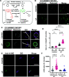
Figure 1
Figure 1
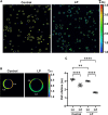
Induction and detection of lipid peroxidation in GPMVs. (A) (i) Cumene hydroperoxide in the presence of transition metal iron Fe(II) produces cumoxyl radicals via the Fenton reaction (a). (ii) Cumoxyl radicals (a) abstract a hydrogen (H) from a polyunsaturated lipid (PUFA-H) generating a lipid radical (PUFA) that reacts immediately with oxygen, generating PUFA peroxy radicals (PUFA-OO). The PUFA peroxy radicals react with C11-BODIPY 581/591, causing deconjugation of C11-BODIPY and a blue shift of the emission wavelength to 510 nm. (B) GPMVs were left untreated or incubated with 50 μM Fe(II) and 500 μM cumene hydroperoxide to induce lipid peroxidation, labeled with 1 μM C11-BODIPY 581/591, allowed to settle for 15–20 min, and imaged at RT using confocal microscopy. Scale bars, 20 μm. (C) Representative images of individual GPMVs labeled with C11-BODIPY 581/591 under control conditions or following lipid peroxidation. GPMVs were co-labeled with DiD to mark the position of the disordered domains. The arrows show examples of the position of lines used to analyze the fluorescence intensity in Lo and Ld domains. Scale bars, 5 μm. (D) Ratio of green (oxidized): red (reduced) BODIPY 581/591 fluorescence in ordered versus disordered domains under control conditions and following lipid peroxidation. Each data point corresponds to an individual GPMV. Error bars show mean ± SD for 27–33 GPMVs. (E) GPMVs were either left untreated or subjected to lipid peroxidation, immunolabeled with an anti-4-HNE antibody and Alexa-488 secondary antibody, and then stained using DiD. Examples of representative GPMVs are shown. Scale bars, 5 μm. (F) Quantification of immunostaining of 4-HNE levels. Fluorescence intensity is reported in arbitrary units. Each data point corresponds to an individual GPMV. Error bars show mean ± SD. P values were determined by unpaired one-way ANOVA with Sidak’s multiple comparison test, α = 0.05 (95% confidence level), ****, P < 0.0001; n.s., not significant. Data in (D, F) were pooled across 2 independent experiments.
Figure 2
Figure 2
Lipid peroxidation increases the percentage of phase-separated vesicles and the area fraction of disordered domains. (A) GPMVs were either left untreated (control) or subjected to lipid peroxidation (LP). They were then labeled sequentially with NBD-DSPE (green) and DiD (magenta) prior to imaging at RT using confocal microscopy. Scale bar: 20 μm. (B) Quantification of the percentage of phase-separated GPMVs for control versus lipid peroxidation conditions. The % of phase-separated GPMVs was calculated using the green channel using VesA software. Data are presented as mean ± SD for >1000 GPMVs per group. Data were pooled across 8 independent experiments. (C, D) Impact of lipid peroxidation on ordered partitioning of Lo (NBD-DSPE) and Ld (DiD) reporter dyes. Points in (C, D) represent >100 GPMVs in each group. ****, P < 0.0001 using unpaired two-tailed t test. Data are representative of 8 independent experiments. Each data point is for an individual field of GPMVs containing >50 GPMVs. (E) Illustration of how the area fraction of ordered (green) and disordered (magenta) domains was quantified for representative GPMVs. Scale bars: 5 μm. (F) Effect of lipid peroxidation on the area fraction of Lo and Ld domains. Each data point corresponds to an individual GPMV. Bars show the mean ± SD for two independent experiments. P values were determined by unpaired one-way ANOVA with Sidak’s multiple comparison test, α = 0.05 (95% confidence level). ****, P < 0.0001. (G) HeLa cells were left untreated (control) or pretreated with lipid peroxidation reagents for 30 min at RT (pre-LP) prior to GPMV preparation. For comparison, a population of control GPMVs and pre-LP GPMVs were subsequently incubated with lipid peroxidation reagents (post LP and pre + post LP, respectively). All GPMVs were then labeled with NBD-DSPE and DiD and imaged using confocal microscopy. The % of phase-separated GPMVs was calculated using the green channel using VesA software. Data are presented as mean ± SD. P values were determined by unpaired one-way ANOVA with Sidak’s multiple comparison test, α = 0.05 (95% confidence level) ****, P < 0.0001; n.s., not significant. Data are representative of 3 independent experiments for >100 GPMVs per group.
Figure 3
Figure 3
Lipid peroxidation decreases the level of lipid packing in both ordered and disordered domains. (A) Fluorescence lifetime imaging microscopy (FLIM) images of GPMVs labeled with Di4 under control conditions and following lipid peroxidation. Lookup table shows the Di4 lifetime (ns). (B) Representative FLIM images of individual GPMVs, highlighting differences in Di4 lifetime in Lo and Ld domains. Lookup table shows Di4 lifetime (ns). (C) Quantification of Di4 lifetimes in the Ld and Lo domains for control GPMVs and GPMVs subjected to lipid peroxidation. For the control sample, individual data points correspond to mean values for a field of >10–15 GPMVs taken from a single lifetime image; total number of control GPMVs = 104. For the LP sample, individual data points correspond to mean values for a field of >50 GPMVs taken from a single lifetime image; total number of LP GPMVs = 433. Error bars show mean ± SD. P values were determined by unpaired one-way ANOVA with Sidak’s multiple comparison test, α = 0.05 (95% confidence level), ****, P < 0.0001; n.s., not significant. Data are representative of two independent experiments.
Figure 4
Figure 4
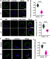
Raft-preferring proteins redistribute to disordered domains in response to lipid peroxidation. (A) Representative images of YFP-GL-GPI in HeLa-cell GPMVs. GPMVs were stained with Fast DiI after lipid peroxidation. (B) Impact of lipid peroxidation on ordered partitioning of YFP-GL-GPI. Each data point corresponds to individual GPMVs. Data are presented as mean ± SD for n = 47–68 GPMVs. (C) Representative images of CTxB-Alexa 555 labeling of COS-7 cell-derived GPMVs. After lipid peroxidation, GPMVs were sequentially labeled with CTxB-Alexa 555, NBD-DSPE, and DiD. (D) Impact of lipid peroxidation on the ordered partitioning of CTxB-Alexa 555. Data are presented as mean ± SD for 37–41 GPMVs. (E) Representative images of PMP22 in HeLa-cell GPMVs. GPMVs were labeled with an Alexa-488-labeled anti-myc antibody, subjected to lipid peroxidation, and then labeled with Fast DiI. (F) Impact of lipid peroxidation on ordered partitioning of PMP22. Data are presented as mean ± SD for 39–56 GPMVs. ****, P < 0.0001 for unpaired two-tailed t test. All data are representative of 2 independent experiments. Scale bars, 5 μm.
Figure 5
Figure 5
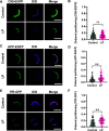
Non-raft proteins remain associated with disordered domains following lipid peroxidation. (A) Representative images of C99-EGFP in HeLa-cell GPMVs. (B) Quantification of ordered partitioning of C99-EGFP across multiple GPMVs. Each data point corresponds to an individual GPMV. Data are presented as mean ± SD for 65–102 GPMVs. (C) Representative images of APP-EGFP in HeLa-cell GPMVs. (D) Quantification of ordered partitioning of APP-EGFP across multiple GPMVs. Data are presented as mean ± SD for 38–54 GPMVs. (E) Representative images of TfR-GFP in HeLa-cell GPMVs. (F) Quantification of ordered partitioning of TfR-GFP across multiple GPMVs. Data are presented as mean ± SD for 35–46 GPMVs. **, P < 0.01 for unpaired two-tailed t test. All data are representative of 2 independent experiments. Scale bars, 5 μm.
Figure 6
Figure 6
Working model for how lipid peroxidation induced via the Fenton reaction impacts ordered and disordered domains in biological membranes. Peroxidized lipids and their bioactive products such as 4-HNE preferentially accumulate in disordered domains. This is accompanied by increases in the relative abundance of the disordered phase, decreased lipid packing in both phases, and changes in protein composition in both phases as the result of the selective redistribution of proteins from the ordered to the disordered phase. SL, saturated lipid; UL, unsaturated lipid; OL, oxidized lipid; chol, cholesterol.

Update of

References

    1. Levental I.; Lyman E. Regulation of membrane protein structure and function by their lipid nano-environment. Nat. Rev. Mol. Cell Biol. 2023, 24 (2), 107–122. 10.1038/s41580-022-00524-4. - DOI - PMC - PubMed
    1. Ammendolia D. A.; Bement W. M.; Brumell J. H. Plasma membrane integrity: implications for health and disease. BMC Biol. 2021, 19 (1), 7110.1186/s12915-021-00972-y. - DOI - PMC - PubMed
    1. Yin H.; Xu L.; Porter N. A. Free radical lipid peroxidation: mechanisms and analysis. Chem. Rev. 2011, 111 (10), 5944–5972. 10.1021/cr200084z. - DOI - PubMed
    1. Niki E.; Yoshida Y.; Saito Y.; Noguchi N. Lipid peroxidation: mechanisms, inhibition, and biological effects. Biochem. Biophys. Res. Commun. 2005, 338 (1), 668–676. 10.1016/j.bbrc.2005.08.072. - DOI - PubMed
    1. Su L. J.; Zhang J. H.; Gomez H.; Murugan R.; Hong X.; Xu D.; Jiang F.; Peng Z. Y. Reactive Oxygen Species-Induced Lipid Peroxidation in Apoptosis, Autophagy, and Ferroptosis. Oxid. Med. Cell Longevity 2019, 2019, 508084310.1155/2019/5080843. - DOI - PMC - PubMed

Publication types