. 2023 Dec;20(4):1358-1379.
doi: 10.14245/ns.2346824.412. Epub 2023 Dec 31.

Mesenchymal Stem Cells Combined With Electroacupuncture Treatment Regulate the Subpopulation of Macrophages and Astrocytes to Facilitate Axonal Regeneration in Transected Spinal Cord

Affiliations

Mesenchymal Stem Cells Combined With Electroacupuncture Treatment Regulate the Subpopulation of Macrophages and Astrocytes to Facilitate Axonal Regeneration in Transected Spinal Cord

Rongyi Zhang et al. Neurospine. 2023 Dec.

Abstract

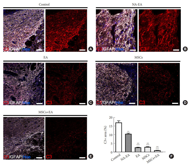
Objective: Herein, we investigated whether mesenchymal stem cells (MSCs) transplantation combined with electroacupuncture (EA) treatment could decrease the proportion of proinflammatory microglia/macrophages and neurotoxic A1 reactive astrocytes and inhibit glial scar formation to enhance axonal regeneration after spinal cord injury (SCI).

Methods: Adult rats were divided into 5 groups after complete transection of the spinal cord at the T10 level: a control group, a nonacupoint EA (NA-EA) group, an EA group, an MSC group, and an MSCs+EA group. Immunofluorescence labeling, quantitative real-time polymerase chain reaction, enzyme-linked immunosorbent assay, and Western blots were performed.

Results: The results showed that MSCs+EA treatment reduced the proportion of proinflammatory M1 subtype microglia/macrophages, but increased the differentiation of anti-inflammatory M2 phenotype cells, thereby suppressing the mRNA and protein expression of proinflammatory cytokines (tumor necrosis factor-α and IL-1β) and increasing the expression of an anti-inflammatory cytokine (interleukin [IL]-10) on days 7 and 14 after SCI. The changes in expression correlated with the attenuated neurotoxic A1 reactive astrocytes and glial scar, which in turn facilitated the axonal regeneration of the injured spinal cord. In vitro, the proinflammatory cytokines increased the level of proliferation of astrocytes and increased the expression levels of C3, glial fibrillary acidic protein, and chondroitin sulfate proteoglycan. These effects were blocked by administering inhibitors of ErbB1 and signal transducer and activator of transcription 3 (STAT3) (AG1478 and AG490) and IL-10.

Conclusion: These findings showed that MSCs+EA treatment synergistically regulated the microglia/macrophage subpopulation to reduce inflammation, the formation of neurotoxic A1 astrocytes, and glial scars. This was achieved by downregulating the ErbB1-STAT3 signal pathway, thereby providing a favorable microenvironment conducive to axonal regeneration after SCI.

Keywords: Axonal regeneration; Electroacupuncture; Macrophages; Mesenchymal stem cells; Microglia; Reactive astrocytes; Spinal cord injury.


Conflict of interest statement

Conflict of Interest

The authors declare no conflict of interest.

Figures

Fig. 1.
Fig. 1.
Activated microglia/macrophages and their subtypes on day 7 post-SCI. (A1, A2) Representative images of CD68+ activated microglia/macrophages (white arrows) in the injured spinal cord in the control group (A1) and MSCs+EA group (A2). (B) A schematic showing the injured spinal cord and the sample sites. (C) Quantitative analysis of CD68+ activated microglia/macrophages in all groups. Data are expressed as means ± standard deviation (SD) (n=5). As compared to the control group, *p < 0.05, **p < 0.01; compared with the NA-EA group, #p < 0.05, ##p < 0.01; compared with the MSCs group, $p < 0.05. (D1–D5) Representative images of CCR7 (red)/CD68 (white) double-labeled proinflammatory M1 macrophages (yellow arrows) in the control (D1), NA-EA (D2), EA (D3), MSCs (D4) and MSCs+EA (D5) groups. (E1–E5) Representative images of CD206 (red)/CD68 (white) double-labeled anti-inflammatory M2 macrophages (yellow arrows) in the control (E1), NA-EA (E2), EA (E3), MSCs (E4), and MSCs+EA (E5) groups. Nuclei were counterstained by Hoechst 33342 (blue). Transplanted MSCs are shown as GFP+ cells (green). Scale bars: A1 and A2=200 μm; D1–D5 and E1–E5=20 μm. (F1, F2) Quantitative analysis of the CCR7+ CD68+ M1 macrophages and the CD206+ CD68+ M2 macrophages. Data are expressed as means ± SD (n=5). As compared to the control group, *p < 0.05, **p < 0.01; compared with the NA-EA group, #p <0.05, ##p < 0.01; compared with the MSCs group, $p < 0.05; compared with the EA group, &p < 0.05. SCI, spinal cord injury; MSC, mesenchymal stem cell; EA, electroacupuncture; NA-EA, nonacupoint EA.
Fig. 2.
Fig. 2.
Western blotting analysis of the pan-macrophage marker CD68, the M1 marker CCR7, and the M2 marker CD206. (A1–A3) Representative Western blots of CD206, CCR7, and CD68 on day 3 (A1), day 7 (A2), and day 14 (A3) after SCI. (B1–B3) Quantitative analysis of CD206, CCR7, and CD68 protein expression on day 3 (B1), day 7 (B2), and day 14 (B3) after SCI. (B4) Quantitative analysis of the ratio of CD206/CCR7 (M2/M1). Data are expressed as means ± SD (n=5). SCI, spinal cord injury; MSC, mesenchymal stem cell; EA, electroacupuncture; NA-EA, nonacupoint EA. Compared with the control group, *p < 0.05, **p < 0.01; compared with the NA-EA group, #p < 0.05, ##p < 0.01; compared with the MSCs group, $p < 0.05; compared with the EA group, &p < 0.05.
Fig. 3.
Fig. 3.
Quantitative real-time polymerase chain reaction (qRT-PCR) and ELISA analysis of the mRNA and protein expression of inflammatory cytokines after SCI. (A1, A2) The mRNA (A1) and protein (A2) levels of TNF-α in the lesion site of the spinal cord tissue as measured by qRT-PCR and ELISA on days 3, 7, and 14 after surgery. (B1, B2) The mRNA and protein levels of IL-1β in the lesion site of spinal cord tissue on days 3, 7, and 14 after SCI. (C1, C2) The mRNA and protein levels of IL-10 in the lesion site of spinal cord tissue on days 3, 7, and 14 after SCI. Data are expressed as means ± standard deviation (n=5). ELISA, enzyme-linked immunosorbent assay; IL, interleukin; TNF, tumor necrosis factor; SCI, spinal cord injury; MSC, mesenchymal stem cell; EA, electroacupuncture; NA-EA, nonacupoint EA. Compared to the control group, *p < 0.05, **p < 0.01; compared with the NAEA group, #p < 0.05, ##p < 0.01; compared with the MSCs group, $p < 0.05, $$p <0.01; compared with the EA group, &p < 0.05, &&p < 0.01.
Fig. 4.
Fig. 4.
The combined MSCs and EA treatment inhibited the expression of GFAP and CSPGs after SCI. (A, B1–B3) The mRNA levels of GFAP (A), NG2 (B1), neurocan (B2), and phosphacan (B3) as measured by qRT-PCR on days 3, 7, and 14 after surgery. (C1–C3) Representative western blots of GFAP and CSPGs on day 3 (C1), day 7 (C2), and day 14 (C3) after SCI. (C4, C5) The quantitative analysis of GFAP and CSPGs Western blots. Data are expressed as means ± standard deviation (n=5). GFAP, glial fibrillary acidic protein; CSPG, chondroitin sulfate proteoglycan; qRT-PCR, quantitative real-time polymerase chain reaction; SCI, spinal cord injury; MSC, mesenchymal stem cell; EA, electroacupuncture; NA-EA, nonacupoint EA. Compared with the Control group, *p < 0.05, **p < 0.01; compared with the NA-EA group, #p < 0.05, ##p < 0.01; compared with the EA group, &&p < 0.01; compared with the MSCs group, $$p < 0.01.
Fig. 5.
Fig. 5.
Immunofluorescence staining showing the expression of GFAP and CSPGs in spinal cord on day 14 after SCI. (A1–E2) Representative images of GFAP and CSPGs immunostaining (red, arrows) in injured spinal cords in the control (A1, A2), NAEA (B1, B2), EA (C1, C2), MSCs (D1, D2), and MSCs+EA (E1, E2) groups. Scale bar=200 μm. ※ indicates the center of the lesion. (F1, F2) Quantitative analysis of the percentage of GFAP and CSPGs immunostaining area in the 5 groups. Data are expressed as means ± standard deviation (n=5). GFAP, glial fibrillary acidic protein; CSPG, chondroitin sulfate proteoglycan; SCI, spinal cord injury; MSC, mesenchymal stem cell; EA, electroacupuncture; NA-EA, nonacupoint EA. Compared with the control group, *p < 0.05, **p < 0.01; compared with the NA-EA group, #p < 0.05, ##p < 0.01; compared with the EA group, &&p < 0.01.
Fig. 6.
Fig. 6.
(A1–A3) Representative images showing the proliferation (Ki67+, red nuclei) of GFAP+ (green color) astrocytes in the 2% fetal bovine serum (FBS) culture medium group, TNF-α (10 ng/mL) group, and TNF-α (10 ng/mL)+IL-1β (10 ng/mL) culture medium for 48 hours. Scale bars=40 μm. (A4, A5) The percentage of Ki67+ or EdU+ astrocytes following different doses of TNF-α (10 ng/mL) and/or IL-1β (1 ng/mL, 10 ng/mL). “+” indicates 2% FBS in DMEM/F12; while “++” indicates 10% FBS in DMEM/F12. Data expressed as means ± standard deviation (SD) (n=3). Compared with 2% FBS group, *p < 0.05, **p < 0.01; compared with IL-1β (1 ng/mL) group, #p < 0.05, ##p < 0.01; compared with IL-1β (10 ng/mL) group, &p < 0.05, &&p < 0.01; compared with TNF-α (10 ng/mL) group, $p < 0.05, $$p < 0.01; compared with TNF-α (10 ng/mL) and IL-1β (1 ng/mL), Δp < 0.05. (B, C1–C3) The mRNA levels of GFAP and CSPGs core proteins (NG2, neurocan, and phosphacan) in cultured astrocytes were measured using qRT-PCR following treatment with TNF-α and/or IL-1β for 24 hours. Data expressed as means ± SD (n=3). Compared with 2% FBS group, *p < 0.05, **p < 0.01; compared with IL-1β (1 ng/mL) group, #p < 0.05, ##p < 0.01; compared with IL-1β (10 ng/mL) group, &p < 0.05, &&p < 0.01; compared with TNF-α (10 ng/mL) group, $p < 0.05, $$p < 0.01. (D1–D3) Representative images of GFAP/ErbB1/p-STAT3 immunofluorescence staining in the 2% FBS group, TNF-α+IL-1β group and TNF-α+IL-1β+IL-10 group. Scale bars=20 μm. (E) Western blotting analysis of GFAP, CSPGs, ErbB1, and p-STAT3 in the 2% FBS group, TNF-α+IL1β group and TNF-α+IL-1β+IL-10 group. Data are representative of 3 experiments and expressed as means ± SD. Compared with the 2% FBS group, *p < 0.05, **p < 0.01. (F) Western blot analysis showing that the addition of inhibitors (AG1478 and AG490) for ErbB1/STAT3 significantly reduced the expression of GFAP, CSPGs, ErbB1, and p-STAT3 when compared with the TNF-α+ IL-1β group. Data expressed as means ± SD (n=3). Compared with the TNF-α+IL-1β group, *p < 0.05, **p < 0.01. TNF, tumor necrosis factor; IL, interleukin; EdU, 5-ethynyl-2’-deoxyuridine; DMEM, Dulbecco’s modified Eagle’s medium; GFAP, glial fibrillary acidic protein; CSPG, chondroitin sulfate proteoglycan; qRT-PCR, quantitative real-time polymerase chain reaction; STAT3, signal transducer and activator of transcription 3.
Fig. 7.
Fig. 7.
The MSCs combined with EA treatment downregulated the ErbB1-STAT3 pathway after SCI. (A, B) Representative images of GFAP/ErbB1 (white arrows) and GFAP/p-STAT3 (white arrows) double-labeled immunofluorescence staining on day 7 after SCI. Scale bars=20 μm. (C) Western blot analysis of ErbB1 and p-STAT3 in injured spinal cord on day 7 after SCI. (D) Western blot analysis of ErbB1 and p-STAT3 in injured spinal cord on day 14 after SCI. Data are expressed as means ± standard deviation. GFAP, glial fibrillary acidic protein; SCI, spinal cord injury; MSC, mesenchymal stem cell; EA, electroacupuncture; NA-EA, nonacupoint EA; STAT3, signal transducer and activator of transcription 3. Compared with the control group, *p < 0.05, **p < 0.01; compared with the NA-EA group, #p < 0.05, ##p < 0.01; compared with the EA group, &p < 0.05; compared with the MSCs group, $p < 0.05, $$p < 0.01.
Fig. 8.
Fig. 8.
The MSCs and EA treatment reduced the number of C3-positive A1 astrocytes. (A–E) Representative images of GFAP (white)/C3 (red) double-labeled immune-positive A1 astrocytes in the control (A), NA-EA (B), EA (C), MSCs (D), and MSCs+EA (E) groups. Scale bar=50 μm. (F) Quantitative analysis of the percentage of C3 immuno-positive area in tissues from the 5 groups. Data are expressed as means ± standard deviation (n=5). GFAP, glial fibrillary acidic protein; SCI, spinal cord injury; MSC, mesenchymal stem cell; EA, electroacupuncture; NA-EA, nonacupoint EA. Compared with the control group, **p < 0.01, ***p < 0.001; compared with the NA-EA group, ###p < 0.001.
Fig. 9.
Fig. 9.
MSCs and EA treatment promoted axonal regeneration at the site of injury on day 14 after SCI. (A–E) Representative images of NF (white arrows)/GFAP immunostaining in the control (A), NA-EA (B), EA (C), MSCs (D), and MSCs+EA (E) groups. In the panels A–E, the scale=200 μm in the low magnification image; the scale=20 μm in the high magnification image. (F, G) Western blot analysis of NF protein expression in the 5 groups. Data are expressed as means ± standard deviation (n=5). GFAP, glial fibrillary acidic protein; SCI, spinal cord injury; MSC, mesenchymal stem cell; EA, electroacupuncture; NA-EA, nonacupoint EA; NF, neurofilament. Compared with the control group, **p < 0.01; compared with the NA-EA group, ##p < 0.01; compared with the EA group, &&p < 0.01; compared with the MSCs group, $$p < 0.01.

Similar articles

Cited by

References

    1. Karimi-Abdolrezaee S, Billakanti R. Reactive astrogliosis after spinal cord injury-beneficial and detrimental effects. Mol Neurobiol. 2012;46:251–64. - PubMed
    1. Trivedi A, Olivas AD, Noble-Haeusslein LJ. Inflammation and spinal cord injury: infiltrating leukocytes as determinants of injury and repair processes. Clin Neurosci Res. 2006;6:283–92. - PMC - PubMed
    1. Gordon S, Taylor PR. Monocyte and macrophage heterogeneity. Nat Rev Immunol. 2005;5:953–64. - PubMed
    1. Martinez FO, Sica A, Mantovani A, et al. Macrophage activation and polarization. Front Biosci. 2008;13:453–61. - PubMed
    1. Kigerl KA, Gensel JC, Ankeny DP, et al. Identification of two distinct macrophage subsets with divergent effects causing either neurotoxicity or regeneration in the injured mouse spinal cord. J Neurosci. 2009;29:13435–44. - PMC - PubMed