. 2024 Jan 2;36(1):90-102.e7.
doi: 10.1016/j.cmet.2023.11.018.

Repression of latent NF-κB enhancers by PDX1 regulates β cell functional heterogeneity

Affiliations

Repression of latent NF-κB enhancers by PDX1 regulates β cell functional heterogeneity

Benjamin J Weidemann et al. Cell Metab. .

Abstract

Interactions between lineage-determining and activity-dependent transcription factors determine single-cell identity and function within multicellular tissues through incompletely known mechanisms. By assembling a single-cell atlas of chromatin state within human islets, we identified β cell subtypes governed by either high or low activity of the lineage-determining factor pancreatic duodenal homeobox-1 (PDX1). β cells with reduced PDX1 activity displayed increased chromatin accessibility at latent nuclear factor κB (NF-κB) enhancers. Pdx1 hypomorphic mice exhibited de-repression of NF-κB and impaired glucose tolerance at night. Three-dimensional analyses in tandem with chromatin immunoprecipitation (ChIP) sequencing revealed that PDX1 silences NF-κB at circadian and inflammatory enhancers through long-range chromatin contacts involving SIN3A. Conversely, Bmal1 ablation in β cells disrupted genome-wide PDX1 and NF-κB DNA binding. Finally, antagonizing the interleukin (IL)-1β receptor, an NF-κB target, improved insulin secretion in Pdx1 hypomorphic islets. Our studies reveal functional subtypes of single β cells defined by a gradient in PDX1 activity and identify NF-κB as a target for insulinotropic therapy.

Keywords: IL-1β; NF-κB; PDX1; chromatin; circadian; diabetes; inflammation; insulin; p65; β cells.


Conflict of interest statement

Declaration of interests M.P. is currently affiliated with Ionis Pharmaceuticals, Inc.

Figures

Figure 1.
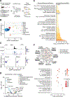
Figure 1.. Single-cell chromatin accessibility sequencing identifies PDX1 and NF-κB enhancer signatures within distinct β-cell populations of the human islet.
(A) Profiling single nuclei and rhythmic bulk chromatin accessibility by ATAC-sequencing in human cadaveric islets. (B) Clustering and dimensional reduction analyses (UMAP) identified 19 distinct subpopulations of cells based on normalized reads at accessible cis-regulatory elements (20,519 single islet nuclei from 3 humans). (C) Aggregate sequencing fragments, segregated by cell-type, at lineage-specific marker genes: GCG (alpha), INS-IGF2 (beta), SST (delta), PDGFRB (stellate), SOX10 (neural crest), CCL4 (lymphatic and myeloid immune), PECAM1 (endothelial), KRT19 (ductal), and REG1A (acinar). Co-accessibility linkage analysis connects individual snATAC-seq peak accessibility scores within networks of coregulated regions. (D) Accessibility at PDX1 (left) and NF-κB (right) motifs was enriched in the β1 and β2 subpopulations of cells, respectively. Individual cell chromVAR z-scores (top) and subpopulation z-score distribution (bottom) across β1 and β2 subpopulations demonstrate significant variation in chromatin accessibility at PDX1 and NF-κB motifs. (E) Differential analysis of TF motif accessibility between β1 and β2 subpopulations revealed enrichment for basic helix-loop-helix (bHLH) and lineage-determining TFs in β1 cells and enrichment of immediate early bZIP and NF-κB TFs in β2 cells. (F) ATAC-seq performed in synchronized whole islets every 4 hrs for 24 hrs following a forskolin shock (n=10 human islet equivalents (IEQs) per sample per time point from 4 humans). Data was analyzed using eJTK_Cycle at a 2-hr resolution, revealing 24-hr patterns in chromatin opening at ATAC-seq peaks (left) and β-cell TF motifs associated with cell identity and rhythmic function (right). See also Figure S1 and Tables S1 and S3.
Figure 2.
Figure 2.. Genetic deficiency of PDX1 leads to de-repression of NF-κB.
(A) Profiling single-cell chromatin accessibility using single-nuclei ATAC-sequencing in islets isolated from control (Pdx1+/) and Pdx1 mutant (Pdx1ΔAIV/) mice (n=8 per genotype). (B) Clustering and dimensional reduction analyses (UMAP) of control and mutant islets cells identified 15 distinct subpopulations of cells based on normalized reads at accessible cis-regulatory elements. (C) Cumulative (top) and single-cell (bottom) accessible reads at the Ins1 gene in pooled control versus Pdx1 mutant β cells. (D) TF binding motifs differentially-enriched within accessible chromatin regions of pooled control versus Pdx1 mutant β cells. Negative values indicate the motif is more enriched in controls, while positive values indicate motif is more enriched in the Pdx1 mutants. (E) Gene ontology analysis of annotated differentially-accessible peaks identified as upregulated in control or Pdx1 mutant β cells. (F) RNA-sequencing reveals dysregulation of circadian, insulin secretion, and NF-κB signaling in Pdx1A4 mutant islets. RNA-sequencing and pathway analyses using pathfindR reveal enrichment for circadian rhythm, insulin secretion, and NF-κB signaling KEGG pathways, as displayed by shared gene enrichment (top) and hierarchal clustering with fold-enrichment and multiple comparison P-values (bottom), among differentially-expressed transcripts (Adjusted P-value < 0.05) in islets isolated from Pdx1A4 (n=5) versus Pdx1 (n=4) heterozygous mice. See also Figure S2.
Figure 3.
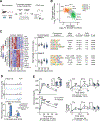
Figure 3.. Circadian analyses reveal rhythmic regulation of NF-κB enhancer activity by PDX1.
(A) Pancreatic islet preparations from control (Pdx1+/) and Pdx1 mutant (Pdx1ΔAIV/) mice were subjected to a circadian synchronization pulse (forskolin), followed by ATAC-seq at two different time points, 36 and 48 hrs post-synchronization (the trough and peak of insulin secretion, respectively) (n=3 per timepoint per genotype). (B) Log2 fold-change in normalized accessible reads with respect to circadian time or genotype following identification of dynamic peaks using likelihood ratio tests. Significant peaks were segregated into different groups of peaks with distinct accessibility patterns using k-means clustering: Group 1 (orange) - sites with increased accessibility in control islets at the 48 hr time point (peak of insulin secretion) but with reduced accessibility in the mutants. Group 2 (green) - sites with increased accessibility at the 36 hr time point (trough of insulin secretion), which are also increased in the mutants. Group 3 sites (yellow) - sites only dependent on circadian time and not genotype. (C) Normalized accessible read Z-scores (left, middle) across all samples reveal increased accessibility nearby insulin secretion genes in control islets (Group 1) and near stress and inflammatory genes in mutant islets (Group 2). Motif analyses (right) within differentially-accessible peaks demonstrate enrichment for β-cell TFs and inflammatory and immediate early TFs in Group 1 and Group 2 chromatin peaks, respectively. (D) Increased normalized chromatin reads in Pdx1ΔAIV/ mutant islets neighboring the TNF-related gene Tnfrsf11b at 36 hrs post-synchronization. (E) Glucose clearance, insulin secretion, C-peptide levels, and area under curves following oral glucose administration (2g/kg) in control (n=14 mice per time point for glucose, n=13 for insulin and C-peptide) and Pdx1 mutant (n=12 mice per time point for glucose, n=9 for insulin and C-peptide) in either the morning (ZT2) or evening (ZT14) analyzed by mixed-effects models. Main and interaction term significance determined by mixed-effects model. *P < 0.05, **P < 0.01, ***P < 0.001, n.s. not significant by Wald (ATAC-seq) or Holm-Šídák test. Data are represented as mean ± SEM. See also Figure S3.
Figure 4.
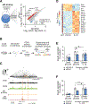
Figure 4.. PDX1 represses NF-κB through long-range chromatin contacts that co-localize with IL-1β-response elements controlling insulin secretion.
(A) p65 ChIP-seq in Beta-TC-6 cells treated with siRNA targeting Pdx1 (siPdx1) reveals increased genome-wide occupancy at inflammatory and circadian (Per2/Hes6) gene regions. (B) Chromatin conformational sequencing coupled with ChIP-seq (HiChIP-seq) targeting PDX1 identifies three-dimensional chromatin contacts involving PDX1 binding sites. (C) Significant chromatin interactions identified between the PDX1-SIN3A binding sites which loop to contact p65 binding sites. siPdx1-treated β cells exhibit increased p65 binding following loss of PDX1 protein, suggesting PDX1-SIN3A chromatin loops normally repress p65 binding near Per2. (D) IL-1β inhibits β-cell gene expression and activates p65-mediated expression of inflammatory genes in Beta-TC-6 cells. (E) Acute (2 hr) administration of IL-1β to β cells inhibits glucose-responsive insulin release specifically in control siRNA-treated cells, whereas siPdx1-treated cells do not respond to IL-1β (n=10 samples per siRNA treatment). Data are represented as mean ± SEM. (F) Antagonism of islet IL-1β signaling using the IL-1β receptor antagonist (IL-1RA) restores glucose-stimulated insulin secretion in Pdx1ΔAIV/ islets (n=8 Pdx1+/ mice, n=5 Pdx1ΔAIV/ mice). Two-way ANOVA, *P < 0.05 by Holm-Šídák test. Data are represented as mean ± SEM. See also Figure S4 and Tables S4–S6.

References

    1. Heinz S, Benner C, Spann N, Bertolino E, Lin YC, Laslo P, Cheng JX, Murre C, Singh H, and Glass CK (2010). Simple combinations of lineage-determining transcription factors prime cis-regulatory elements required for macrophage and B cell identities. Mol Cell 38, 576–589. - PMC - PubMed
    1. Wortham M, and Sander M (2021). Transcriptional mechanisms of pancreatic beta-cell maturation and functional adaptation. Trends Endocrinol Metab 32, 474–487. - PMC - PubMed
    1. Chiou J, Zeng C, Cheng Z, Han JY, Schlichting M, Miller M, Mendez R, Huang S, Wang J, Sui Y, et al. (2021). Single-cell chromatin accessibility identifies pancreatic islet cell type- and state-specific regulatory programs of diabetes risk. Nat Genet 53, 455–466. - PMC - PubMed
    1. Chiou J, Geusz RJ, Okino ML, Han JY, Miller M, Melton R, Beebe E, Benaglio P, Huang S, Korgaonkar K, et al. (2021). Interpreting type 1 diabetes risk with genetics and single-cell epigenomics. Nature 594, 398–402. - PMC - PubMed
    1. Fasolino M, Schwartz GW, Patil AR, Mongia A, Golson ML, Wang YJ, Morgan A, Liu C, Schug J, Liu J, et al. (2022). Single-cell multi-omics analysis of human pancreatic islets reveals novel cellular states in type 1 diabetes. Nat Metab 4, 284–299. - PMC - PubMed

Publication types