Review
. 2024 Jan 3;44(1):e1051232024.
doi: 10.1523/JNEUROSCI.1051-23.2023.

Cortical Reorganization after Limb Loss: Bridging the Gap between Basic Science and Clinical Recovery

Affiliations
Review

Cortical Reorganization after Limb Loss: Bridging the Gap between Basic Science and Clinical Recovery

Tawnee Sparling et al. J Neurosci. .

Abstract

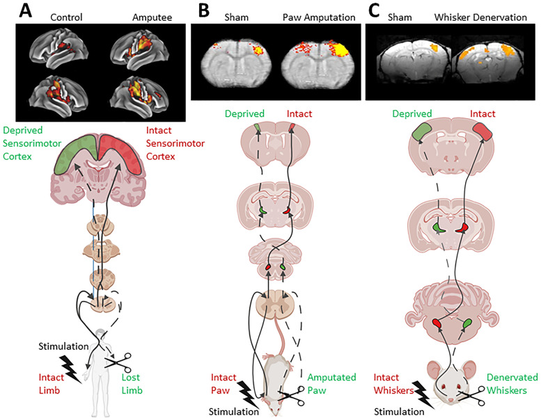
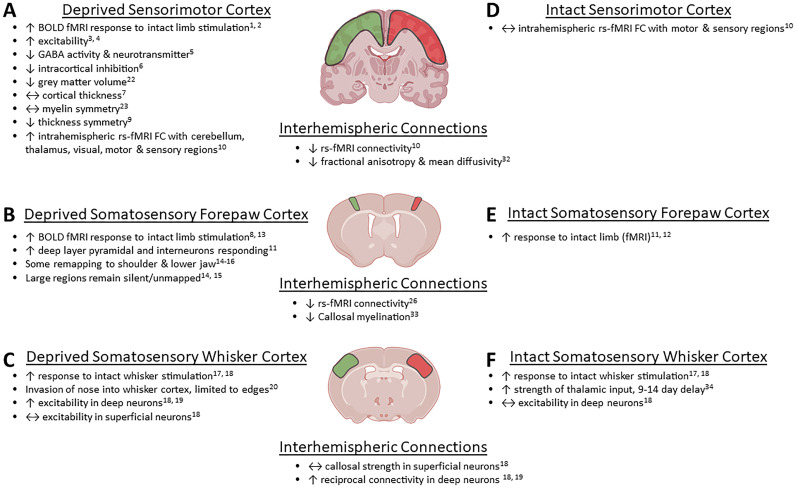
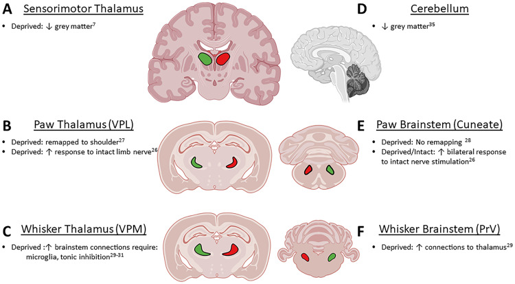
Despite the increasing incidence and prevalence of amputation across the globe, individuals with acquired limb loss continue to struggle with functional recovery and chronic pain. A more complete understanding of the motor and sensory remodeling of the peripheral and central nervous system that occurs postamputation may help advance clinical interventions to improve the quality of life for individuals with acquired limb loss. The purpose of this article is to first provide background clinical context on individuals with acquired limb loss and then to provide a comprehensive review of the known motor and sensory neural adaptations from both animal models and human clinical trials. Finally, the article bridges the gap between basic science researchers and clinicians that treat individuals with limb loss by explaining how current clinical treatments may restore function and modulate phantom limb pain using the underlying neural adaptations described above. This review should encourage the further development of novel treatments with known neurological targets to improve the recovery of individuals postamputation.Significance Statement In the United States, 1.6 million people live with limb loss; this number is expected to more than double by 2050. Improved surgical procedures enhance recovery, and new prosthetics and neural interfaces can replace missing limbs with those that communicate bidirectionally with the brain. These advances have been fairly successful, but still most patients experience persistent problems like phantom limb pain, and others discontinue prostheses instead of learning to use them daily. These problematic patient outcomes may be due in part to the lack of consensus among basic and clinical researchers regarding the plasticity mechanisms that occur in the brain after amputation injuries. Here we review results from clinical and animal model studies to bridge this clinical-basic science gap.

Keywords: amputation; amputee rehabilitation; cortical reorganization; limb loss; neural circuits; prosthetics.


Figures

Figure 1.
Figure 1.
Diagram depicting the similar BOLD fMRI responses in people after limb loss (A), rodents after paw amputation (B), and rodents after whisker denervation (C). All three instances have increased responses to intact limb/paw/whisker stimulation in bilateral sensorimotor cortices. fMRI images reused with permission from (A) Valyear et al. (2020), (B) Pelled et al. (2009), and (C) Petrus et al. (2019). BOLD fMRI, blood oxygenation level-dependent functional magnetic resonance imaging.
Figure 2.
Figure 2.
Diagram depicting the cortical adaptations after unilateral limb loss/paw amputation/whisker denervation. Deprived (green—A) and intact (red—D) sensorimotor cortices in upper and lower limb amputees have many adaptations noted after injury. Deprived (green—C) and intact (red—E) somatosensory forepaw cortex adaptations after unilateral paw amputation. Deprived (green—C) and intact (red—F) somatosensory whisker cortical adaptations after unilateral whisker denervation. Interhemispheric connections are related to observations between the two hemispheres and mediated by the corpus callosum. BOLD fMRI, blood oxygenation level-dependent functional magnetic resonance imaging; FC, functional connectivity; rs-fMRI, resting-state functional magnetic resonance imaging; S2, secondary somatosensory area; M2, secondary motor area.
Figure 3.
Figure 3.
Diagram depicting the subcortical brain regions where adaptations are observed in models of unilateral limb/paw/whisker amputation. A, Deprived (green) sensorimotor thalamus in upper and lower limb amputees has reduced grey matter volume. D, Cerebellum also has reduced grey matter volume after amputation. B, Deprived (green) thalamus and E, brainstem experience some adaptations after unilateral paw amputation in rodents. C, Unilateral whisker denervation drives changes in the thalamus and brainstem F, VPL, ventral posterolateral nucleus; VPM, ventral posteromedial nucleus (sensory thalamus); PrV, principal nucleus of the fifth trigeminal nerve.

Similar articles

Cited by

References

    1. Abbass K (2012) Effica de gabapentina en el tratamiento de pacientes adultos con color en el miembro fantasma. Ann Pharmacother 46:1707–1711. 10.1345/aph.1Q793 - DOI - PubMed
    1. Adcock KS, et al. (2022) Vagus nerve stimulation does not improve recovery of forelimb motor or somatosensory function in a model of neuropathic pain. Sci Rep 12:1–7. 10.1038/s41598-022-13621-3 - DOI - PMC - PubMed
    1. Ahmed A, Bhatnagar S, Mishra S, Khurana D, Joshi S, Ahmad S (2017) Prevalence of phantom limb pain, stump pain, and phantom limb sensation among the amputated cancer patients in India: a prospective, observational study. Indian J Palliat Care 23:24–35. 10.4103/IJPC.IJPC_28_17 - DOI - PMC - PubMed
    1. Ahmed MA, Mohamed SA, Sayed D (2011) Long-term antalgic effects of repetitive transcranial magnetic stimulation of motor cortex and serum beta-endorphin in patients with phantom pain. Neurol Res 33:953–958. 10.1179/1743132811Y.0000000045 - DOI - PubMed
    1. Alviar MJM, Hale T, Dungca M (2016) Pharmacologic interventions for treating phantom limb pain. Cochrane Database Syst Rev 2016:CD006380. 10.1002/14651858.CD006380.pub3 - DOI - PMC - PubMed

Publication types