Review
. 2024 Jan 2;83(1):63-81.
doi: 10.1016/j.jacc.2023.10.025.

Novel Techniques in Imaging Congenital Heart Disease: JACC Scientific Statement

Affiliations
Review

Novel Techniques in Imaging Congenital Heart Disease: JACC Scientific Statement

Ritu Sachdeva et al. J Am Coll Cardiol. .

Abstract

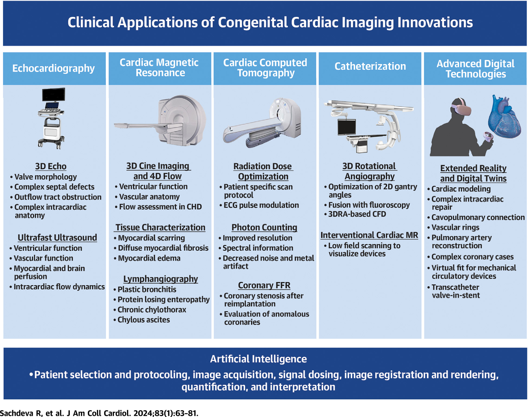
Recent years have witnessed exponential growth in cardiac imaging technologies, allowing better visualization of complex cardiac anatomy and improved assessment of physiology. These advances have become increasingly important as more complex surgical and catheter-based procedures are evolving to address the needs of a growing congenital heart disease population. This state-of-the-art review presents advances in echocardiography, cardiac magnetic resonance, cardiac computed tomography, invasive angiography, 3-dimensional modeling, and digital twin technology. The paper also highlights the integration of artificial intelligence with imaging technology. While some techniques are in their infancy and need further refinement, others have found their way into clinical workflow at well-resourced centers. Studies to evaluate the clinical value and cost-effectiveness of these techniques are needed. For techniques that enhance the value of care for congenital heart disease patients, resources will need to be allocated for education and training to promote widespread implementation.

Keywords: angiography; artificial intelligence; cardiac computed tomography; cardiac magnetic resonance; digital twin technology; echocardiography.


Conflict of interest statement

Funding Support and Author Disclosures Dr Armstrong is a consultant for Edwards Lifesciences, Medtronic, Cook Medical, Abbott, and Starlight Cardiovascular; and receives research support from Siemens Healthineers and Renata Medical. Dr Grosse-Wortmann is a consultant for Siemens Healthineers. Dr Powell is a consultant for Siemens Healthineers. All other authors have reported that they have no relationships relevant to the contents of this paper to disclose.

Figures

CENTRAL ILLUSTRATION
CENTRAL ILLUSTRATION. Innovations in Cardiac Imaging: A Novel Paradigm in CHD Evaluation
The novel imaging techniques in echocardiography (echo), cardiac magnetic resonance, cardiac computed tomography, catheterization, and advanced digital technologies have a broad range of clinical applications in congenital heart disease (CHD). In addition, artificial intelligence is being applied to different steps in the imaging process across all the cardiac imaging modalities. 3DRA = 3-dimensional rotational angiography; CFD = computational fluid dynamics; ECG = electrocardiogram; FFR = fractional flow reserve; MR = magnetic resonance.
FIGURE 1
FIGURE 1. 3D Transthoracic Echocardiography of the Mitral Valve With Transillumination
Comparison of 3D echocardiographic rendering of a mass on the mitral valve leaflet (white arrow) with traditional imaging (left) vs transillumination (right), which offers better delineation of borders and depth perception.
FIGURE 2
FIGURE 2. 3D Transesophageal Echocardiography of the Tricuspid Valve With Transillumination
3D transesophageal echocardiographic image of the tricuspid valve with transillumination to highlight the leaflet edges and commissures (top left: view from the right atrium; top right: view from the right ventricle). Markers have been placed to indicate the leaflets. Dual crop feature allows for rapid and simultaneous visualization from multiple perspectives.
FIGURE 3
FIGURE 3. Shear-Wave Imaging
(Left) SWI based on acoustic radiation force, which allows exact timing of the shear wave at end-systole or end-diastole. (Right) SWI of natural mechanical waves associated with cardiac mechanical events such as valve closure. CA = cardiac amyloidosis; HV = healthy volunteers; SWI = shear-wave imaging; UF = ultrafast. Adapted from Tamborini et al with permission.
FIGURE 4
FIGURE 4. 3D Cine Magnetic Resonance Imaging
Steady-state free precession sequence with a temporal resolution of 20 phases per cardiac cycle obtained after administration of a gadolinium-based contrast agent in a 2-year-old girl with mesocardia, congenitally corrected transposition of the great arteries, and ventricular septal defect who underwent surgical pulmonary artery band placement. AAo = ascending aorta; LV = left ventricle; MPA = main pulmonary artery; RV = right ventricle.
FIGURE 5
FIGURE 5. Vascular 4D Flow
The 4D phase contrast acquisition in a patient after the Fontan procedure demonstrates preferential blood flow from the inferior vena cava (IVC) to the left pulmonary artery (LPA) and from the superior vena cava (SVC) to the right pulmonary artery (RPA). Image provided by F. Rijnberg, Leiden, the Netherlands.
FIGURE 6
FIGURE 6. Intracardiac 4D Flow
A 4D flow study with retrospective tracking of the valve plane in a patient after repair of an atrioventricular septal defect demonstrates left atrioventricular valve (A) inflow and (B) regurgitation. LA = left atrium; LAVV = left atrioventricular valve; LV = left ventricle. Images provided by J. Westenberg, Leiden, the Netherlands.
FIGURE 7
FIGURE 7. Magnetic Resonance Lymphangiography
(A) 3D T2-weighted turbo spin echo acquisition and (B) still frame from a 3D T1-weighted sequence 14 minutes after injection during a dynamic contrast-enhanced magnetic resonance lymphangiography (DCMRL) acquisition in a 4-year-old patient with failing Fontan, plastic bronchitis, and chronic chylous effusions. The static T2-weighted images show severely abnormal thoracic lymphatics with right pulmonary lymphangiectasia (asterisk), as well as a right-side pleural effusion (solid arrow) and ascites (dashed arrow). The DCMRL shows retrograde lymphatic flow to both lungs, especially to the right (asterisk), as well as a subhepatic peritoneal lymphatic leak (arrowhead). Images provided by C. Lam, Toronto, Canada.
FIGURE 8
FIGURE 8. CCT Images With the Use of Low Radiation
Electrocardiography-gated CCT images using single heart beat with low radiation. (A) Intraluminal view of the right ventricle (RV) showing a muscular ventricular septal defect (arrows) posterior to the septomarginal trabeculation (***). (B) External 3D reconstruction of venous (*) and arterial (**) ventricular assist device cannula in a patient with single-ventricle congenital heart disease. Narrowing of the innominate artery (arrow) is noted proximal to the systemic-to-pulmonary artery shunt (***) insertion. CCT = cardiac computed tomography.
FIGURE 9
FIGURE 9. Computed Tomographic Fractional Flow Reserve (CT FFR)
(A) 2D short-axis CCT image of anomalous aortic origin of the right coronary artery from the ascending aorta just above the left sinus of Valsalva with an intramural proximal course (arrow) and normalization of vessel size as it leaves the aortic wall. The scan was performed using a dual source scanner with diastolic acquisition after administration of sublingual nitroglycerin. (B) CT FFR shows significant limitation to flow in the right coronary artery with a measurement of 0.59 at 2 cm distal to the anomalous origin.
FIGURE 10
FIGURE 10. Interventional CMR Imaging
CMR still frame showing 20 mm × 3 cm balloon inflated with 1% gadolinium and 1 mm magnetic resonancevisible markers (white arrow) easily distinguished from the implanted stent (red arrow) in the inferior vena cava.
FIGURE 11
FIGURE 11. Advanced Digital Technology 3D Outputs in Congenital Heart Disease Imaging
Reconstructed from stacks of 2D source data including echocardiographic (Echo), computed tomographic (CT), and cardiac magnetic resonance (MR) images (teal background), standard 3D volume outputs have become universally adopted for advanced 3D visualization in echocardiography, CT and MR (purple background). Exponential growth in higher quality and more interactive 3D outputs has created a wealth of new display options, including 3D printing to digital twin and virtual reality (VR)based game engine simulation, particularly for hemodynamic predictions and procedural planning (green background). AR = augmented reality.

References

    1. Dey D, Arnaout R, Antani S, et al. Proceedings of the NHLBI Workshop on Artificial Intelligence in Cardiovascular Imaging: state-of-the-art review. J Am Coll Cardiol Img 2023;16(9):1209–1223. - PMC - PubMed
    1. Simpson J, Lopez L, Acar P, et al. Three-dimensional echocardiography in congenital heart disease: an expert consensus document from the European Association of Cardiovascular Imaging and the American Society of Echocardiography. J Am Soc Echocardiogr 2017;30(1):1–27. - PubMed
    1. Shah SS, A D, Madan N, et al. Initial experience with Pediatric 3D Transesophageal Echo Probe and Imaging System. Abstract presented at: 8th World Congress of Pediatric Cardiology and Cardiac Surgery; August 27–September 1, 2023; Washington, DC.
    1. Genovese D, Addetia K, Kebed K, et al. First clinical experience with 3-dimensional echocardiographic transillumination rendering. J Am Coll Cardiol Img 2019;12(9):1868–1871. - PMC - PubMed
    1. Sun F, Sun A, Chen Y, et al. Novel TrueVue series of 3D echocardiography: revealing the pathological morphology of congenital heart disease. Front Physiol 2022;13:1000007. - PMC - PubMed

Publication types

MeSH terms