. 2024 Jan 3;14(1):328.
doi: 10.1038/s41598-023-48652-x.

PRMT5 inhibition shows in vitro efficacy against H3K27M-altered diffuse midline glioma, but does not extend survival in vivo

Affiliations

PRMT5 inhibition shows in vitro efficacy against H3K27M-altered diffuse midline glioma, but does not extend survival in vivo

Elizabeth J Brown et al. Sci Rep. .

Abstract

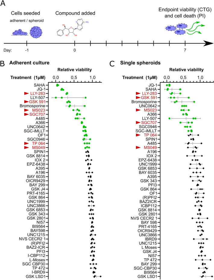
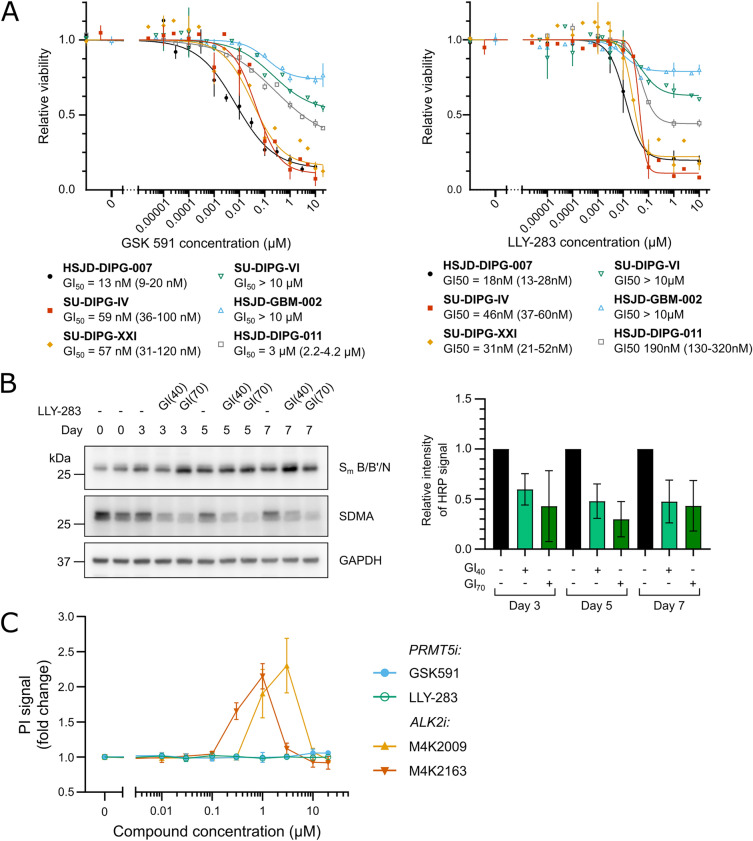
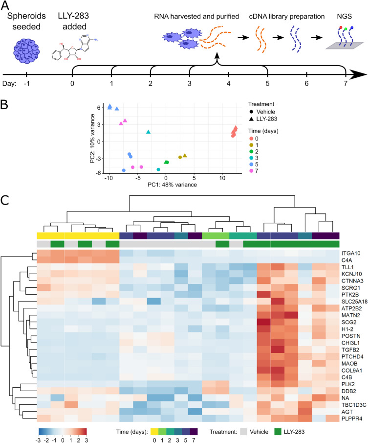
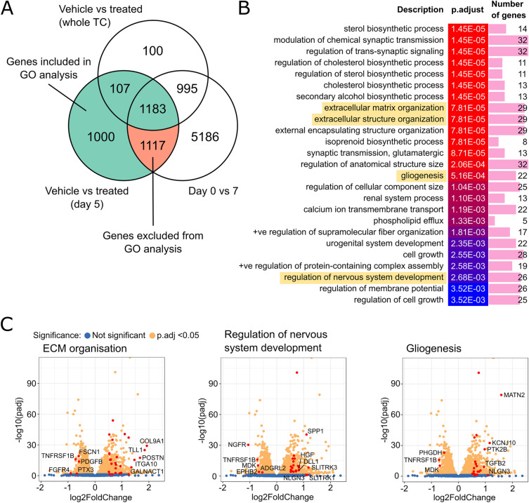
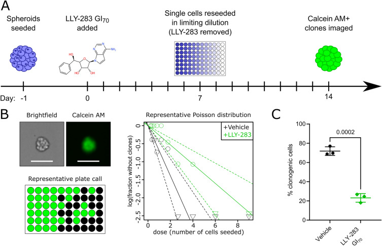
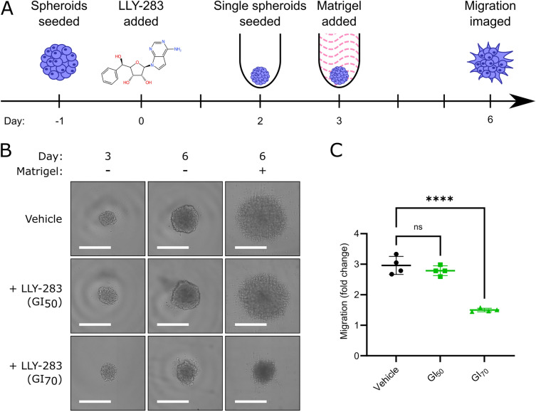
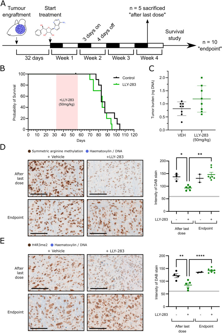
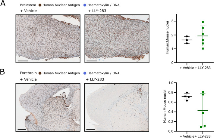
H3K27-altered Diffuse Midline Glioma (DMG) is a universally fatal paediatric brainstem tumour. The prevalent driver mutation H3K27M creates a unique epigenetic landscape that may also establish therapeutic vulnerabilities to epigenetic inhibitors. However, while HDAC, EZH2 and BET inhibitors have proven somewhat effective in pre-clinical models, none have translated into clinical benefit due to either poor blood-brain barrier penetration, lack of efficacy or toxicity. Thus, there remains an urgent need for new DMG treatments. Here, we performed wider screening of an epigenetic inhibitor library and identified inhibitors of protein arginine methyltransferases (PRMTs) among the top hits reducing DMG cell viability. Two of the most effective inhibitors, LLY-283 and GSK591, were targeted against PRMT5 using distinct binding mechanisms and reduced the viability of a subset of DMG cells expressing wild-type TP53 and mutant ACVR1. RNA-sequencing and phenotypic analyses revealed that LLY-283 could reduce the viability, clonogenicity and invasion of DMG cells in vitro, representing three clinically important phenotypes, but failed to prolong survival in an orthotopic xenograft model. Together, these data show the challenges of DMG treatment and highlight PRMT5 inhibitors for consideration in future studies of combination treatments.


Conflict of interest statement

The authors declare no competing interests.

Figures

Figure 1
Figure 1
An epigenetic probe screen identified the PRMT family as necessary for DMG viability. (A) An overview of the method for viability screening of the epigenetic probe set. HSJD-DIPG-007 cells were seeded into one of two screens (either to grow adherently or as spheroids). The following day each inhibitor was added to a final concentration of 1 µM, alongside a DMSO vehicle control. After 7 days of treatment, the relative viability was measured with the CellTiter-Glo assay. (B,C) Relative viability of HSJD-DIPG-007 adherent cell cultures after 7 days of growth in the presence of 1 µM of the indicated epigenetic inhibitor. Data points are the mean of 3 (B) or 4 (C) technical repeats annotated with the SD. Data points for probes that significantly reduced HSJD-DIPG-007 viability are depicted as green circles (multiple two-tailed t-tests, FDR = 1%). The names of PRMT family probes are highlighted in red and marked with a triangle.
Figure 2
Figure 2
PRMT5 inhibition potently reduces the viability of multiple DMG cell lines. (A) The relative viability of DMG single spheroids formed from the indicated DMG cell line* after a 7 day treatment with the PRMT5 inhibitors GSK591 (left) or LLY-283 (right). GI50 values were interpolated from each growth inhibition curve. (* except for SU-DIPG-IV which grows as a monolayer). (B) Left, the dimethylation of Sm B/B’/N after treatment with the 40% growth inhibitory concentration (GI40) of LLY-283 visualised by immunoblot (representative of 3 independent repeats). The cropped images span the entire width of the membrane and retain all samples included on the gel. To allow visualisation of multiple post translational modifications and proteins of different molecular weights, membranes were stripped and re-stained with different antibodies as described in the methods. Uncropped Western blots are shown in Supplementary Fig. 2. Right, quantification of band intensity of all 3 independent repeats. (C) SU-DIPG-IV cells were treated with the indicated concentration of the PRMT5 inhibitors GSK591 or LLY-283, or ACVR1 inhibitors M4K2009 or M4K2163 for 7 days in the presence of 1 µg/mL PI. Fluorescence signal was normalised to the vehicle control for each compound. Data points represent the mean of 3 independent repeats ± SD.
Figure 3
Figure 3
PRMT5 inhibition altered gene expression after 3 days of treatment. (A) HSJD-DIPG-007 cells were seeded as spheroids and treated with the LLY-283 GI40 the following day. Total cellular RNA was collected after a 0, 1, 2, 3, 5, 7 or 10 days treatment with LLY-283. RNA was collected and sequenced for 3 technical repeats for days 0 and 5, 2 technical repeats for day 7 and 1 technical repeat for days 1, 2 and 3. (B) The clustering of samples according to the 1st and 2nd principal components. Gene expression changes over time and diverges after 3 days along PC2 according to treatment group. (C) The relative expression of the 25 genes with the highest PC2 weighting (scaled by row). Column clustering shows the similarity between day 0 and vehicle samples, and the differential expression within later treated samples.
Figure 4
Figure 4
PRMT5 inhibition alters ECM organisation and differentiation of DMG cells. (A) The overlap between the genes differentially expressed between treatment groups for day 5 samples only, between treatment groups over the whole time course, or between day 0 and day 7. (B) The top 25 significant gene ontology (GO) terms enriched in the list of genes that were differentially expressed between treatment groups at day 5 (excluding those that were differentially expressed between days 0 and 7, but not the full time course. Similar terms have been removed). Terms selected for further validation are highlighted in yellow. (C) Volcano plots of differentially expressed genes at day 5 of the LLY-283 treatment where significantly changed genes are coloured yellow (padj < 0.05). Genes associated with the indicated GO terms are highlighted in red.
Figure 5
Figure 5
PRMT5 inhibition impairs clonogenicity of DMG cells. (A) HSJD-DIPG-007 cells were seeded as loose spheroids. The following day the LLY-283 GI70 was added to the growth medium. After a 7-day treatment with LLY-283 GI70 spheroids were split and re-seeded as single cells in a titration (6 technical repeats per seeding density) which allows derivation of the fraction of clonogenic cells in the population. After 7 days calcein AM was added to the growth medium and viable clones were imaged. (B) Left to right: Representative brightfield and fluorescent images of a HSJD-DIPG-007 clone, an overview of calls (green where at least one clone is present) of one 96 well plate and a Poisson distribution calculated from one independent limiting dilution assay. (C) The percentage of clonogenic cells, + /− a 7-day pre-treatment with LLY-283 GI70. Data points are the estimated stem cell fraction from each independent repeat, annotated with mean and SD. Statistical significance (p = 0.0002) was calculated by unpaired t-test.
Figure 6
Figure 6
PRMT5 inhibition reduced invasion of DMG cells into Matrigel (A) HSJD-DIPG-007 cells were seeded as loose spheroids and the following day were treated with a vehicle control, the LLY-283 GI40 or GI70. After 2 days HSDJ-DIPG-007 cells were dissociated, counted and re-seeded into ultra-low-attachment U-bottomed 384 well plates (generating single spheroids) in medium containing the same concentration of vehicle/LLY-283. The following day half of the spheroids were embedded in 4.65 mg/mL Matrigel (again maintaining vehicle/LLY-283) and the other half remained as controls for undisturbed spheroid growth. Invasion into Matrigel was imaged by brightfield after a further 3 days for a total treatment period of 6 days. (B) Representative images of spheroids before Matrigel addition (day 3) and at 3 days + /− Matrigel addition (day 6) showing characteristic outward invasion of HSJD-DIPG-007 cells. Scale bars represent 500 µm. (C) Quantification of HSJD-DIPG-007 invasion into the surrounding Matrigel expressed as the fold change in overhead cross-sectional area compared to the without Matrigel spheroid growth control. ****p < 0.0001 by ordinary one-way ANOVA with Dunnett’s multiple comparisons test.
Figure 7
Figure 7
PRMT5 inhibition did not improve survival in a patient dervied xenograft model of DMG (A) Athymic nude mice were engrafted with HSJD-DIPG-007 cells. Tumours were allowed to initiate for 32 days before vehicle (n = 15) or LLY-283 (n = 15) was administered at 50 mg/kg by oral gavage, 3 days on, 4 days off for 4 weeks. After 4 weeks 5 mice from each group were sacrificed, and the remaining were used to measure survival (endpoint criteria: 20% weight loss due to disease). (B) Survival of mice after 4 week treatment with vehicle or LLY-283. The treatment period is highlighted in red. Median survival for each group was compared by log-rank (Mantel-Cox test, p = 0.2) (C) Tumour burden as indicated by the amount of R206H ACVR1 DNA present in the brains of mice sacrificed immediately after administration of the final LLY-283 dose. (D) Left: Representative images of mouse brainstems harvested after the last dose of LLY-283 or at the endpoint, + /− 50 mg/kg LLY-283 that have been stained for symmetric arginine dimethylation (D) or H4R3me2 (E) (counterstained with haematoxylin). Scale bars represent 100 µm. Right: Quantification of the intensity of DAB staining in the nuclei within the brainstem. Dashed line indicates the intensity of staining in the IgG isotype control. Significance was determined by one way ANOVA with Tukey’s multiple comparison test (* p < 0.05; ** p < 0.01; **** p < 0.0001).
Figure 8
Figure 8
PRMT5 inhibition impaired migration of DMG cells into the mouse forebrain. Left: Representative images of sectioned mouse brainstems (A) or forebrains (B) harvested immediately after the final dose of vehicle/LLY-283 (50 mg/kg) immunohistochemically stained for human nuclear antigen (counterstained with haematoxylin). Scale bars represent 500 µm. Right: Quantification of the ratio of human:mouse cells in the brainstem (A) or forebrain (B). Each data point represents one field of view from each biological repeat and each treatment group is annotated with the mean and SD.

Similar articles

Cited by

References

    1. Pritchard-Jones K. Childhood cancer in Britain: Incidence, survival and mortality. Br. J. Cancer. 2007;96(12):1927. doi: 10.1038/SJ.BJC.6603800. - DOI
    1. Wong JF, Brown EJ, Williams E, Bullock AN. Fostering open collaboration in drug development for paediatric brain tumours. Biochem. Soc. Trans. 2019;47(5):1471–1479. doi: 10.1042/BST20190315. - DOI - PMC - PubMed
    1. Prajapati HP, Kannaujia SK. A simplified overview of the World Health Organization classification of central nervous system tumors 2021. Surg. Neurol. Int. 2022;13:252. doi: 10.25259/SNI_102_2022. - DOI - PMC - PubMed
    1. Jansen MHA, van Vuurden DG, Vandertop WP, Kaspers GJL. Diffuse intrinsic pontine gliomas: A systematic update on clinical trials and biology. Cancer Treat Rev. 2012;38(1):27–35. doi: 10.1016/J.CTRV.2011.06.007. - DOI - PubMed
    1. Caretti V, et al. Subventricular spread of diffuse intrinsic pontine glioma. Acta Neuropathol. 2014;128(4):605–607. doi: 10.1007/s00401-014-1307-x. - DOI - PMC - PubMed

Substances