. 2024 Jan 3;14(1):425.
doi: 10.1038/s41598-023-50710-3.

Mesenchymal-epithelial transition and AXL inhibitor TP-0903 sensitise triple-negative breast cancer cells to the antimalarial compound, artesunate

Affiliations

Mesenchymal-epithelial transition and AXL inhibitor TP-0903 sensitise triple-negative breast cancer cells to the antimalarial compound, artesunate

Mirko Terragno et al. Sci Rep. .

Abstract

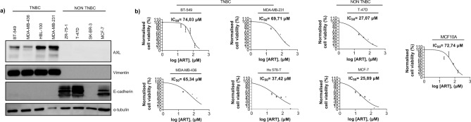
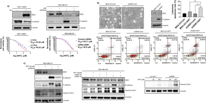
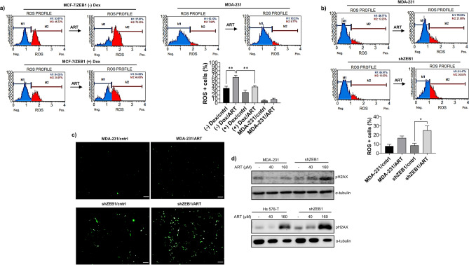
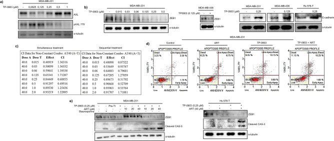
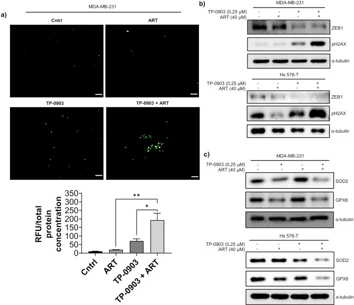
Triple-negative breast cancer (TNBC) is a difficult-to-treat, aggressive cancer type. TNBC is often associated with the cellular program of epithelial-mesenchymal transition (EMT) that confers drug resistance and metastasis. EMT and reverse mesenchymal-epithelial transition (MET) programs are regulated by several signaling pathways which converge on a group of transcription factors, EMT- TFs. Therapy approaches could rely on the EMT reversal to sensitise mesenchymal tumours to compounds effective against epithelial cancers. Here, we show that the antimalarial ROS-generating compound artesunate (ART) exhibits higher cytotoxicity in epithelial than mesenchymal breast cancer cell lines. Ectopic expression of EMT-TF ZEB1 in epithelial or ZEB1 depletion in mesenchymal cells, respectively, reduced or increased ART-generated ROS levels, DNA damage and apoptotic cell death. In epithelial cells, ZEB1 enhanced expression of superoxide dismutase 2 (SOD2) and glutathione peroxidase 8 (GPX8) implicated in ROS scavenging. Although SOD2 or GPX8 levels were unaffected in mesenchymal cells in response to ZEB1 depletion, stable ZEB1 knockdown enhanced total ROS. Receptor tyrosine kinase AXL maintains a mesenchymal phenotype and is overexpressed in TNBC. The clinically-relevant AXL inhibitor TP-0903 induced MET and synergised with ART to generate ROS, DNA damage and apoptosis in TNBC cells. TP-0903 reduced the expression of GPX8 and SOD2. Thus, TP-0903 and ZEB1 knockdown sensitised TNBC cells to ART, likely via different pathways. Synergistic interactions between TP-0903 and ART indicate that combination approaches involving these compounds can have therapeutic prospects for TNBC treatment.


Conflict of interest statement

The authors declare no competing interests.

Figures

Figure 1
Figure 1
TNBC cell lines were more resistant to the treatment with ART. (a) Characterisation of breast cancer cell lines. Immunoblots show expression of mesenchymal proteins vimentin and AXL and epithelial marker E-cadherin in breast cancer cell lines. While TNBC-derived cell lines expressed AXL and vimentin, non-TNBC-derived cells expressed epithelial marker E-cadherin. In HER + cell line SK-BR3, E-cadherin-encoding CDH1 gene carries a large homozygous deletion. Anti- Tubulin antibody was used to confirm equal loading; (b) MTS assay (BT-549) and MTT assay (all remaining cell lines) demonstrated that IC50 values for ART were higher in mesenchymal cells (between 37.42 μM and 74.03 μM) than in MCF-7 or T-47D cell lines (25.89 μM and 27.07 μM respectively). Note that MCF-10A cells displayed higher resistance to ART than most breast cancer cell lines. ART IC50 for MCF-7 and MDA-MB-231 are extracted from the MTT data on MCF- 7/ZEB1 (-) Dox and MDA-MB-231 siRNA cntrl presented in Fig. 2a. MTT results are expressed as mean ± SEM of six technical replicates (except for MCF-10A analysed in triplicate) while MTS results are expressed as mean ± SEM of three technical replicates. IC50 values were determined using MTT assay as described in Materials and Methods. In the immunoblots shown in figure a, the membranes were cut into fragments containing proteins of known molecular weights before incubating with the antibodies. This also applies to the blots presented in Figs. 2, 3d, 4a,b,e, 5b,c.
Figure 2
Figure 2
Differentiation status of breast cancer cells determined their response to ART treatment. (a) EMT was induced by ectopic expression of ZEB1 in MCF-7 cells (left panel) or reverted by ZEB1 depletion in MDA-231 cells (right panel). Western blots demonstrate that modulation ZEB1 expression affected E-cadherin levels. Whereas inducing EMT in MCF-7 cells increased viability of ART-treated cells, activating partial MET in MDA-231 cells by ZEB1 KD sensitized cells to ART. IC50 values were determined using MTT assay as described in Materials and Methods. MTT results are expressed as mean ± SEM of six technical replicates; (b) Reduced viability of epithelioid cells in response to ART associated with increased rate of apoptosis. shRNA-mediated depletion of ZEB1 in MDA-MB-231 cells led to a partial MET as evidenced by the analysis of cell morphology and reactivation of P-cadherin. ZEB1 depletion increases the proportion of Annexin V- positive cells treated with 160 μM ART for 48 h. Graph shows results of four independent measurements (mean ± SEM). A one-way Anova test followed by a post hoc Tukey’s multiple comparisons test were used to check significance among groups; *p < 0.05; ** p < 0.01; *** p < 0.001; (c) ZEB1 depletion in mesenchymal breast cancer cell lines resulted in the cleavage of caspase-3. ZEB1 KD was carried out by siRNA (in MDA-MB-231 or MDA-MB-436 cells) or using shRNA (in Hs 587-T cells) and expression of ZEB1 cleaved caspase-3 and cadherins was analysed by immunoblotting as indicated. Tubulin was used as a loading control.
Figure 3
Figure 3
ZEB1 expression impact on ART-generated ROS levels and DNA damage. (a) ZEB1-induced EMT reduced the proportion of ROS-positive cells induced by ART treatment. Treatment of MDA-MB-231 cells with ART in these experiments illustrated low levels of ROS generation in ART- resistant cells; (b) ZEB1 depletion in MDA-MB-231 cells increased the ROS production in ART-treated or untreated cells. (a, b) Proportion of ROS-positive cells was determined using Muse Oxidative assay which detects superoxide anion O2-. Blue and red peaks reflect proportions of ROS- negative and ROS-containing cells, respectively. The experiments demonstrated that ZEB1 expression reduced proportion of superoxide positive cells in response to ART treatment. Graphs show the results expressed as mean ± SEM of three (a) and four (b) independent experiments. A one- way Anova test followed by a post hoc Tukey’s multiple comparisons test were used to check significance among groups; * p < 0.05; ** p < 0.01; *** p < 0.001; (c) MDA-MB-231 control or ZEB1 KD cells were treated with ART or left untreated. The amount of cells with high general oxidant levels was assessed using the fluorescent reporter CM-H2DCFDA; scale bar 100 μm; (d) ZEB1 protected DNA from ART-induced DNA damage. Control or ZEB1-depleted MDA-MB-231 or Hs 578-T cells were treated with the indicated concentrations of ART and the level of DNA damage evaluated using an anti-gH2AX antibody.
Figure 4
Figure 4
AXL inhibitor TP-0903 promoted MET and potentiated pro-apoptotic effects of ART. (a) TP-0903 effectively inhibited AXL phosphorylation. Total and phosphorylated AXL protein was detected by immunoblotting in cells treated with the indicated concentration of TP-0903; (b) Prolonged treatment with TP-0903 led to the partial restoration of epithelial phenotypes in TNBC cells. MDA-MB-231, Hs 578-T or MDA-MB-436 cells were maintained with indicated concentrations of the inhibitor for 48 h and expression of ZEB1 or E-cadherin analyzed in Western blotting; (c) A representative CompuSyn report after simultaneous/sequential treatment of MDA-MB-231 cells with TP-0903 at different concentrations plus ART 40 μM for 96 h. The number of CIs values showing synergism was higher in the sequential treatment. Dose A = ART; Dose T = TP-0903; Effect = growth inhibition; CI = Combination Index. CI < 1 indicates synergism. Effect and CI were calculated after performing the MTT assay on four technical replicates; (d) MDA-MB-231 cells were cultured with or without TP-0903 0.25 μM and ART 40 μM, and apoptosis was analysed using fluorescent annexin V apoptosis assay; (e) Caspase 3 cleavage was tested in ART and TP-0903-treated cells by Western blotting with a cleaved caspase-3 specific antibody.
Figure 5
Figure 5
Combined treatment with ART and TP-0903 stimulated total ROS production, gH2AX expression and repressed SOD2 and GPX8. (a) MDA-MB-231 cells were treated with ART and TP-0903 as indicated or left untreated. Fluorescence intensity was calculated by the ImageJ software. Results are expressed as mean ± SEM of three different microscopic fields. One-way Anova test followed by a post hoc Tukey’s multiple comparisons test were used to analyse significance between control and treatment groups. * p < 0.05; ** p < 0.01; *** p < 0.001; scale bar 100 μm; (b, c) Expression of indicated proteins was analysed by Western blotting as shown.

References

    1. Andreopoulou E, et al. Therapies for triple negative breast cancer. Expert Opin. Pharmacother. 2015;16:983–998. doi: 10.1517/14656566.2015.1032246. - DOI - PMC - PubMed
    1. De Ruijter TC, et al. Characteristics of triple-negative breast cancer. J. Cancer Res. 2011;137:183–192. doi: 10.1007/s00432-010-0957-x. - DOI - PMC - PubMed
    1. Lehmann B, et al. Identification of human triple-negative breast cancer subtypes and preclinical models for selection of targeted therapies. J. Clin. Invest. 2011;121:2750–67. doi: 10.1172/JCI45014. - DOI - PMC - PubMed
    1. Wang DY, et al. Molecular stratification within triple-negative breast cancer subtypes. Sci. Rep. 2019;9:19107. doi: 10.1038/s41598-019-55710-w. - DOI - PMC - PubMed
    1. Casalino L, et al. FRA-1 as a Regulator of EMT and Metastasis in Breast Cancer. Int. J. Mol. Sci. 2023;24:8307. doi: 10.3390/ijms24098307. - DOI - PMC - PubMed

MeSH terms