. 2024 Jan 3;14(1):443.
doi: 10.1038/s41598-024-51198-1.

Distolingual root prevalence in mandibular first molar and complex root canal morphology in incisors: a CBCT analysis in Indian population

Affiliations

Distolingual root prevalence in mandibular first molar and complex root canal morphology in incisors: a CBCT analysis in Indian population

Komal Sheth et al. Sci Rep. .

Abstract

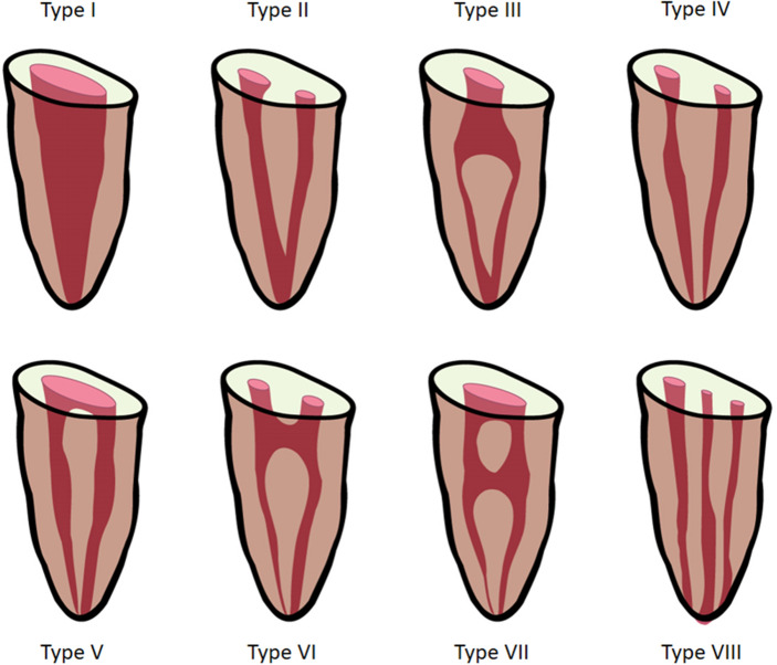
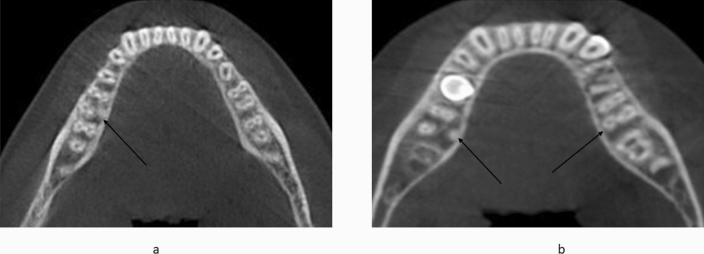
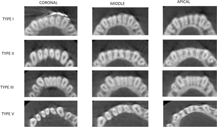
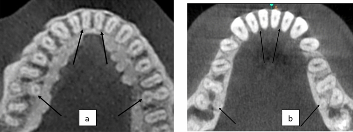
Cone-beam computed tomography was used to understand the possible correlation between the prevalence of distolingual root (DLR) in permanent mandibular first molars (MFMs) and the associated complicated mandibular incisor's root canal morphology (MIs) in an Indian population. A total of 400 scans were evaluated for MFMs and MIs. The prevalence of DLRs and root canal anatomy of MIs were assessed based on Vertucci's classification, and then the sample were grouped according to age, sex and side. Statistical analysis was used to evaluate the possible correlation between the presence of DLRs in the first molar and root canal morphology of incisors. Chi square test was used to evaluate the correlation between the root canal configurations of MIs with the existence of DLRs in MFMs. There was no statistically significant difference between sexes or ages for the prevalence of DLRs in the first molars (p > 0.05), which was 6.62%, with the right side having a greater frequency of DLRs (7.8%) than the left (5.5%). Vertucci Type I canal configuration was most common for the mandibular central (66.75%) and lateral incisors (58.62%). Vertucci Type III was the most common complicated canal morphology, followed by Types V, II, and IV for MIs, with no statistically significant difference in the studied sample's age and sex. (p < 0.05). No association was observed between the presence of DLRs in first molars and complicated root canal configurations in MIs. Taken together, the possibility of complicated root canal configuration in MIs was lesser in the presence of DLRs in MFMs among the Indian population.


Conflict of interest statement

The authors declare no competing interests.

Figures

Figure 1
Figure 1
Carlsen and Alexandersen’s classification of radix entomolaris (RE) based on its location of cervical part. (a) Type A: the RE is located lingually to the distal root complex which has two cone-shaped macrostructures. (b) Type B: the RE is located lingually to the distal root complex which has one cone-shaped macrostructures. (c) Type C: the RE is located lingually to the mesial root complex. (d) Type AC: the RE is located lingually between the mesial and distal root complexes.
Figure 2
Figure 2
Ribeiro and Consolaro’s classification of Radix entomolaris based on its bucco-lingual orientation. (a) Type 1: a straight root or root canal. (b) Type 2: a curved coronal third which becomes straighter in the middle and apical third. (c) Type 3: an initial curve in the coronal third with a second buccally oriented curve which begins in the middle or apical third.
Figure 3
Figure 3
Vertucci’s root canal configuration from pulp chamber to the root apex.
Figure 4
Figure 4
CBCT scans showing DLR in MFMs through various sections from coronal to apical. (a) Sagittal section of mandibular first molar. (b) Coronal third section of the axial view. (c) Middle third section of the axial view. (d) Apical third section of the axial view.
Figure 5
Figure 5
CBCT images showing (a) Unilateral and (b) Bilateral distribution of DLR in MFMs.
Figure 6
Figure 6
CBCT scans showing root canal morphology according to Vertucci’s classification in Mandibular incisors.
Figure 7
Figure 7
Axial section of CBCT scans showing a) Complicated and b) Simple root canal configuration of Mandibular Incisors in presence of DLR in MFMs.

References

    1. Chandra SS, Chandra S, Shankar P, Indira R. Prevalence of radix entomolaris in mandibular permanent first molars: A study in a South Indian population. Oral Surg. Oral Med. Oral Pathol. Oral Radiol. Endodontol. 2011;112(3):e77–82. doi: 10.1016/j.tripleo.2011.02.016. - DOI - PubMed
    1. Calberson FL, De Moor RJ, Deroose CA. The radix entomolaris and paramolaris: Clinical approach in endodontics. J. Endod. 2007;33(1):58–63. doi: 10.1016/j.joen.2006.05.007. - DOI - PubMed
    1. Kokate SR, Pawar AM, Hegde VR. Contemporary approach in successful endodontic intervention in ‘Radix Entomolaris’. World J. Dent. 2013;4(3):208–213. doi: 10.5005/jp-journals-10015-1233. - DOI
    1. Carlsen OL, Alexandersen V. Radix entomolaris: Identification and morphology. Eur. J. Oral Sci. 1990;98(5):363–373. doi: 10.1111/j.1600-0722.1990.tb00986.x. - DOI - PubMed
    1. Corzon ME. Miscegenation and the prevalence of three-rooted mandibular first molars in the Baffin Eskimo. Commun. Dent. Oral Epidemiol. 1974;2(3):130–131. doi: 10.1111/j.1600-0528.1974.tb01669.x-i1. - DOI - PubMed

MeSH terms