

SARS-CoV and SARS-CoV-2 display limited neuronal infection and lack the ability to transmit within synaptically connected axons in stem cell-derived human neurons
- PMID: 38172412
- PMCID: PMC11035468
- DOI: 10.1007/s13365-023-01187-3
SARS-CoV and SARS-CoV-2 display limited neuronal infection and lack the ability to transmit within synaptically connected axons in stem cell-derived human neurons
Abstract
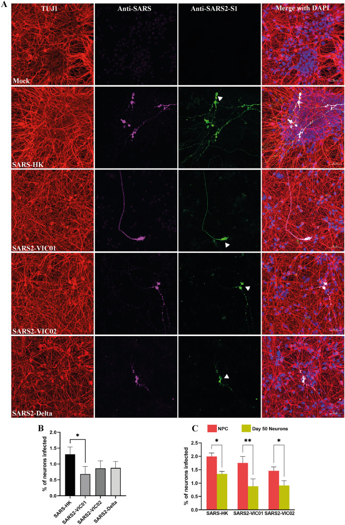
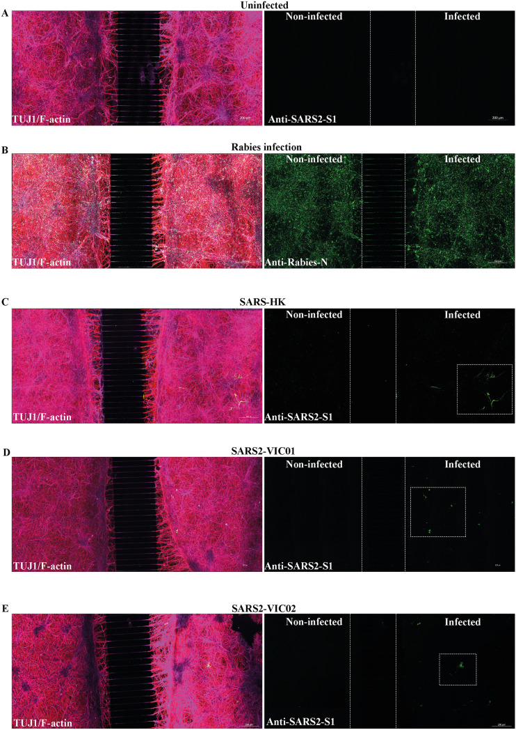
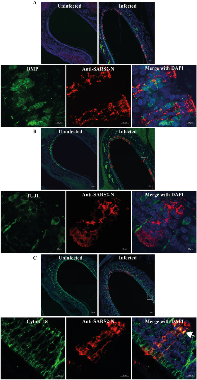
Sarbecoviruses such as SARS and SARS-CoV-2 have been responsible for two major outbreaks in humans, the latter resulting in a global pandemic. While sarbecoviruses primarily cause an acute respiratory infection, they have been shown to infect the nervous system. However, mechanisms of sarbecovirus neuroinvasion and neuropathogenesis remain unclear. In this study, we examined the infectivity and trans-synaptic transmission potential of the sarbecoviruses SARS and SARS-CoV-2 in human stem cell-derived neural model systems. We demonstrated limited ability of sarbecoviruses to infect and replicate in human stem cell-derived neurons. Furthermore, we demonstrated an inability of sarbecoviruses to transmit between synaptically connected human stem cell-derived neurons. Finally, we determined an absence of SARS-CoV-2 infection in olfactory neurons in experimentally infected ferrets. Collectively, this study indicates that sarbecoviruses exhibit low potential to infect human stem cell-derived neurons, lack an ability to infect ferret olfactory neurons, and lack an inbuilt molecular mechanism to utilise retrograde axonal trafficking and trans-synaptic transmission to spread within the human nervous system.
Keywords: Axonal trafficking; Neurotropism; SARS; SARS-CoV-2; Sarbecovirus; Stem cell–derived neurons; Trans-synaptic transmission.
© 2023. The Author(s).
Conflict of interest statement
The authors declare no competing interests.
Figures
Similar articles
-
Replication Kinetics, Cell Tropism, and Associated Immune Responses in SARS-CoV-2- and H5N1 Virus-Infected Human Induced Pluripotent Stem Cell-Derived Neural Models.mSphere. 2021 Jun 30;6(3):e0027021. doi: 10.1128/mSphere.00270-21. Epub 2021 Jun 23. mSphere. 2021. PMID: 34160239 Free PMC article.
-
Human Pluripotent Stem Cell-Derived Neural Cells and Brain Organoids Reveal SARS-CoV-2 Neurotropism Predominates in Choroid Plexus Epithelium.Cell Stem Cell. 2020 Dec 3;27(6):937-950.e9. doi: 10.1016/j.stem.2020.09.016. Epub 2020 Sep 21. Cell Stem Cell. 2020. PMID: 33010822 Free PMC article.
-
Microglia Do Not Restrict SARS-CoV-2 Replication following Infection of the Central Nervous System of K18-Human ACE2 Transgenic Mice.J Virol. 2022 Feb 23;96(4):e0196921. doi: 10.1128/jvi.01969-21. Epub 2021 Dec 22. J Virol. 2022. PMID: 34935438 Free PMC article.
-
Infectivity, virulence, pathogenicity, host-pathogen interactions of SARS and SARS-CoV-2 in experimental animals: a systematic review.Vet Res Commun. 2020 Nov;44(3-4):101-110. doi: 10.1007/s11259-020-09778-9. Epub 2020 Jul 10. Vet Res Commun. 2020. PMID: 32651761 Free PMC article.
-
Development of variant-proof severe acute respiratory syndrome coronavirus 2, pan-sarbecovirus, and pan-β-coronavirus vaccines.J Med Virol. 2023 Jan;95(1):e28172. doi: 10.1002/jmv.28172. Epub 2022 Oct 6. J Med Virol. 2023. PMID: 36161303 Free PMC article. Review.
Cited by
-
Neuroimmune pathophysiology of long COVID.Psychiatry Clin Neurosci. 2025 Jun 19:10.1111/pcn.13855. doi: 10.1111/pcn.13855. Online ahead of print. Psychiatry Clin Neurosci. 2025. PMID: 40536011 Free PMC article.
-
Inflammatory Response and Defects on Myelin Integrity in the Olfactory System of K18hACE2 Mice Infected with SARS-CoV-2.eNeuro. 2024 Jun 17;11(6):ENEURO.0106-24.2024. doi: 10.1523/ENEURO.0106-24.2024. Print 2024 Jun. eNeuro. 2024. PMID: 38834299 Free PMC article.
-
The Neurological Implications of COVID-19: A Comprehensive Narrative Review.Cureus. 2024 May 15;16(5):e60376. doi: 10.7759/cureus.60376. eCollection 2024 May. Cureus. 2024. PMID: 38887342 Free PMC article. Review.
References
Publication types
MeSH terms
Grants and funding
LinkOut - more resources
Full Text Sources
Medical
Miscellaneous
