. 2024 Feb;56(1):142-155.
doi: 10.1038/s12276-023-01125-7. Epub 2024 Jan 4.

Vitamin D ameliorates age-induced nonalcoholic fatty liver disease by increasing the mitochondrial contact site and cristae organizing system (MICOS) 60 level

Affiliations

Vitamin D ameliorates age-induced nonalcoholic fatty liver disease by increasing the mitochondrial contact site and cristae organizing system (MICOS) 60 level

Gyu Hee Kim et al. Exp Mol Med. 2024 Feb.

Abstract

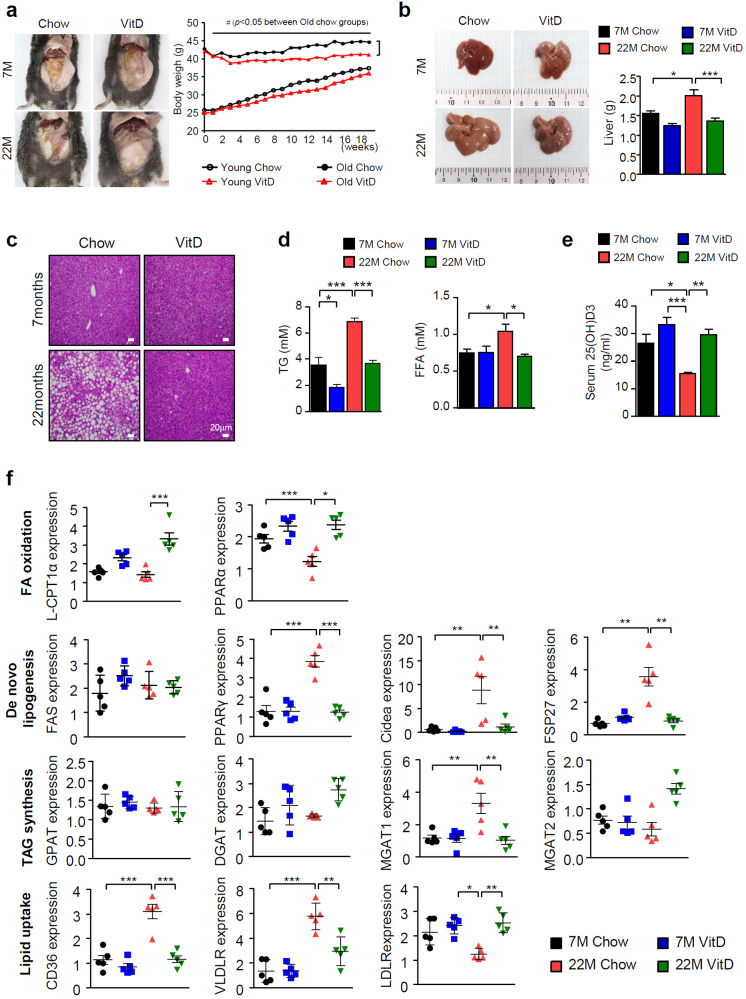
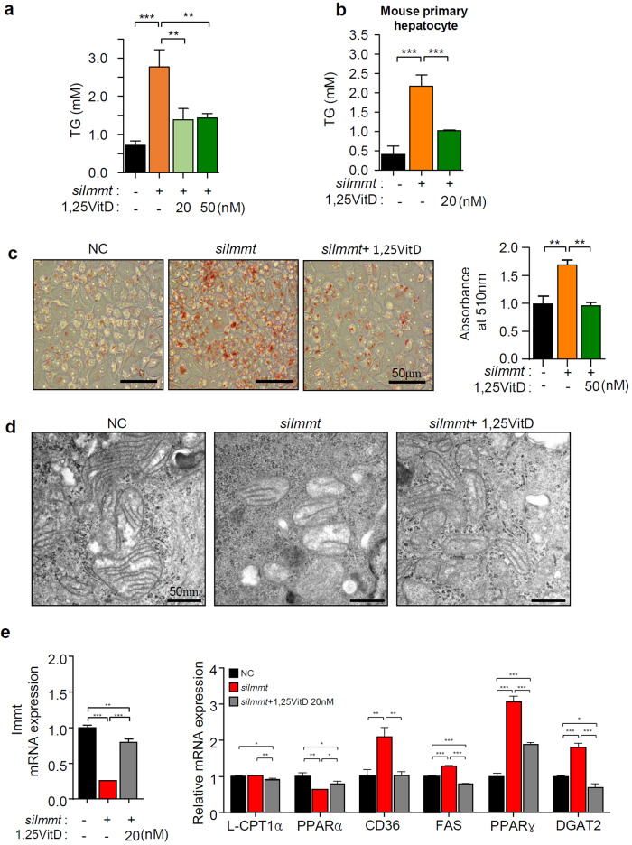
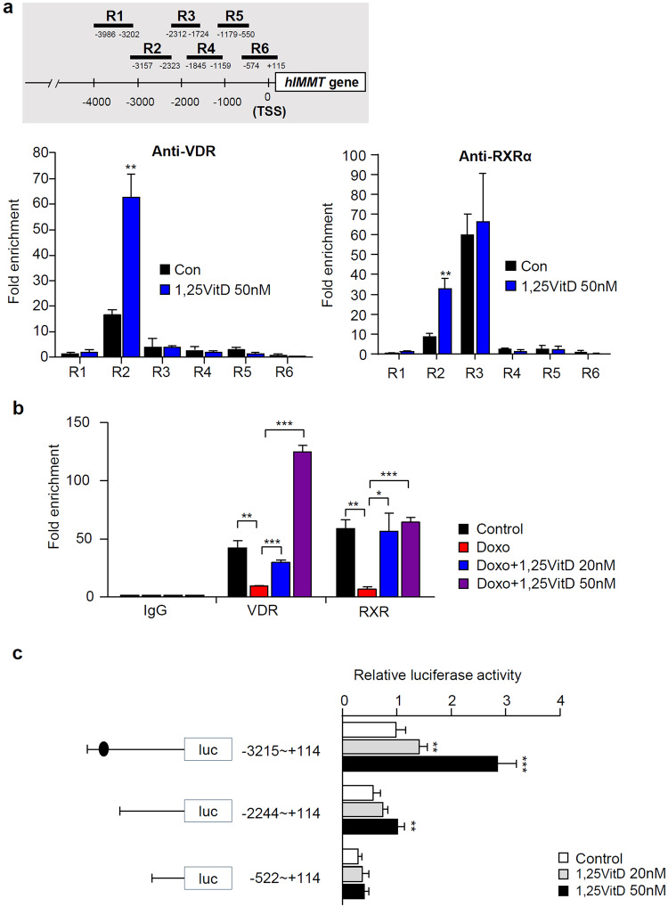
Nonalcoholic fatty liver disease (NAFLD) is the most common liver disease. Despite intensive research, considerable information on NAFLD development remains elusive. In this study, we examined the effects of vitamin D on age-induced NAFLD, especially in connection with mitochondrial abnormalities. We observed the prevention of NAFLD in 22-month-old C57BL/6 mice fed a vitamin D3-supplemented (20,000 IU/kg) diet compared with mice fed a control (1000 IU/kg) diet. We evaluated whether vitamin D3 supplementation enhanced mitochondrial functions. We found that the level of mitochondrial contact site and cristae organizing system (MICOS) 60 (Mic60) level was reduced in aged mice, and this reduction was specifically restored by vitamin D3. In addition, depletion of Immt, the human gene encoding the Mic60 protein, induced changes in gene expression patterns that led to fat accumulation in both HepG2 and primary hepatocytes, and these alterations were effectively prevented by vitamin D3. In addition, silencing of the vitamin D receptor (VDR) decreased the Mic60 levels, which were recovered by vitamin D treatment. To assess whether VDR directly regulates Mic60 levels, we performed chromatin immunoprecipitation and reporter gene analysis. We discovered that VDR directly binds to the Immt 5' promoter region spanning positions -3157 to -2323 and thereby upregulates Mic60. Our study provides the first demonstration that a reduction in Mic60 levels due to aging may be one of the mechanisms underlying the development of aging-associated NAFLD. In addition, vitamin D3 could positively regulate Mic60 expression, and this may be one of the important mechanisms by which vitamin D could ameliorate age-induced NAFLD.


Conflict of interest statement

The authors declare no competing interests.

Figures

Fig. 1
Fig. 1. Vitamin D3 specifically prevents the development of NAFLD in aged mice.
a Body weight changes in 3 month- or 18-month-old mice during the 18-week study period (n = 10–12). b The weight of livers extracted from mice as indicated. c Representative images of H&E stained liver tissue sections of 7- or 22-month-old mice. Scale bar = 30 μm. d Tissue TG and serum FFA levels were measured using ELISA kits according to the manufacturer’s instructions. Each value was normalized to that of the protein concentration (n = 5). e Serum vitamin D3 levels (n = 5). f Quantitative real-time PCR of genes related to lipid metabolism (n = 5). Statistical analyses were performed via one-way ANOVA with Tukey’s post hoc test for multiple comparisons; *p < 0.05, **p < 0.01, ***p < 0.001.
Fig. 2
Fig. 2. Alterations in MICOS and mitochondrial dynamics-associated protein levels in aged mice.
Changes in the levels of proteins involved in either mitochondrial dynamics (a) or MQC (b) (n = 3). Relative values compared to the value of young mice fed a chow diet are presented as the means ± SEMs (n = 3). c Protein levels of MICOS subunits (left panel) and quantitative real-time PCR analysis of MICOS subunits (right panel, n = 5). d Immunofluorescence images of mouse liver tissue obtained using an anti-Mic60 antibody as indicated. Scale bars = 50 μm. Statistical analyses were performed via one-way ANOVA with Tukey’s post hoc test for multiple comparisons; *p < 0.05, **p < 0.01.
Fig. 3
Fig. 3. The knockdown of Mic60 specifically leads to TG accumulation.
a Immunoblot analysis after transfection of individual siRNAs as indicated. b Alterations in the protein levels of MICOS proteins and proteins involved in mitochondrial dynamics after transfection of siImmt or siLonP1 are shown. c Immunoblots showing the Sam50 and SLC25A46 levels in the liver tissue obtained from the animals as indicated (n = 3). d The TG level in HepG2 cells was measured with a TG assay kit following the manufacturer’s instruction (n = 3). e Genes related to lipid metabolism was measured by qPCR after the transfection of either siImmt or siLonP1. Statistical analyses were performed via one-way ANOVA with Tukey’s post hoc test for multiple comparisons; *p < 0.05, **p < 0.01, ***p < 0.001 (n = 3). Alterations in TG accumulation (f) and the expression of genes related to lipid metabolism (g) after the transfection of each siRNA into primary hepatocytes purified from mice were analyzed as described in Materials and Methods.
Fig. 4
Fig. 4. Mic60 expression is correlated with Vitamin D level.
HepG2 cells (a) or mouse primary hepatocyte cells (b) were treated with 1,25VitD3 24 h after siImmt transfection. Immunoblot analyses were performed using antibodies as indicated. c The mitochondrial membrane potential was measured with a JC-1 assay kit (Abcam, left panel), and cellular ATP amounts (right panel) were measured with ATP colorimetric/fluorometric assay kit (Abcam), according to the manufacturer’s instructions. d Intracellular ROS were quantified by 2’,7’-dichlorofluorescin after incubation for 20 min in serum-free media containing 10 µM dichlorofluorescin diacetate (DCF-DA, Invitrogen). e The oxygen consumption rate (OCR) in live cells was measured using Seahorse XF HS Mini (Agilent Technologies, CA, USA) according to the manufacturer’s protocol. The OCR levels were normalized to the amount of protein in each sample. f The amount of reduced GSH was determined using a colorimetric assay kit (EZ-glutathione assay kit, DG-GLU200, DogenBio, Seoul, Korea) according to the manufacturer’s instructions (reduced GSH = GSH-2GSSG).
Fig. 5
Fig. 5. Alterations in the expression levels of VDR and its related genes in aged mice.
a Changes in VDR levels in aged mice with or without vitamin D3 supplementation. Left panel: Western blot; right panel: qPCR (n = 5). b Alterations in the expression patterns of representative genes involved in VDR-mediated transcriptional networks in each animal groups (n = 5). c VDR-dependent alterations in the levels of MICOS-related proteins in HepG2 cells. Western blotting was performed 24 h after siVDR transfection.
Fig. 6
Fig. 6. Vitamin D3 supplementation prevents TG accumulation by restoring the Mic60 level.
TG levels in either HepG2 cells (a) or mouse primary hepatocyte cells (b) (n = 3). c Cellular lipid droplets were visualized by Oil red O staining (left panel) and quantified (right panel) (n = 3). d Electron microscopy images of HepG2 cells after the indicated treatment. Scale bars = 50 nm. e Recovery of the Mic60 level (left panel) and alterations in the levels of representative genes involved in lipid metabolism (right panel) in siImmt-transfected-mouse primary hepatocyte cells by 1,25VitD3 treatment.
Fig. 7
Fig. 7. Induction of cellular senescence triggered a reduction in the Mic60 level.
a Measurement of the cellular cytotoxicity of doxorubicin at different concentrations for 24 h using an MTT assay. b The MICOS protein levels were altered by increasing the concentration of doxorubicin. c Western blot analysis after induction of senescence in the presence or absence of 20 nM 1,25VitD3. d Measurement of Immt expression by qRT‒PCR (n = 5). e Analysis of the effects of Dox treatment on the mitochondrial dynamics, as determined by immunoblotting, as indicated. f Changes in TG accumulation by doxorubicin in the presence or absence of 1,25VitD3. g The silencing of Immt, but not LonP1, promoted the expression of cell senescence markers (n = 3).
Fig. 8
Fig. 8. VDR-RXR upregulated Immt expression by directly binding to the Immt gene.
a Chromatin immunoprecipitation (ChIP) assay was performed using either anti-VDR or anti-RXR antibodies as described in Materials and Methods. b ChIP of control vehicle or 500 µM doxorubicin treatment in HepG2 cells in combination with 20 nM or 50 nM 1,25VitD3 using the R2 region of the Immt promoter. The recruitment of VDR or RXRα to the position R2 ranging from −3157 to −2323 was the representative value for comparisons to the IgG control (three sets per group). c Luciferase reporter gene analysis of whole Immt (−3215 to +114) or deletion mutants from −2244 (−2244 to +114) and −522 (−522 to +114) (n = 3). Statistical analyses were performed via one-way ANOVA with Tukey’s post hoc test for multiple comparisons; *p < 0.05, **p < 0.01, ***p < 0.001.

References

    1. Maurice J, Manousou P. Non-alcoholic fatty liver disease. Clin. Med. 2018;18:245–250. doi: 10.7861/clinmedicine.18-3-245. - DOI - PMC - PubMed
    1. Ipsen DH, Lykkesfeldt J, Tveden-Nyborg P. Molecular mechanisms of hepatic lipid accumulation in non-alcoholic fatty liver disease. Cell Mol. Life Sci. 2018;75:3313–3327. doi: 10.1007/s00018-018-2860-6. - DOI - PMC - PubMed
    1. Li Y, Adeniji NT, Fan W, Kunimoto K, Torok NJ. Non-alcoholic Fatty Liver Disease and Liver Fibrosis during Aging. Aging Dis. 2022;13:1239–1251. doi: 10.14336/AD.2022.0318. - DOI - PMC - PubMed
    1. Gong Z, Tas E, Yakar S, Muzumdar R. Hepatic lipid metabolism and non-alcoholic fatty liver disease in aging. Mol. Cell Endocrinol. 2017;455:115–130. doi: 10.1016/j.mce.2016.12.022. - DOI - PubMed
    1. Barzilai N, Huffman DM, Muzumdar RH, Bartke A. The critical role of metabolic pathways in aging. Diabetes. 2012;61:1315–1322. doi: 10.2337/db11-1300. - DOI - PMC - PubMed