Review
. 2024 Feb;56(1):40-50.
doi: 10.1038/s12276-023-01137-3. Epub 2024 Jan 4.

Relevance of the endoplasmic reticulum-mitochondria axis in cancer diagnosis and therapy

Affiliations
Review

Relevance of the endoplasmic reticulum-mitochondria axis in cancer diagnosis and therapy

Garam An et al. Exp Mol Med. 2024 Feb.

Abstract

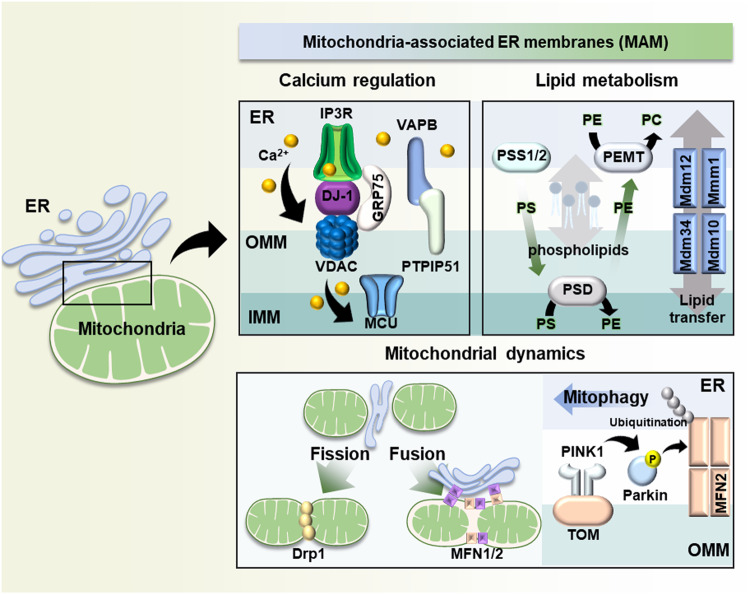
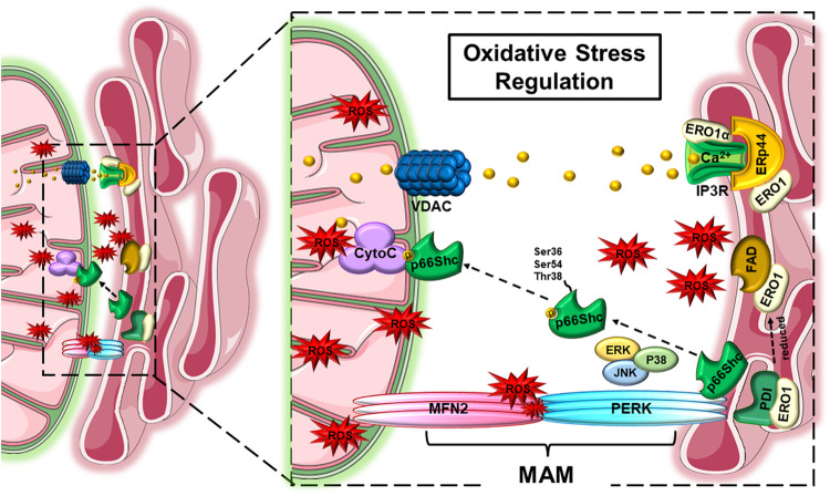
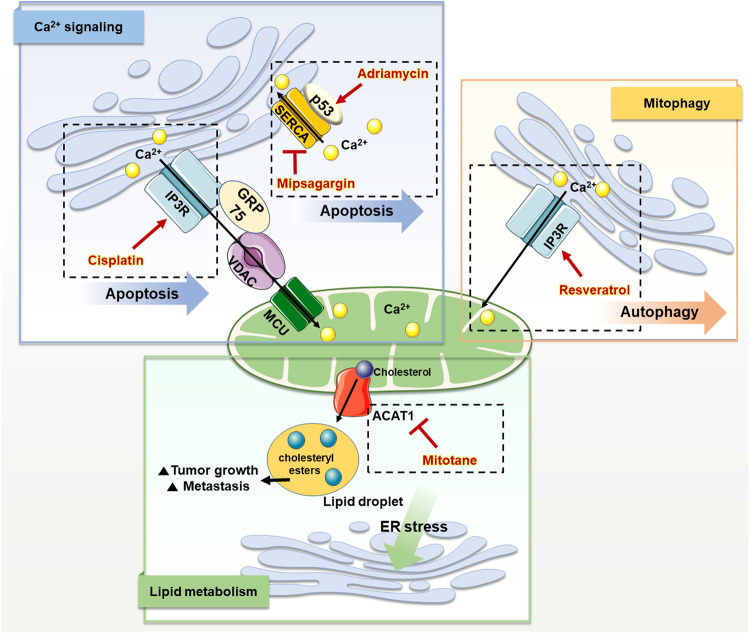
Dynamic interactions between organelles are responsible for a variety of intercellular functions, and the endoplasmic reticulum (ER)-mitochondrial axis is recognized as a representative interorganelle system. Several studies have confirmed that most proteins in the physically tethered sites between the ER and mitochondria, called mitochondria-associated ER membranes (MAMs), are vital for intracellular physiology. MAM proteins are involved in the regulation of calcium homeostasis, lipid metabolism, and mitochondrial dynamics and are associated with processes related to intracellular stress conditions, such as oxidative stress and unfolded protein responses. Accumulating evidence has shown that, owing to their extensive involvement in cellular homeostasis, alterations in the ER-mitochondrial axis are one of the etiological factors of tumors. An in-depth understanding of MAM proteins and their impact on cell physiology, particularly in cancers, may help elucidate their potential as diagnostic and therapeutic targets for cancers. For example, the modulation of MAM proteins is utilized not only to target diverse intracellular signaling pathways within cancer cells but also to increase the sensitivity of cancer cells to anticancer reagents and regulate immune cell activities. Therefore, the current review summarizes and discusses recent advances in research on the functional roles of MAM proteins and their characteristics in cancers from a diagnostic perspective. Additionally, this review provides insights into diverse therapeutic strategies that target MAM proteins in various cancer types.


Conflict of interest statement

The authors declare no competing interests.

Figures

Fig. 1
Fig. 1. Representative roles of mitochondria-associated ER membranes (MAMs).
The figure shows representative MAM proteins and their mechanisms of regulation of multiple cellular functions. (1) Calcium regulation is modulated mainly by the IP3R-GRP75-VDAC-MCU and VAPB-PTPIP51 complexes in MAMs. (2) Lipid synthesis and transfer are mediated by the enzymes PSS1/2, PSD, and PEMT and the Mdm12-Mmm1-Mdm34-Mdm10 complex. (3) Drp1 and MFN1/2 are involved in mitochondrial fission and fusion, respectively. The PINK1/Parkin pathway mediates mitophagy. ER endoplasmic reticulum, OMM outer mitochondrial membrane, IMM inner mitochondrial membrane. IP3R inositol 1,4,5-triphosphate receptor, GRP75 glucose-related regulated protein 75, VDAC voltage-dependent anion channel, MCU mitochondria calcium uniporter, VAPB vesicle-associated membrane protein B, PTPIP51 protein tyrosine phosphatase-interacting protein-51, PS phosphatidylserine, PE phosphatidylethanolamine, PC phosphatidylcholine, PSS1/2 phosphatidylserine synthase 1/2, PSD phosphatidylserine decarboxylase, PEMT phosphatidylethanolamine-N-methyltransferase, MFN1/2 mitofusin 1/2, PINK1 PTEN-induced putative kinase 1, TOM mitochondrial translocase of the outer membrane 70.
Fig. 2
Fig. 2. Mitochondria-associated ER membranes (MAMs) regulate oxidative stress.
The figure shows the regulation of reactive oxygen species (ROS) production by several proteins present in the mitochondrial ER membrane. Reduced ERO1 generates ROS through an interaction with FAD. ERO1a, one of the ERO1 isoforms, regulates ROS production through an interaction with IP3R by releasing calcium ions into mitochondria, which induces chronic ER stress. Additionally, p66Shc modulates ROS production via phosphorylation at Ser36, Ser54, and Thr38 by ERK, JNK, and P38. Finally, MFN2 regulates ROS production in a manner dependent on its expression level through an interaction with PERK. ERO1 endoplasmic reticulum oxidoreductase 1, ERp44 endoplasmic reticulum protein 44, IP3R inositol 1,4,5-triphosphate receptor, GRP75 glucose-related regulated protein 75, VDAC voltage-dependent anion channel, CytoC cytochrome c, FAD flavin adenine dinucleotide, PERK protein kinase R-like endoplasmic reticulum kinase.
Fig. 3
Fig. 3. Representative characteristics of mitochondria-associated ER membranes (MAMs) in cancer and their therapeutic targets.
The figure shows representative alterations in MAMs in cancer cells from three perspectives (Ca2+ signaling, mitophagy, and lipid metabolism) and the therapeutic drugs that target them. In cancer, the function of the IP3R-GRP75-VDAC complex is impaired, thus limiting Ca2+ trafficking to mitochondria and inducing resistance to mitochondrial apoptosis. Cisplatin targets IP3R and promotes the activity of its complex, which activates the influx of Ca2+ into mitochondria and induces apoptosis. Additionally, p53 mutations have been detected in various cancers, and these mutations result in the inhibition of Ca2+ influx into the ER and thus in cell death. Adriamycin increases p53 levels in MAMs and facilitates Ca2+ influx into the ER through SERCA, promoting apoptosis in cancer. Mipsagargin inhibits SERCA activity and increases the intracellular Ca2+ level, which can trigger cancer cell death. Resveratrol promotes Ca2+ signaling through IP3R, resulting in autophagy-induced cancer cell death. ACAT-1 generates cholesteryl esters that induce the accumulation of lipid droplets, resulting in tumor growth and metastasis. Mitotane inhibits ACAT-1 and causes free cholesterol accumulation in the ER, leading to ER stress-mediated apoptosis in cancer cells.

References

    1. Bernhard W, Rouiller C. Close topographical relationship between mitochondria and ergastoplasm of liver cells in a definite phase of cellular activity. J. Biophys. Biochem. Cytol. 1956;2:73–78. doi: 10.1083/jcb.2.4.73. - DOI - PMC - PubMed
    1. Phillips MJ, Voeltz GK. Structure and function of ER membrane contact sites with other organelles. Nat. Rev. Mol. Cell Biol. 2016;17:69–82. doi: 10.1038/nrm.2015.8. - DOI - PMC - PubMed
    1. Zhang, Y., Yao, J., Zhang, M., Wang, Y. & Shi, X. Mitochondria-associated endoplasmic reticulum membranes (MAMs): possible therapeutic targets in heart failure. 10, 10.3389/fcvm.2023.1083935 (2023). - PMC - PubMed
    1. Liu J, Yang J. Mitochondria-associated membranes: a hub for neurodegenerative diseases. Biomed. Pharmacother. 2022;149:112890. doi: 10.1016/j.biopha.2022.112890. - DOI - PubMed
    1. Cheng, H. et al. The molecular mechanisms underlying mitochondria-associated endoplasmic reticulum membrane-induced insulin resistance. 11, 10.3389/fendo.2020.592129 (2020). - PMC - PubMed