. 2024 Jul;20(7):877-884.
doi: 10.1038/s41589-023-01521-0. Epub 2024 Jan 3.

The eRF1 degrader SRI-41315 acts as a molecular glue at the ribosomal decoding center

Affiliations

The eRF1 degrader SRI-41315 acts as a molecular glue at the ribosomal decoding center

João P L Coelho et al. Nat Chem Biol. 2024 Jul.

Abstract

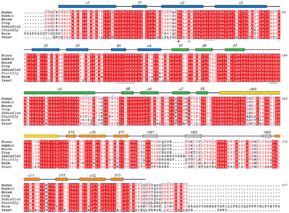
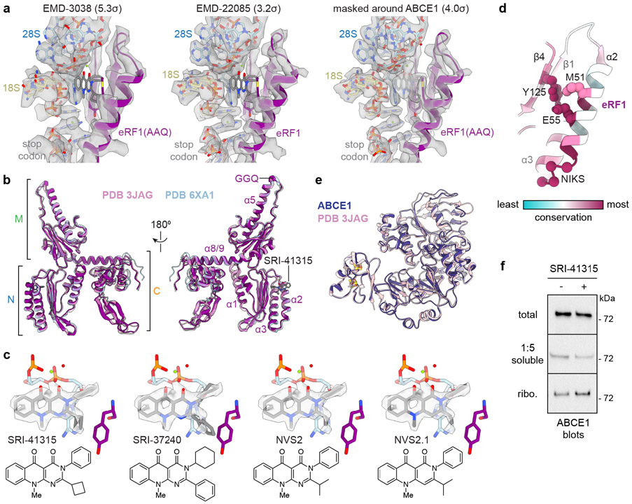
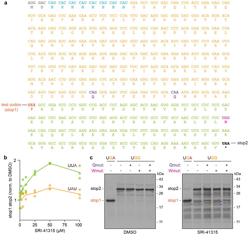
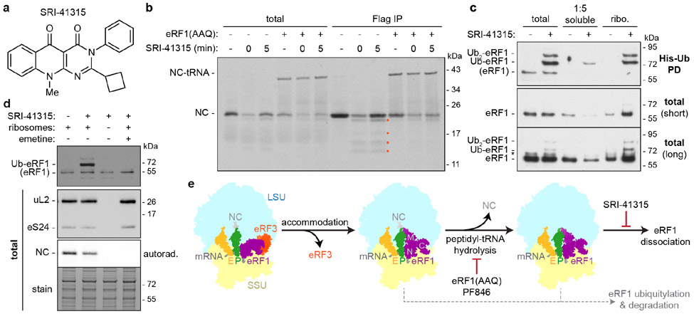
Translation termination is an essential cellular process, which is also of therapeutic interest for diseases that manifest from premature stop codons. In eukaryotes, translation termination requires eRF1, which recognizes stop codons, catalyzes the release of nascent proteins from ribosomes and facilitates ribosome recycling. The small molecule SRI-41315 triggers eRF1 degradation and enhances translational readthrough of premature stop codons. However, the mechanism of action of SRI-41315 on eRF1 and translation is not known. Here we report cryo-EM structures showing that SRI-41315 acts as a metal-dependent molecular glue between the N domain of eRF1 responsible for stop codon recognition and the ribosomal subunit interface near the decoding center. Retention of eRF1 on ribosomes by SRI-41315 leads to ribosome collisions, eRF1 ubiquitylation and a higher frequency of translation termination at near-cognate stop codons. Our findings reveal a new mechanism of release factor inhibition and additional implications for pharmacologically targeting eRF1.


Figures

Extended Data Figure 1 ∣
Extended Data Figure 1 ∣. Characterization of SRI-41315 in an in vitro translation system.
a, Effect of SRI-41315 on protein synthesis. Representative titrations of SRI-41315 into in vitro translation reactions of a radiolabeled 3xFlag-tagged model protein ending with the indicated stop codon or a nonstop (NS) control, analyzed by SDS-PAGE and autoradiography on film (top) or by phosphorimaging (bottom). NC, expected nascent protein chain; NC-tRNA, nonhydrolyzed peptidyl-tRNA adduct. Orange dots, smaller products specifically observed with SRI-41315, representative of 5 replicates with similar results. b, SRI-41315 does not trap nascent proteins on ribosomes. In vitro translation reactions as in panel a without or with 0.5 μM eRF1(AAQ) and/or 100 μM SRI-41315 added after 5 min were size fractionated on 10-50% sucrose gradients. The total (T) and eleven fractions collected from the top of each gradient were analyzed by SDS-PAGE and autoradiography. Note: eRF1(AAQ) but not SRI-41315 retains NC-tRNAs and NCs hydrolyzed from tRNAs during SDS-PAGE in ribosomal fractions, representative of 3 replicates with similar results.
Extended Data Figure 2 ∣
Extended Data Figure 2 ∣. SRI-41315 traps ubiquitylated eRF1 on ribosomes.
a, RNF14 levels in different mammalian lysates. Two-fold dilutions of rabbit reticulocyte lysate (RRL) or Flp-In 293 T-REx cell lysate analyzed by SDS-PAGE and immunoblotting. Note: low levels of RNF14 in RRL relative to GCN1 and ribosomal proteins, representative of 2 replicates with similar results. b, RNF14 mediates eRF1 ubiquitylation with SRI-41315. In vitro translation reactions containing 10 μM His-tagged ubiquitin without or with 50 nM wildtype (WT) or catalytically inactive (C417A) recombinant RNF14 (rRNF14) and 100 μM SRI-41315 were analyzed directly (total) or after denaturing pulldowns of His-tagged ubiquitin (His-Ub PD) by SDS-PAGE and immunoblotting. Note: WT but not C417A rRNF14 enhances SRI-41315-dependent ubiquitylation of eRF1, representative of 3 replicates with similar results. Residual eRF1 ubiquitylation is likely due to endogenous RNF14 in RRL. c, Titration of SRI-41315 into in vitro translation reactions containing 50 nM recombinant RNF14 analyzed directly (total) or after His-Ub PD by SDS-PAGE and immunoblotting, representative of 3 replicates with similar results. d, eRF1 ubiquitylation is slower than translation. Timecourses assaying eRF1 ubiquitylation in the presence of 50 nM recombinant RNF14, 10 μM His-tagged ubiquitin, and 25 μM SRI-41315 (top) compared to timecourses of radiolabeled nascent protein (NC) synthesis (bottom) in in vitro translation reactions, representative of 2 replicates with similar results. e, SRI-41315 traps eRF1 on ribosomes. Translation reactions as in Fig. 1c were size fractionated over 10-50% sucrose gradients. A total (T) sample and eleven fractions collected from the top of each gradient were analyzed directly or after His-Ub PD by SDS-PAGE and immunoblotting for eRF1. Note: SRI-41315 retains both unmodified and ubiquitylated eRF1 in ribosomal fractions, representative of 3 replicates with similar results.
Extended Data Figure 3 ∣
Extended Data Figure 3 ∣. Effects of SRI-41315 in cells.
a,b, Lysates of Flp-In 293 T-REx cells treated without or with 30 μM SRI-41315, 1.8 μM emetine, a concentration of the translation elongation inhibitor demonstrated to cause ribosome collisions, and/or 1 μM MLN-7243, an inhibitor of the E1 ubiquitin-activating enzyme, for 2 hr were analyzed by SDS-PAGE and immunoblotting a, directly or b, after size fractionation on sucrose gradients, representative of 2 replicates with similar results. Note: ubiquitylated eRF1 (Ub-eRF1) is detected specifically with SRI-41315 and suppressed by MLN-7243. SRI-41315 and the low dose of emetine both lead to the recruitment of the ribosome collision sensor EDF1 to ribosomal fractions. c, SRI-41315 induces the recruitment of EDF1 to ribosomes in vitro. SDS-PAGE and immunoblotting for the ribosome collision sensor EDF1 in total, soluble, or ribosomal fractions from translation reactions containing the indicated concentrations of SRI-41315, representative of 2 replicates with similar results. d, SRI-41315 induces eRF1 degradation in Flp-In 293 T-REx cells after 20 hr, representative of 3 replicates with similar results.
Extended Data Figure 4 ∣
Extended Data Figure 4 ∣. Cryo-EM data processing.
a, Ribosome-nascent protein complexes (RNCs) from translation reactions containing 0.5 μM eRF1(AAQ) and 100 μM SRI-41315 added at 5 min were affinity purified via the 3xFlag tag encoded in the nascent chain (NC) and analyzed by SDS-PAGE and Coomassie staining (top) or immunoblotting (bottom). NC-tRNA, nonhydrolyzed peptidyl-tRNA adduct. b, Representative cryo-EM micrograph of RNCs from panel a. c, Summary of cryo-EM data processing and classification strategy.
Extended Data Figure 5 ∣
Extended Data Figure 5 ∣. Quality of cryo-EM maps and model.
a, Fourier shell correlation (FSC) vs. resolution (1/Å) curves for the indicated cryo-EM maps. b, The indicated cryo-EM map colored by local resolution. c, Model vs. map FSC curves. d, Density of the defined nascent protein sequence in the ribosomal exit tunnel in the sharpened cryo-EM map contoured at 2.8σ. e, The cryo-EM map of the SRI-41315 binding site as in Fig. 2b colored by local resolution (left) or by entity (right). Magnesium ions are in green. f, Coordination of the magnesium ion at the SRI-41315 binding site compared to density in the sharpened cryo-EM map contoured at 4.0σ.
Extended Data Figure 6 ∣
Extended Data Figure 6 ∣. eRF1 secondary structure and alignments.
Sequence alignments of eRF1 from the indicated species (H. sapiens UniProt P62495; O. cuniculus UniProt P62497; M. musculus UniProt Q8BWY3; X. laevis UniProt P35615; D. rerio UniProt Q803E5; D. melanogaster UniProt Q9VPH7; C. elegans UniProt O16520; S. cerevisiae UniProt P12385) with secondary structure designations above colored based on their presence in the N (blue), M (green), and C (orange) domains of eRF1. In the accommodated state of eRF1, α8 of the M domain and α9 of the C domain form a continuous helix (yellow). eRF1 also contains a minidomain insertion (gray) in the C domain that is not present in structurally similar decoding factors. In the N domain, Met51 (purple arrowhead) involved in SRI-41315 binding, as well as the NIKS and YxCxxxF motifs (gray lines) and Glu55 (gray arrowhead) involved in stop codon recognition, are indicated below. Note: human, rabbit, and mouse eRF1 are 100% identical. Part of the C-terminal extension in C. elegans eRF1 is not shown.
Extended Data Figure 7 ∣
Extended Data Figure 7 ∣. Structural comparisons.
a, Validation of SRI-41315 density. The model of the SRI-41315 binding site as in Fig. 2b docked into cryo-EM maps of the rabbit ribosome bound to eRF1(AAQ) without SRI-41315 (EMD-3038; left), of the human ribosome bound to eRF1 trapped by PF846 (EMD-22085; middle), or generated from particles selected for occupancy of the recycling factor ABCE1, contoured at the indicated levels. Note: the left and middle maps lack SRI-41315, while the right map retains strong density corresponding to SRI-41315 coexisting with ABCE1 binding. b, The overall conformation of accommodated eRF1 is unchanged with SRI-41315. The model of eRF1(AAQ) bound to SRI-41315 (purple) aligned to eRF1(AAQ) without SRI-41315 (pink; PDB 3JAG) or eRF1 trapped on ribosomes by PF846 (blue; 6XA1). The N, M, and C domains (left), the GGQ motif, and the SRI-41315 binding site (right) are indicated. c, Docking of related small molecule eRF1 degraders in the SRI-41315 binding site. d, SRI-41315 binding region of eRF1 (PDB 1DT9) colored by conservation. Note: Met51 is less conserved than Tyr125 and other residues required for stop codon decoding. e, SRI-41315 does not change ABCE1 conformation. Alignment of ABCE1 on termination complexes without (pink; PDB 3JAG) or with (dark blue) SRI-41315. Iron-sulfur clusters are colored by heteroatoms. f, ABCE1 is slightly stabilized on ribosomes with SRI-41315. Total, soluble, and ribosomal fractions of translation reactions without or with 100 μM SRI-41315 as in Fig. 1c were analyzed by SDS-PAGE and immunoblotting for ABCE1, representative of 3 replicates with similar results.
Extended Data Figure 8 ∣
Extended Data Figure 8 ∣. Flow cytometry analysis.
a, Cells were initially gated by FSC-A vs. SSC-A to exclude debris. b, Cells from the gate in panel a were gated by FSC-A vs. FSC-W to exclude doublets. c, Cells from the gate in panel b were gated by GFP positivity (left) as judged by comparison to an untransduced reference (right) to exclude untransduced cells from analysis.
Extended Data Figure 9 ∣
Extended Data Figure 9 ∣. Cryptic stop codon analysis.
a, Transcript (top line) and polypeptide (bottom line) sequence of the reporter designed to test translation termination at cryptic stop codons. The reporter contains an N-terminal 8xHis tag (blue), a modified calmodulin sequence (light orange), the test codon position (dark orange; stop1), a modified sequence encoding the autonomously folding villin headpiece (VHP) domain followed by the cytosolic domain of Sec61β (light green), and the UAA stop codon (black; stop2). The reporter sequence was modified to remove all codons, except for one UGG codon (pink), that start with a U and contain a purine (A/G) in the second and/or third positions. Additional cryptic codons that start with a C and contain purines in the second and third positions (purple) were tested by mutations (see panel c). b, SRI-41315 induces translation termination at specific cryptic stop codons. Quantification of the ratio of stop1 to stop2 products from reporters containing UUA (green) or UAU (orange) in the test codon position synthesized in vitro with increasing concentrations of SRI-41315. The normalized average (line) and individual ratios (dots) from three independent experiments are shown. c, Mutagenesis of additional cryptic stop codons. Assays as in Fig. 4a with UGA (lane 1) or UGG (lanes 2-5) in the test codon position and additional mutation of CAG and CAA codons (Qmut; purple) upstream of the stop1 position and/or of the UGG codon (Wmut; pink) downstream of the stop1 position as indicated in panel a. Radiolabeled reporter products generated without (left) or with (right) 100 μM SRI-41315 are shown. Note: changes in products upon mutation of these codons occur specifically with SRI-41315, representative of 3 replicates with similar results. Purple dots denote products abolished by mutating the CAG/CAA codons; pink dot denotes the lower band of a doublet abolished by mutating the UGG codon. Yellow dot denotes a band with increased intensity after mutation of the CAG/CAA codons.
Figure 1 ∣
Figure 1 ∣. SRI-41315 inhibits protein synthesis and induces eRF1 ubiquitylation in vitro.
a, Chemical structure of SRI-41315. b, SRI-41315 inhibits protein synthesis. In vitro translation reactions of a radiolabeled 3xFlag-tagged nascent chain (NC) without or with 0.5 μM eRF1(AAQ) and/or 100 μM SRI-41315 added at the beginning (0 min) or 5 min into the reaction were analyzed directly or after denaturing anti-Flag immunoprecipitations (IP) by SDS-PAGE and autoradiography. Orange dots indicate products specifically observed with SRI-41315, representative of 5 replicates with similar results. NC-tRNA, nascent chain-tRNA adduct. c, SRI-41315 traps eRF1 on ribosomes and induces eRF1 ubiquitylation. Total, soluble, and ribosomal fractions of translation reactions containing 50 nM RNF14 and 10 μM His-tagged ubiquitin (His-Ub) without or with100 μM SRI-41315 were analyzed directly or after denaturing His-Ub pulldowns (PD) by SDS-PAGE and immunoblotting for eRF1. Unmodified eRF1 and ubiquitylated eRF1 (Ub-eRF1, Ub2-eRF1) are indicated, representative of 3 replicates with similar results. *, non-specific band. d, eRF1 ubiquitylation requires translation. Translation reactions containing 10 μM His-Ub were performed as usual or with translation mix that had been centrifuged to remove ribosomes (lane 3). 25 μM SRI-41315 or a high dose (100 μM) of the translation elongation inhibitor emetine were included as indicated. His-Ub PD (top) reveals that eRF1 ubiquitylation requires SRI-41315, ribosomes, and active translation (lane 2), representative of 2 replicates with similar results. e, Mechanisms of eRF1 inhibition. Scheme of translation termination showing ribosomal delivery of eRF1 in an inactive conformation by eRF3, followed by accommodation of the eRF1 M domain into the peptidyl transferase center after eRF3 dissociation to catalyze release of the nascent protein chain (NC). PF846 and mutation of the eRF1 GGQ motif to AAQ [eRF1(AAQ)] prevents peptidyl-tRNA hydrolysis. SRI-41315 prevents eRF1 dissociation from ribosomes after this step. The large ribosomal subunit (LSU; light blue), small ribosomal subunit (SSU; light yellow), and P- (green) and E-site (light orange) tRNAs are indicated.
Figure 2 ∣
Figure 2 ∣. Cryo-EM structure of SRI-41315 on a terminating ribosome.
a, Cryo-EM map (left) and structural model (right) of a terminating rabbit ribosome with eRF1(AAQ) (purple) in the accommodated conformation, the recycling factor ABCE1 (dark blue), and SRI-41315. The mRNA (gray) and P- (green) and E-site (orange) tRNAs are indicated. LSU, large ribosomal subunit (light blue); SSU, small ribosomal subunit (yellow). b, Model and map (transparent), contoured at 4.8σ, of the SRI-41315 (dark gray) binding site showing interactions with eRF1(AAQ) (purple), 28S rRNA (light blue), and 18S rRNA (yellow). The second (+2) and third (+3) nucleotides of the UAA stop codon (light gray) are indicated. Magnesium ions are green.
Figure 3.
Figure 3.. Impact of SRI-41315 on eRF1 and the decoding center.
a, Effect of SRI-41315 on eRF1. Structures of ribosomes-bound eRF1(AAQ) (purple) with SRI-41315 (dark gray), eRF1(AAQ) without SRI-41315 (pink; PDB 3JAG), or eRF1 trapped by PF846 (blue; PDB 6XA1), aligned on eRF1. The decoding center containing the UAA stop codon (light gray) and 18S rRNA bases involved in decoding (yellow) is shown. Met51 of eRF1 would clash with the cyclobutyl group of SRI-41315 and must move out of the way (pink arrow) to accommodate and stabilize SRI-41315 binding. Glu55 and Tyr125 involved in stop codon recognition are indicated. b, Effect of SRI-41315 on the ribosome. Alignment of the decoding center and the SRI-41315 binding site with the corresponding region of the human ribosome bound to eRF1 and PF846 (6XA1; transparent model) shows no significant changes in the positions or conformations of rRNA or mRNA. c, Met51 mutations affect the potency of SRI-41315-induced eRF1 degradation. HeLa RNF14 rescue or knockout (KO) cells were transduced with wildtype (WT), M51A, or M51R 3xFlag-eRF1-IRES-GFP constructs. After treatment with the indicated concentrations of SRI-41315 for 4 hours, cells were fixed, permeabilized, stained with APC-conjugated anti-Flag, and analyzed by flow cytometry. The median Flag/GFP ratio for each SRI-41315-treated cell population was normalized to DMSO-treated samples (n=3; two outliers in the RNF14 KO measurements are not shown).
Figure 4 ∣
Figure 4 ∣. SRI-41315 induces translation termination at cryptic stop codons.
a, SRI-41315 enhances translation termination at near-cognate stop codons. Scheme (top) of a reporter encoding an N-terminal 8xHis tag (blue), modified (mod.) calmodulin (light orange), a test codon position (stop1), the autonomously folding villin headpiece (VHP) domain, the cytosolic domain of Sec61β (light green), and the UAA stop codon (stop2). Reporters containing the indicated codon at the stop1 position were synthesized in vitro without or with 25 μM SRI-41315. The radiolabeled protein products were enriched by denaturing His tag pulldowns and analyzed by SDS-PAGE and autoradiography. Products of translation termination at the stop1 (orange dots) or stop2 positions are indicated. b, The ratio of the stop1 product generated with 25 μM SRI-41315 relative to DMSO was quantified for each reporter in three experiments as in (A). Average (lines) and individual replicate values (dots) are shown. R, purine; Y, pyrimidine.

References

    1. Hellen CUT Translation Termination and Ribosome Recycling in Eukaryotes. CSH Perspect Biol 10, a032656 (2018). - PMC - PubMed
    1. Frolova L et al. A highly conserved eukaryotic protein family possessing properties of polypeptide chain release factor. Nature 372, 701–703 (1994). - PubMed
    1. Brown A, Shao S, Murray J, Hegde RS & Ramakrishnan V Structural basis for stop codon recognition in eukaryotes. Nature 524, 493–496 (2015). - PMC - PubMed
    1. Matheisl S, Berninghausen O, Becker T & Beckmann R Structure of a human translation termination complex. Nucleic Acids Res 43, 8615–8626 (2015). - PMC - PubMed
    1. Shao S et al. Decoding Mammalian Ribosome-mRNA States by Translational GTPase Complexes. Cell 167, 1229–1240.e15 (2016). - PMC - PubMed

Methods References

    1. Rohou A & Grigorieff N CTFFIND4: Fast and accurate defocus estimation from electron micrographs. J Struct Biol 192, 216–221 (2015). - PMC - PubMed
    1. Kimanius D, Dong L, Sharov G, Nakane T & Scheres SHW New tools for automated cryo-EM single-particle analysis in RELION-4.0. Biochem J 478, 4169–4185 (2021). - PMC - PubMed
    1. Sanchez-Garcia R et al. DeepEMhancer: a deep learning solution for cryo-EM volume post-processing. Commun Biol 4, 874 (2021). - PMC - PubMed
    1. Chandrasekaran V et al. Mechanism of ribosome stalling during translation of a poly(A) tail. Nat Struct Mol Biol 26, 1132–1140 (2019). - PMC - PubMed
    1. Brown A, Baird MR, Yip MC, Murray J & Shao S Structures of translationally inactive mammalian ribosomes. eLife 7, e40486 (2018). - PMC - PubMed

MeSH terms

LinkOut - more resources